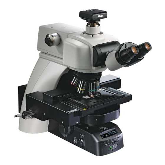
Nikon Eclipse Ni-E Instruction Manual
Upright motorized microscope (focusing nosepiece system)
Hide thumbs
Also See for Eclipse Ni-E:
- Instruction manual (57 pages) ,
- Instruction manual (72 pages) ,
- Instruction manual (186 pages)

Subscribe to Our Youtube Channel
Related Manuals for Nikon Eclipse Ni-E


Summary of Contents for Nikon Eclipse Ni-E
- Page 1 M572 E 11.8.NF.1 (1/2) *M572EN01* Upright Motorized Microscope (Focusing Nosepiece System) Instruction Manual Operation...
-
Page 3: Introduction
● No part of this manual may be reproduced or transmitted in any form without prior written permission from Nikon. ● The contents of this manual are subject to change without notice. -
Page 4: Contents Of The Manual
Introduction Contents of the Manual The instruction manual for ECLIPSE Ni-E (Focusing Nosepiece System) is provided in two volumes. Operation (This manual) Assembly/Maintenance Safety Precautions Assembly Components Troubleshooting Microscopy Operations Maintenance and Storage Before Microscopy Specifications Operation Flowchart Before reading the “Assembly/Maintenance” manual, IR-DIC (Infrared Ray Differential Interference read the “Safety Precautions”... -
Page 5: Summary Of Contents
Summary of Contents Summary of Contents (See the next page for detailed contents.) Safety Precautions Introduction Notes on Handling the Product Contents of the Manual Symbols Used in This Manual Components (System Configuration and Controls) Before Microscopy (Information Setting for Motorized Operation) Operation Flowchart IR-DIC (Infrared Ray Differential Interference Contrast) Microscopy... -
Page 6: Table Of Contents
Contents Contents Introduction ......................... i Contents of the Manual ....................ii Symbols Used in This Manual ..................ii Summary of Contents ....................... iii Safety Precautions......................vi WARNING and CAUTION Symbols ................vi Meaning of Symbols Used on the Product ..............vi WARNING ........................ vii CAUTION ........................ix Notes on Handling the Product.................. - Page 7 Contents Adjusting the Field Diaphragm ................61 Operating the Nosepiece (objective operation)............62 11.1 Objective Vertical Movement Lever (objective switching, dipping) ....62 11.2 Parfocal Correction of Objective ..............63 11.3 Centering the Objective..................64 11.4 Inserting/Removing the DIC Slider..............64 11.5 Single nosepiece ....................
-
Page 8: Safety Precautions
Safety Precautions Safety Precautions To ensure correct and safe operation, read this manual before using this product. WARNING and CAUTION Symbols Although this product is designed and manufactured to be completely safe during use, incorrect usage or failure to follow the safety instructions provided may cause personal injury or property damage. - Page 9 ● To avoid the risk of fire, do not place fabric, must be observed. paper or highly flammable volatile materials Safety is a top design priority for Nikon products. such as gasoline, petroleum benzine, paint Safety is ensured as long as the user observes all...
- Page 10 Safety Precautions WARNING Hazards of mercury lamps Hazardous sample handling (when using the NI-FLEI Epi-fluorescence This product is intended primarily for microscopic attachment) observations and image capture of samples The light source used with the epi-fluorescence (biological tissue, brain slice, etc.) in chambers. attachment (HG Precentered Fiber Illuminator) Check to determine whether a sample is requires special care during handling because of...
- Page 11 If water or lamphouse. Using unspecified products may a chemical solution enters this product, stop using cause malfunctions. the product, and contact your nearest Nikon Lamphouse model representative. ● Specified lamp: 12V 100W halogen lamp PHILIPS 7724 or...
- Page 12 Safety Precautions CAUTION Cautions on assembling and installing the product ● Take care to avoid pinching your fingers or hands during product assembly and installation. ● Scratches or fouling optical components (such as lens and filters) with fingerprints, etc. will degrade microscope images.
-
Page 13: Notes On Handling The Product
Notes on Handling the Product Notes on Handling the Product ● To avoid splashes, do not use this product near Handling the product carefully water. This product is a precision instrument. Avoid ● Make sure the ambient temperature is 0 to subjecting it to sudden impacts and shocks. - Page 14 Unpacking and unlocking 1) Check the package contents. ● ECLIPSE Ni-E x1 ● Hex driver X3 Hex wrench X2 ● Toolbox X1 ● Accessory sticker X2 types...
-
Page 15: Components
Figures are examples of the basic system configuration. Devices other than those shown in the figures are also available. For a list of components used for ECLIPSE Ni-E microscope (focusing nosepiece system), refer to Chapter 1, “2 Components List” in the separately provided “Assembly/Maintenance” instruction manual. -
Page 16: Ni-E Focusing Nosepiece System Basic Configuration
Chapter 1 Components Ni-E Focusing Nosepiece System Basic Configuration Components Motorized quadrocular tilting tube (Model: NI-TT-E) Motorized DSC zooming port for quadrocular tube (Model: NI-RPZ-E) Epi-fluorescence attachment (Model: NI-FLEI) Camera head Contact arm (Model: NIE-CAM) HG adapter (Model: C-HGFIB) HG fiber (Model: C-HGFIF 15/30) Eyepiece Arm spacer 49 mm... -
Page 17: Controls
Chapter 1 Components Controls FL/DIC analyzer slider NI-FA (used for epi-fluorescence/ differential interference Diopter contrast microscopy) adjustment ring Field diaphragm lever Aperture diaphragm lever A. ST OP F. ST OP Aperture diaphragm centering screws Field diaphragm centering screws FN -S2 JAPA Slide click release button Condenser centering... -
Page 18: Operation Buttons
Chapter 1 Components Operation Buttons FUNCTION buttons (Factory settings: Button 1, 2: Condenser CCW, CW rotations Button 3, 4: Excitation filter wheel CCW, CW rotations Button 5, 6: Barrier filter wheel Display panel CCW, CW rotations) brightness control button Z-RESET button DISPLAY Previous button DISPLAY Next button DISPLAY switch button... -
Page 19: Connectors
(Connect to ergo controller (Connect to MICROSCOPE for the joystick controller) motorized MODEL ECLIPSE Ni-E NIKON CORPORATION TOKYO, JAPAN 9 1 0 0 0 1 M A D E I N J A P A N This device complies with Part 15 of the FCC Rules. Operation is subject to the following two conditions:... - Page 20 Chapter 1 Components...
-
Page 21: Microscopy Operations
Chapter Microscopy Operations This chapter describes the following microscopy operation procedures. In the examples, the basic system configurations shown in the previous chapter are used. Operation procedure 1: IR-DIC (Infrared Ray Differential Interference Contrast) microscopy Operation procedure 2: Epi-fluorescence microscopy (including cautions on switching between or in conjunction with IR-DIC microscopy) The procedures described herein assume that all required components are attached to the microscope, with all necessary cables properly connected, and that information registration for motorized operation has been completed. -
Page 22: Before Microscopy
Chapter 2 Microscopy Operations Before Microscopy Information for motorized operation of the microscope has been set upon shipment. If you start using the microscope with the motorized settings of factory default, turn ON the switch of the Control Box A after the assembly of the microscope to use. However, information on optical elements such as optional filter cube are not set a factory shipment. - Page 23 Chapter 2 Microscopy Operations Setting Default setting Where to see in this manual (DS-L3 for details) Configured DS-L3 (See DS-L3 Chapter 2 “3.1 →DS-L3 Chapter 6 “4.1.1 Selecting the Buttons Buttons displayed on the [MICROSCOPE [MICROSCOPE CONTROL] to be Displayed” CONTROL] screen Screen (Ni-E)”.) Configured...
-
Page 24: Operation Flowchart
Chapter 2 Microscopy Operations Operation Flowchart The microscopy operation flow is shown below. See the procedure for each microscopy operation in the next and subsequent sections for details. IR-DIC (Infrared Ray Differential Interference Contrast) Microscopy Procedure IR-DIC (Infrared Ray Differential Interference Contrast) Microscopy Procedure Preparation Preparation Turn on the power. - Page 25 Chapter 2 Microscopy Operations Epi-fluorescence Microscopy Procedure Epi-fluorescence Microscopy Procedure Find the target in the specimen with dia-illumination, Preparation Preparation Turn off the dia-illumination lamp. p.29 Close the shutter and block the illumination path. p.30 Bring the filter cube into the optical path. p.31 p.31 Fully open the field diaphragm and aperture diaphragm of the epi-fluorescence attachment...
-
Page 26: Ir-Dic (Infrared Ray Differential Interference Contrast) Microscopy Procedure
Chapter 2 Microscopy Operations IR-DIC Microscopy Preparation Focus and Orientation Microscopy Procedure 9 10 11 12 13 14 15 16 17 18 19 20 21 22 23 ■ IR-DIC (Infrared Ray Differential Interference Contrast) Microscopy Procedure This section describes the IR-DIC microscopy procedure which is performed using the microscopes controls and buttons. The microscope can also be controlled from DS-L3 if a DS-L3 DS Camera Control Unit is connected to the microscope. -
Page 27: Turn On The Dia-Illumination Lamp
Chapter 2 Microscopy Operations IR-DIC Microscopy Preparation Focus and Orientation Microscopy Procedure 9 10 11 12 13 14 15 16 17 18 19 20 21 22 23 ■ Display at power on N i - E V x . x x _ x x x x . x x x x . F 1 When the power is turned on, operation progress D a t a L o a d i n g . -
Page 28: Lower The Condenser Slightly From The Uppermost Position
Chapter 2 Microscopy Operations IR-DIC Microscopy Preparation Focus and Orientation Microscopy Procedure 9 10 11 12 13 14 15 16 17 18 19 20 21 22 23 ■ Adjust optical path in tube to direct 100% of light to binocular. Press the BINO optical path button on the front of the main body. -
Page 29: Fully Open The Field Diaphragm And Aperture Diaphragm
Chapter 2 Microscopy Operations IR-DIC Microscopy Preparation Focus and Orientation Microscopy Procedure 9 10 11 12 13 14 15 16 17 18 19 20 21 22 23 ■ Fully open the field diaphragm and aperture diaphragm. Press the left side of the DIA field diaphragm button ( mark side) and fully open the field diaphragm. - Page 30 Chapter 2 Microscopy Operations IR-DIC Microscopy Preparation Focus and Orientation Microscopy Procedure 9 10 11 12 13 14 15 16 17 18 19 20 21 22 23 ■ Move the epi-fluorescence cube turret to empty position, and remove the DC slider (objective side) and polarizer from the optical path. Press the FL CUBE CW/CCW button and bring the epi-fluorescence cube turret’s [OPEN] (empty position) into the optical path.
-
Page 31: Focus And Orientation
Chapter 2 Microscopy Operations IR-DIC Microscopy Preparation Focus and Orientation Microscopy Procedure 9 10 11 12 13 14 15 16 17 18 19 20 21 22 23 ■ Focus and orientation Bring the 4x or 10x objective into the optical path. Move the objective vertical movement lever to the center. -
Page 32: Focus On The Sample
Chapter 2 Microscopy Operations IR-DIC Microscopy Preparation Focus and Orientation Microscopy Procedure 9 10 11 12 13 14 15 16 17 18 19 20 21 22 23 ■ Focus on the sample. Look into the eyepiece, and adjust the brightness appropriately by turning the dia-illumination brightness control knob. -
Page 33: Adjust The Interpupillary Distance
Chapter 2 Microscopy Operations IR-DIC Microscopy Preparation Focus and Orientation Microscopy Procedure 9 10 11 12 13 14 15 16 17 18 19 20 21 22 23 ■ Perform diopter adjustment. Look into the right eyepiece with your right eye and the left eyepiece with your left eye. -
Page 34: Focus And Center The Condenser
Chapter 2 Microscopy Operations IR-DIC Microscopy Preparation Focus and Orientation Microscopy Procedure 9 10 11 12 13 14 15 16 17 18 19 20 21 22 23 ■ Focus and center the condenser. Press the DIA field diaphragm button to close the field diaphragm to a minimum and look into the eyepiece. -
Page 35: Adjust The Aperture Diaphragm
70 to 80% of the numerical aperture of the objective. Since an excessively small Adjusting aperture diaphragm aperture diaphragm opening will degrade the image resolution, Nikon does not recommend Aperture setting the aperture diaphragm to less than 60% diaphragm image of the numerical aperture of the objective. -
Page 36: Adjust The Field Diaphragm
Chapter 2 Microscopy Operations IR-DIC Microscopy Preparation Focus and Orientation Microscopy Procedure 9 10 11 12 13 14 15 16 17 18 19 20 21 22 23 ■ Adjust the field diaphragm. Press the DIA field diaphragm button to adjust the field diaphragm so that it circumscribes the field of view. - Page 37 Chapter 2 Microscopy Operations IR-DIC Microscopy Preparation Focus and Orientation Microscopy Procedure 9 10 11 12 13 14 15 16 17 18 19 20 21 22 23 ■ Adjust the brightness. Adjust the brightness of the image using the ND filter. Press the ND filter IN/OUT switch to the [IN] (lower) side to bring the filter into the optical path.
-
Page 38: Adjust The Orientation (Vibration Direction) Of The Polarizer And Analyzer
Chapter 2 Microscopy Operations IR-DIC Microscopy Preparation Focus and Orientation Microscopy Procedure 9 10 11 12 13 14 15 16 17 18 19 20 21 22 23 ■ Adjust the orientation (vibration direction) of the polarizer and analyzer. This adjustment is performed to determine the basic performance of differential interference contrast method. - Page 39 Chapter 2 Microscopy Operations IR-DIC Microscopy Preparation Focus and Orientation Microscopy Procedure 9 10 11 12 13 14 15 16 17 18 19 20 21 22 23 ■ Viewing the sample Attach the DIC slider (objective side) to the slider nosepiece. Turn the objective vertical movement lever counterclockwise to lower the objective.
-
Page 40: Observe A Sample Using A Microscope
Chapter 2 Microscopy Operations IR-DIC Microscopy Preparation Focus and Orientation Microscopy Procedure 9 10 11 12 13 14 15 16 17 18 19 20 21 22 23 ■ Observe a sample using a microscope. • Use ND filters to adjust the image brightness. •... - Page 41 Chapter 2 Microscopy Operations IR-DIC Microscopy Preparation Focus and Orientation Microscopy Procedure 9 10 11 12 13 14 15 16 17 18 19 20 21 22 23 ■ Turn off the power. Turn off the power switches (press to the “O” position) on the control box A and connected motorized devices.
-
Page 42: Diagonal Illumination
Chapter 2 Microscopy Operations IR-DIC Microscopy Preparation Focus and Orientation Microscopy Procedure 9 10 11 12 13 14 15 16 17 18 19 20 21 22 23 ■ Diagonal illumination Using diagonal illumination, contrast can be applied to almost transparent samples thus making it possible to observe them. -
Page 43: Epi-Fluorescence Microscopy Procedure
Chapter 2 Microscopy Operations Preparation Focusing Microscopy Epi-fluorescence Microscopy Procedure 9 10 11 12 13 14 ■ ■ Epi-fluorescence Microscopy Procedure This section describes the epi-fluorescence microscopy procedure which is performed using the microscope’s controls and buttons. Precautions on switching between epi-fluorescence microscopy IR-DIC microscopy are also described. Find the target in the sample with dia-illumination as described in “3 IR-DIC (Infrared Ray Differential Interference Contrast) Microscopy Procedure”... -
Page 44: Close The Shutter And Block The Illumination Path
Chapter 2 Microscopy Operations Preparation Focusing Microscopy Epi-fluorescence Microscopy Procedure 9 10 11 12 13 14 ■ ■ Close the shutter and block the illumination path. Press the [FL SHUTTER] button and close the shutter in the motorized fluorescence cube turret. The shutter is closed when the shutter button is lit. -
Page 45: Bring The Filter Cube Into The Optical Path
Chapter 2 Microscopy Operations Preparation Focusing Microscopy Epi-fluorescence Microscopy Procedure 9 10 11 12 13 14 ■ ■ Bring the filter cube into the optical path. Press the FL CUBE CW/CCW button to bring the desired filter cube into the optical path. Pressing the FL CUBE CW button turns the turret by one address in the clockwise direction (as viewed from above), while the CCW button turns the turret in the... -
Page 46: Turn On The Mercury Lamp
Chapter 2 Microscopy Operations Preparation Focusing Microscopy Epi-fluorescence Microscopy Procedure 9 10 11 12 13 14 ■ ■ Turn on the mercury lamp. See your illuminator’s manual for details. IN TE NS ILI GH T C- H G FI E Power indicator (green) ON LA MP... -
Page 47: Open The Shutter
Chapter 2 Microscopy Operations Preparation Focusing Microscopy Epi-fluorescence Microscopy Procedure 9 10 11 12 13 14 ■ ■ Open the shutter. Press the [FL SHUTTER] button and open the shutter in the motorized epi-fluorescence cube turret which is closed in procedure 2. When the shutter is opened, the button illumination will turn off. - Page 48 Chapter 2 Microscopy Operations Preparation Focusing Microscopy Epi-fluorescence Microscopy Procedure 9 10 11 12 13 14 ■ ■ Focusing and Optical System Adjustment Focus on the sample. Focus on the sample by rotating the focus knob. (See Chapter 3, “4.1 Proper Focusing Procedure”.) Z-RESET The Z-axis coordinate value can be reset to zero.
- Page 49 Chapter 2 Microscopy Operations Preparation Focusing Microscopy Epi-fluorescence Microscopy Procedure 9 10 11 12 13 14 ■ ■ Adjust the field diaphragm of the epi-fluorescence attachment. Use the field diaphragm lever of the epi-fluorescence attachment to adjust the field diaphragm so that it circumscribes the field of view.
-
Page 50: Turn Off The Mercury Lamp
For fluorescence observations, press the microscope’s dia-illumination ON/OFF switch to “OFF” to make the diascopic image disappear. Bright ambient lights will make it difficult to view the image. Nikon recommends keeping the room dark during fluorescence observations. FL/DIC analyzer slider NI-FA/dummy slider... - Page 51 Chapter 2 Microscopy Operations Preparation Focusing Microscopy Epi-fluorescence Microscopy Procedure 9 10 11 12 13 14 ■ ■ Turn off the power. Turn off the power switches (press to the “O” position) on the control box A and connected motorized devices. (Each Power LED OFF power LED turns off.) Power switch...
- Page 52 Chapter 2 Microscopy Operations Preparation Focusing Microscopy Epi-fluorescence Microscopy Procedure 9 10 11 12 13 14 ■ ■ Switching from IR-DIC (infrared ray differential interference contrast) microscopy to epi-fluorescence microscopy Epi-fluorescence and differential interference contrast methods can be used concurrently to cover the shortcomings of each.
- Page 53 Chapter 2 Microscopy Operations Preparation Focusing Microscopy Epi-fluorescence Microscopy Procedure 9 10 11 12 13 14 ■ ■ • Fully open the field diaphragm and aperture diaphragm of the epi-fluorescence attachment. A. ST OP F. ST OP Turn on the power of the HG precentered fiber illuminator and continue from procedure 6 of the Aperture Field diaphragm...
- Page 54 Chapter 2 Microscopy Operations...
-
Page 55: Individual Operations
Chapter Individual Operations This chapter provides detailed descriptions on how to use the Ni-E microscope display panel, control buttons, each component and each device mainly used with the Ni-E focusing nosepiece system. -
Page 56: Display Panel Details
Chapter 3 Individual Operations Display Panel Details The 2-line, 24-character fluorescent display panel on the front of the main body can display the following information: Power ON display When the power is turned on, operation progress is N i - E V x . - Page 57 Chapter 3 Individual Operations Microscope state display Pattern 1 Objective, Z-axis position (unit: um) _ _ _ _ _ _ _ _ _ 0 . 0 0 0 u m Bottom Dia-illumination status, field diaphragm status (unit: D I A : O N _ _ _ _ _ _ _ _ _ _ F S 3 0 .
- Page 58 Chapter 3 Individual Operations Contents of the “microscope state” display are as follows. Microscope State Display Contents Objective ]: Blank: Achromat 0 0 0 x ]: Apo TIRF A T 0 0 0 x ]: Plan 0 0 0 x ]:...
-
Page 59: Using Operation Buttons On Ni-E
Chapter 3 Individual Operations Using Operation Buttons on Ni-E Various operation buttons are provided on the front, right, and left of the Ni-E main body. The following describes the function of each button (factory default setting): Front Operation Button Front Operation Button Name Function Display panel... -
Page 60: Right Operation Button
Chapter 3 Individual Operations Right Operation Button Right Operation Button Name Function FL CUBE CW button Rotate the 1st layer motorized epi-fluorescence cube turret by one address clockwise (as viewed from above). FL CUBE CCW button Rotate the 1st motorized epi-fluorescence cube turret by one address counterclockwise (as viewed from above). -
Page 61: Left Operation Button
Chapter 3 Individual Operations Left Operation Button Left Operation Button Name Function OBJ CW button Rotate the motorized nosepiece by one address clockwise (as viewed from above). (The motorized nosepiece is not used in the focusing nosepiece. Change the function of the button to a different operation.) OBJ CCW button Rotate the motorized nosepiece by one address counterclockwise (as viewed from above). -
Page 62: Adjusting The Brightness Of A Diascopic Image
Chapter 3 Individual Operations Adjusting the Brightness of a Diascopic Image The brightness of a diascopic image can be adjusted by changing the lamp voltage or by using ND filters. Adjustment by Lamp Voltage Turning the dia-illumination brightness control knob changes the voltage of the lamp to change the brightness of the diascopic image. -
Page 63: Adjustment With Nd Filters
Chapter 3 Individual Operations Adjustment with ND Filters There are two methods for adjusting the brightness with ND filters: bringing/removing the main body’s internal ND filters in/from the optical path, or operating the motorized ND filter wheel attached between the main body and dia-illumination. Using ND filters in the main body ND filters are used to adjust light intensity. -
Page 64: Focusing On The Sample (Vertical Objective Movement)
Chapter 3 Individual Operations Focusing on the Sample (Vertical Objective Movement) Focus adjustment is performed using the focus knobs on the Ni-E main body, ergo controller, or focus knob on joystick. The following instructions assume the use of the focus knobs on the main body. See the Ni-E (Focusing Stage System) instruction manual for information on using the ergo or joystick controller. -
Page 65: Focus Knobs On The Main Body
Chapter 3 Individual Operations Enabling/disabling the operation of the elevating section You can disable the operation of the elevating section. As factory default setting, a focus knob is used to drive the elevating section, but you can “disable” the setting when an application software such as NIS-E is used to control the elevating section or you wan to prevent unintended behavior of the elevating section, including an accidental contact of the focus knob of Ni-E microscope or ergo controller (or joystick). -
Page 66: Resetting The Z-Axis Coordinate
Chapter 3 Individual Operations Position exchange of the fine focus knob The fine focus knobs, located on both sides of the main body, are flat on one end, and convex on the other. The fine focus knob on each side is attached with a magnet, and can easily be swapped. -
Page 67: Refocusing
Chapter 3 Individual Operations Refocusing The objective can be raised to a preset retracting position, and then restored back to the original by refocusing operation. When the Escape button is pressed, the objective is raised to the preset retracting position while saving the current position. Pressing the Escape button once more returns the objective to the original position. -
Page 68: Bringing The Target Into The Optical Path (Horizontal Stage Movement)
Chapter 3 Individual Operations Bringing the Target into the Optical Path (Horizontal Stage Movement) When using the standard stage FN-3PS2, bring the target into optical path using the stage X, Y knobs. (So that the sample is illuminated.) When using a motorized XY stage, use the ergo controller or joystick to control the horizontal stage movement. Standard Stage Operation Notes on moving the stage Avoid the following actions, which can cause equipment... -
Page 69: Operating The Motorized Xy Stage
Chapter 3 Individual Operations Operating the Motorized XY Stage In order to use the motorized XY stage, the motorized XY stage Movable claw must be connected to the motorized XY stage controller and the Motorized XY motorized XY stage controller must be connected to the stage connector box. -
Page 70: Adjusting The Diopter
Chapter 3 Individual Operations Adjusting the Diopter The diopter adjustment ring on an eyepiece can be adjusted to match the eyesight of your right and left eyes. A properly adjusted diopter compensates for differences in visual acuity between the right and left eyes of a person, making binocular observation easier. -
Page 71: Focusing And Centering The Condenser
Chapter 3 Individual Operations Focusing and Centering the Condenser Adjust the condenser position so that the light passing through the condenser forms an image at the correct position (center of the optical path) on the surface of the sample. Follow Steps 1 through 12 in Chapter 2, “3 IR-DIC (Infrared Ray Differential Interference Contrast) Microscopy Procedure”... -
Page 72: Adjusting The Aperture Diaphragm
70 to 80% of the numerical aperture of the objective. It provides satisfactory images with suitable contrast. Since an excessively small aperture diaphragm opening will degrade image resolution, Nikon does not recommend setting the aperture diaphragm to less than 60% of the Right size of the aperture diaphragm numerical aperture of the objective. -
Page 73: Using The Condenser
Chapter 3 Individual Operations Using the Condenser The focusing nosepiece system uses the FN-C LWD condenser appropriate for dia-illumination bright-field microscopy (objective 4x to 100x), dia-illumination differential interference contrast microscopy (infrared ray range, visible range) and diagonal illumination microscopy. ■ Optical module The turret of the LWD condenser has one empty hole and three holes for attaching optical modules. - Page 74 Chapter 3 Individual Operations ■ Aperture diaphragm A condenser is equipped with an aperture diaphragm to adjust the illumination angular aperture. Turning the aperture diaphragm lever changes the size of the aperture diaphragm. Turn the diaphragm clockwise to open and counterclockwise to close. A scale indicating the numerical aperture of the aperture diaphragm of the condenser is displayed at the top of the aperture diaphragm lever.
-
Page 75: Adjusting The Field Diaphragm
Chapter 3 Individual Operations Adjusting the Field Diaphragm The field diaphragm is used to restrict illumination to the area of the sample being viewed. Operate the DIA field diaphragm button to change the size of the field diaphragm. Press the left side ( mark) of the DIA field diaphragm button to open, and the right side ( mark) to close the diaphragm. -
Page 76: Operating The Nosepiece (Objective Operation)
Chapter 3 Individual Operations Operating the Nosepiece (objective operation) Sliding nosepiece A slider nosepiece that can mount two objectives or a single nosepiece that can mount only single objective can be used with Slide click the Ni-E focusing nosepiece system. release button To use the sliding nosepiece, attach the objectives to the front Objective... -
Page 77: Parfocal Correction Of Objective
Chapter 3 Individual Operations Move the vertical movement lever at the left end to the Slide click release button center to raise the front objective. With the front objective raised, hold the grip while pressing Focusing nosepiece FN -S2 JAPA the slide click release button and pull the sliding nosepiece movement to the front. -
Page 78: Centering The Objective
Chapter 3 Individual Operations Centering the Objective 11.3 With the sliding nosepiece, you can center the objective to Centering screws minimize the misalignment of the center of the field of view on the front and rear objectives. Adjust the three centering screws in such a way that the center of the field of view of the rear objective and the center of the field of view of the front objective match. -
Page 79: Using The Polarizer Turret
Chapter 3 Individual Operations Using the Polarizer Turret The polarizer turret is attached to the field lens and used for A . S differential interference contrast microscopy. The turret has four holes with the FN-P polarizer for visible range (λ=430 nm to 770 nm) and the FN-IRP polarizer for infrared ray (λ=850 nm to 950 nm) attached. -
Page 80: Adjusting The Binocular Section
When observing a position slightly deeper than the surface (about 0.2 mm, depending on the state of the specimen) of a specimen such as a brain slice, Nikon recommends using infrared ray microscopy or a water immersion objective with a correction ring (a Plan100xW). -
Page 81: Differential Interference Contrast Microscopy
Chapter 3 Individual Operations Differential Interference Contrast Microscopy An analyzer, a polarizer, an object lens side DIC slider, and a condenser side DIC module are required for differential interference contrast microscopy, which observes clear colorless specimen or undyed specimen at high contrast. An Ni-E focusing nosepiece system can be used for differential interference contrast microscopy in the infrared region (λ=850 nm to 950 nm) and in the visible light region (λ=430 nm to 770 nm). -
Page 82: Using Optical Elements
Chapter 3 Individual Operations Using Optical Elements 16.2 ■ DIC module for the condenser There are three types of DIC modules to be attached to the LWD condenser: [D-C DIC N1 DRY], [D-C DIC N2 DRY] and [D-C DIC NR DRY]. Note that an inappropriate combination may result in an inability to produce an interference contrast image, or a significant degradation of contrast. -
Page 83: Epi-Fluorescence Microscopy
Chapter 3 Individual Operations Epi-fluorescence Microscopy An epi-fluorescence microscopy used for the observation of a fluorescence image requires a brighter illuminator including such an optical element as a filter or mercury lamp. Ni-E uses an epi-fluorescence attachment with a fluorescence cube turret to which a filter cube is attached (motorized, intelligent, or manual), HG precentered fiber illuminator using a mercury lamp as an illuminator (motorized or manual), ND filters, an aperture diaphragm and a field diaphragm. - Page 84 Chapter 3 Individual Operations Using double layered epi-fluorescence cube turrets Two epi-fluorescence cube turrets can be used by layering them on top of each other. Attach an attachment using a laser as an illuminator on the 1st layer, which should be used for light stimulation when using a double-decked turret.
-
Page 85: Selecting Filters
Chapter 3 Individual Operations Selecting Filters 17.2 A filter cube consists of three types of optical components: an Barrier filter U V - 2 A excitation filter (EX filter), a barrier filter (BA filter), and a dichroic E X 33 0- 38 0 D M 40 B A 42... - Page 86 – for example, TRITC in the case of FITC and TRITC. This will result in very dark TRITC fluorescent images. For such cases, Nikon recommends using Wavelength multiband filters. Long-pass filter (Both FITC and TRITC images are visible.)
-
Page 87: Protecting The Sample And Preventing It From Decoloration (Using The Shutter)
Chapter 3 Individual Operations Protecting the Sample and Preventing It from Decoloration (Using the Shutter) 17.3 If the sample is continuously exposed to the strong light of the mercury lamp used for the epi-fluorescence attachment, it may become damaged or decolorized. Be sure to close the shutter when suspending microscopy or when pausing epi-fluorescence microscopy to perform microscopy with diascopic light. - Page 88 Chapter 3 Individual Operations Motorized Shutter Connector There are two connectors for connecting the motorized shutter cable: “EPI SHUTTER” on the rear of the main body and “DIA SHUTTER” on the connector box. Connect the EPI motorized shutter on the epi-fluorescence side to the “EPI SHUTTER”...
-
Page 89: Adjusting The Brightness Of The Fluorescent Image (Using Nd Filters And The Aperture Diaphragm)
Chapter 3 Individual Operations Adjusting the Brightness of the Fluorescent Image (Using ND Filters and the Aperture 17.4 Diaphragm) The brightness of the epi-fluorescence image can be adjusted by using the ND filters in the epi-fluorescence attachment and the motorized HG precentered fiber illuminator. ND filters are used to adjust light intensity. The higher filter number, the lower transmittance, and darker images are produced when placed in the optical path. -
Page 90: Restricting The Illumination To The Area Of The Sample Being Viewed (Centering And Adjusting The Field Diaphragm)
Chapter 3 Individual Operations Aperture diaphragm The aperture diaphragm is used to adjust the numerical aperture of the illumination system. Under epi-fluorescence microscopy, it is also used to adjust the brightness of the image, as well as the size of stray light. Diaphragm A . -
Page 91: Changing The Waveform Characteristics Of The Excitation Light (D-Fb Excitation Balancer)
Chapter 3 Individual Operations Changing the Waveform Characteristics of the Excitation Light (D-FB Excitation Balancer) 17.6 By attaching the D-FB Excitation Balancer to the epi-fluorescence attachment, the waveform characteristics of the excitation light can be changed arbitrarily. The balancer is to be used together with a dual-band filter cube. -
Page 92: Other Notes On Epi-Fluorescence Microscopy
Chapter 3 Individual Operations ■ Objectives The excitation balancer should be used in combination with the following objectives. Use of other objectives may result in unevenness in the field of view. Plan fluor 40x/0.75 40xH/1.3 100xH/1.3 S fluor 40x/0.9 40xH/1.3 100xH/1.3 Plan apochromat 40x/0.95... -
Page 93: Useful Functions
Chapter 3 Individual Operations Useful Functions Mode Function 18.1 “MODE” saves and loads a “microscopy state” (position and status of motorized accessories) and is useful for an observation when switching between different microscopy state. To use this function, you must have registered a set of motorized units for loading in each mode. More than one motorized unit can be registered for one mode with up to 8 modes available for setting. - Page 94 Chapter 3 Individual Operations Now MODE1 has been registered and the registered mode name is displayed on the [MODE1] button. [SAVE MODE1] button is shown on the right of the mode name button. Repeat the same procedure for MODE2 registration. (4) MODE1 and MODE2 have been registered Save the mode.
- Page 95 Chapter 3 Individual Operations Load the mode. Press [FUNCTION1] and [FUNCTION2] buttons alternately. This assumes that a load function for MODE 1 and MODE 2 is allocated to FUNCTION 1 button and FUNCTION 2 button respectively. The MODE load function is not set to a FUNCTION button in Ni-E Loading a mode as default.
-
Page 96: Interlocking Function
Chapter 3 Individual Operations Motorized Unit that Save/Loads the State with MODE Function Setting items Stored Information CONDEN. Motorized universal condenser’s condenser module currently in the optical path (not supported for focusing nosepiece) (The motorized condenser is not used in the focusing nosepiece system.) FL TURRET Filter cube of the motorized epi-fluorescence cube turret in the optical path FL [SHUTTER]... -
Page 97: Capturing Images
Chapter 3 Individual Operations Capturing Images A microscope image can be captured with a DS camera head DS camera attached to a tube or zooming port. head Use DS-L3 or DS-U3 DS Camera Control Unit. DS-L3 is connected to the USB connector, DSC1 connector at A. - Page 98 Adjust the camera’s white balance. To adjust the white balance, press the WB button while capturing an image of a clear section of the specimen slide. (For fluorescent photomicrography, Nikon recommends adjusting the white balance under normal bright-field microscopy conditions before capturing...
- Page 99 Chapter 3 Individual Operations NCB filter The Ni-E’s filter cassette is equipped with an NCB filter for color temperature conversion. Align the specimen. Readjust the focus onto the target. Adjust the image brightness using the camera exposure compensation function. Check the image using the DS-L3 [FREEZE] button. (10) If the image is acceptable, press the CAPTURE button to save the image.
- Page 100 Even if the overall magnification is the same on the monitor, the combination of objective and camera zoom can result in significant variations in exposure time. Nikon recommends increasing the magnification of the objective rather than of the zoom. (Generally, the numerical aperture of the objective increases with magnification. The larger the numerical aperture, the brighter the resulting image.)
-
Page 101: Operation On Ds-L3
Chapter 3 Individual Operations Operation on DS-L3 Setting Up the Microscope 20.1 If you use the microscope as it is set up as default, a specific setup is not required, however, if you want to change the setting to meet your needs, you want to use useful functions, or you want to set the information on optical elements, use DS-L3 for the operation below. - Page 102 Chapter 3 Individual Operations Shown below is the actual settings. See (1) through (7). Saving the setting After completing the setting, be sure to press the [SAVE] button on the [MICROSCOPE SETUP MENU] screen to save the settings. If the DS-L3 is turned off without saving settings, previous values are restored.
- Page 103 Chapter 3 Individual Operations (2) Configuring the Device Connection Information (See DS-L3 Chapter 6 “3 Setting the Connections of Motorized Units”.) (2-1) Connecting the digital camera (See DS-L3 Chapter 6 “3.1 Configuring the Connection of Digital Camera”.) Perform this configuration when a camera control unit is connected to the main body using a trigger cable to capture an image by sending trigger signal from the DSC connector on the main body.
- Page 104 Chapter 3 Individual Operations (3) Configuring the Button Functions (See DS-L3 Chapter 6 “4 Configuring the Functions of Buttons”.) [SETUP MENU] → [BUTTON FUNC] Change the function of buttons equipped with the Ni-E microscope and the ergo controller. The function of buttons located on the DS-L3 screen can be selected as desired here.
- Page 105 Chapter 3 Individual Operations Units Supported for Buttons Operation Button Target Unit [INTSL] Shutter built in the HG precentered fiber illuminator [FL TURRET] Shutter built in the epi-fluorescence cube turret on the 1st layer (default setting) [FL 2nd] Shutter built in the epi-fluorescence cube turret on the 2nd layer FL SHUTTER Button [EPI/DIA/AUX] Motorized shutter connected to the EPI SHUTTER connector at the position set in “(2)
- Page 106 Chapter 3 Individual Operations ■ Enabling operation button →(See DS-L3 Chapter 6 “4.2.5 Enabling/Disabling the Button Operation”.) Enable/disable the buttons located on the front and lateral right/left on the base of the Ni-E microscope. The buttons can only be configured as a group (front, lateral left, lateral right) not individually. <Factory setting>...
- Page 107 Chapter 3 Individual Operations ■ SLEEP button (See DS-L3 Chapter 6 “4.1.2 Showing/Hiding the [SLEEP] Button”.) Set to show or hide the [SLEEP] button on the [MICROSCOPE CONTROL] screen to turn ON/OFF the sleep mode on the DS-L3. The sleep state is the state where the power supply to motorized devices is stopped to minimize the generation of noise.
- Page 108 Chapter 3 Individual Operations (4) Configuring the Movement (See DS-L3 Chapter 6 “5 Configuring the Movement of Motorized Devices”.) Set the movement of the Ni-E microscope and motorized accessories. (4-1) Configuring Interlocking (See DS-L3 Chapter 6 “5.1 Configuring Interlocked Operation”.) See Chapter 3 “18.2 Interlocking Function”...
- Page 109 Chapter 3 Individual Operations (4-2) Escape distance (See DS-L3 Chapter 6 “5.2 Setting the Retracting Amount of the Elevating Section”.) Set the distance the objective rises from the current position when the microscope’s [ESCAPE] button is pressed. <Factory setting> “SOFTWARE LIMIT” For software limit, see “(6) Configuring Other Functions - (6-4) Software Limit”...
- Page 110 Chapter 3 Individual Operations (5) Setting Mode Function (See DS-L3 Chapter 5 “2 Using the MODE Function”.) In order to use the MODE function, “register” the motorized devices for the modes and “save” the status of the registered motorized device. “Load” the saved mode on the [MICROSCOPE CONTROL] screen. See Chapter 3 “18.1 Mode Function”...
- Page 111 Chapter 3 Individual Operations (6) Configuring Other Functions (See DS-L3 Chapter 6 “6 Configuring Other Functions”.) (6-1) Ni-E display panel (See DS-L3 Chapter 6 “6.1 Setting the Display of the Ni-E Front Display Panel”.) Set a display pattern to be displayed on the display panel of Ni-E microscope at the system startup. The display pattern depends on the type of the motorized unit connected and is inclusive of 9 patterns.
- Page 112 Chapter 3 Individual Operations (6-4) Software limit (See DS-L3 Chapter 6 “6.4 Setting the Software Limits”.) Programmatically set the limit position of the microscope’s elevating section and the motorized XY stage movement. In order to set the limit in XY direction, the motorized XY stage must be connected. Setting items Default setting Configurable range...
- Page 113 Chapter 3 Individual Operations (7) Maintenance (7-1) FL turret drive speed (See DS-L3 Chapter 6 “6.5 Setting the Driving Speed of the Epi-Fluorescence Cube Turret”.) Set the drive speed of the epi-fluorescence cube turret to High or Low. When a thick dichroic mirror is used for a filter cube, set the drive speed to “Low”. If you are using two layered epi-fluorescence cube turrets, configure 1st and 2nd layers separately.
-
Page 114: Microscope Control
(1) What in Ni-E can be controlled by DS-L3 Required setting Device Operation available on DS-L3 (Reference in DS-L3 Instruction Manual) ECLIPSE Ni-E main body ON/OFF and lamp brightness control of None dia-illumination Adjustment of DIA field diaphragm Retraction and restoration... - Page 115 Chapter 3 Individual Operations (2) Buttons on the [MICROSCOPE CONTROL] screen In order to control the microscope from DS-L3, press the [MIC] or [CAM-MIC] button on the [MICROSCOPE CONTROL] top screen to display the [MICROSCOPE CONTROL] or [CAM-MIC CONTROL] screen. The same operation is available from either screen, but the [MICROSCOPE CONTROL] screen is used as an example to summarize the operation in this document.
- Page 116 Chapter 3 Individual Operations (3) Controlling Ni-E with buttons Pressing a button allows you to control the following parts of Ni-E or motorized accessories: See the list for the function and descriptions on the buttons. A. ST OP F. ST OP JAPA FN- S2N IN TE NS...
- Page 117 Chapter 3 Individual Operations Reference for Buttons with their Functions and Operation Procedures Operation Operation Button Function Reference in DS-L3 Instruction Manual Chapter 5, “1.1 Switching Switch the objective. the Objective (Motorized Nosepiece)” [NOSEPIECE] [Objective (Address)] Switch the optical path of the motorized quadrocular tilting tube to the binocular section.
- Page 118 Chapter 3 Individual Operations Operation Operation Button Function Reference in DS-L3 Instruction Manual Chapter 5 “1.10 Adjusting Adjust the aperture diaphragm the DIA Aperture Diaphragm diameter of a motorized universal (Motorized Universal condenser. Condenser)” [A. STOP] Switch the ND of the HG precentered fiber illuminator.
- Page 119 Chapter 3 Individual Operations Operation Operation Button Function Reference in DS-L3 Instruction Manual Open/close the built-in shutter in Chapter 5 “1.12.1 the HG precentered fiber Opening/Closing the illuminator. (Equivalent to the Motorized HG Precentered [SHUTTER INTSL] button on the Fiber Illuminator’s Built-in [SHUTTER INTSL] [INTSL] sub screen.) Shutter”...
- Page 120 Chapter 3 Individual Operations Operation Operation Button Function Reference in DS-L3 Instruction Manual Chapter 5, “3 Entering the Enter the sleep state to reduce Sleep State (Noise noise. Reduction)” [SLEEP] Chapter 5 “4 Changing the Change the background color for Background Color of the [MICROSCOPE CONTROL].
Need help?
Do you have a question about the Eclipse Ni-E and is the answer not in the manual?
Questions and answers