Table of Contents
Advertisement
Advertisement
Table of Contents

Related Manuals for Planmeca ProMax 3D Mid


Summary of Contents for Planmeca ProMax 3D Mid
- Page 1 Planmeca ProMax 3D Mid user's manual 2D imaging...
-
Page 3: Table Of Contents
TEMPOROMANDIBULAR JOINT (TMJ) EXPOSURE ........41 Double TMJ exposure (lateral, PA or lateral-PA) ............41 Multi-angle TMJ exposure (PA or lateral) ..............48 SINUS EXPOSURE ..................53 10.1 Patient positioning ......................54 10.2 Taking an exposure ..................... 56 Planmeca ProMax 3D Mid 1 User’s Manual (2D) - Page 4 IEC 60364 - equipment is used according to the operating instructions Planmeca pursues a policy of continual product development. Although every effort is made to produce up-to-date product documentation this publication should not be regarded as an infallible guide to current specifications. We reserve the right to make changes without prior notice.
-
Page 5: Introduction
Romexis software version 3.7.0.r or later. NOTE Refer to the User’s Manual for 3D imaging for details on information displays and for any general advice about the X-ray unit. Planmeca ProMax 3D Mid 1 User’s Manual (2D) -
Page 6: Main Parts
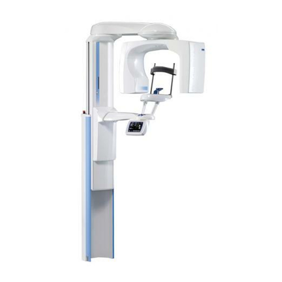
MAIN PARTS MAIN PARTS NOTE Refer to the User’s Manual for 3D imaging for a general view of the X-ray system. Sensors Dimax sensor 3D sensor 3D Mid ProFace sensor 2 Planmeca ProMax 3D Mid User’s Manual (2D) -
Page 7: Patient Supports
MAIN PARTS Patient supports Bite piece Chin cup Chin support Temple supports Chin rest Adapter Planmeca ProMax 3D Mid 3 User’s Manual (2D) -
Page 8: Exposure Switch
C-arm will stop moving and a help message will appear on the control panel display. Guide the patient away from the X-ray unit. Then clear the help message by touching OK. 4 Planmeca ProMax 3D Mid User’s Manual (2D) -
Page 9: Emergency Stop Button
X-ray unit will carry out a self-test which will last a few seconds. The X-ray unit is then ready for use. NOTE To prolong the lifetime of the Planmeca ProMax 3D Mid On / off switch X-ray unit, always switch the X-ray unit off when it is not in active use. -
Page 10: Programs
2D imaging system (optional) Basic panoramic Standard panoramic 3D, 3D Mid ProFace programs (standard) or Dimax Lateral double TMJ PA double TMJ PA rotational sinus 3D or Standard panoramic + TMJ 3D Mid ProFace 6 Planmeca ProMax 3D Mid User’s Manual (2D) - Page 11 Bitewing panoramic 3D, 3D Mid ProFace programs (optional) or Dimax Horizontal and vertical segmenting Cephalostat Scanning Ceph Dimax (optional) Planmeca ProCeph ProCeph Dynamic Exposure Panoramic DEC Dimax Control (DEC) Cephalometric DEC (optional) Planmeca ProMax 3D Mid 7 User’s Manual (2D)
-
Page 12: Panoramic Programs
1 cm NOTE This program is optimized for interproximal imaging and a shadow of the opposite side teeth may therefore be visible in the radiograph. 8 Planmeca ProMax 3D Mid User’s Manual (2D) - Page 13 TMJ exposures. The exposure is taken in two stages. Teeth are imaged during the first exposure (1/2) and both TMJs during the second exposure (2/2). layer laser beam 1 cm Planmeca ProMax 3D Mid 9 User’s Manual (2D)
-
Page 14: Temporomandibular Joint (Tmj) Programs
(2/2) temporomandibular joints. The imaging angle is adjustable (default angle: 17°). PA exposures are taken perpendicular to the long axis of the condyle (90° - 17° = 73°). The condyle angle is shown as default angle. 1 cm 10 Planmeca ProMax 3D Mid User’s Manual (2D) - Page 15 Three lateral multi-angle left-hand or right-hand side TMJ exposures. 1 2 3 The imaging angle for image no. 2 is adjustable (three imaging angles: 17° ±7° by default). The selected imaging angle is in image no. 2. 1 cm Planmeca ProMax 3D Mid 11 User’s Manual (2D)
-
Page 16: Sinus Programs
X-ray X-ray beam. 1 cm Lateral non-rotational (L / R) Lateral non-rotational exposure of the left or right sinus area. X-ray (L) 1 cm 12 Planmeca ProMax 3D Mid User’s Manual (2D) - Page 17 PROGRAMS Midsagittal non-rotational (L / R) Lateral non-rotational sinus exposure in the middle of the jaw. Exposure can be taken from the left or right jaw side. X-ray (L) 1 cm Planmeca ProMax 3D Mid 13 User’s Manual (2D)
-
Page 18: Control Panel
NOTE The exposure time indicated on the display (before an exposure is taken) is an estimated value. The real value is shown after the exposure. Variation in exposure time is caused by mA automation. 14 Planmeca ProMax 3D Mid User’s Manual (2D) -
Page 19: General Settings
The preset exposure values are average values and they are only meant to guide the user. To change the preset exposure values, touch the kV / mA field on the main display. Planmeca ProMax 3D Mid 15 User’s Manual (2D) - Page 20 8 “PANORAMIC EXPOSURE” on page 32, 9 “TEMPOROMANDIBULAR JOINT (TMJ) EXPOSURE” on page 41 and 10 “SINUS EXPOSURE” on page 53. Confirm your selection and return to the main display by touching OK. 16 Planmeca ProMax 3D Mid User’s Manual (2D)
- Page 21 NOTE If needed, the Go Entry 1 function can be disabled on information display i230. This might be necessary if there is no space for the C-arm to move back. Planmeca ProMax 3D Mid 17 User’s Manual (2D)
-
Page 22: Selecting Panoramic Program
NOTE Program type Standard + TMJ is not available for X-ray units with Dimax sensor. When you have selected the program the main display will be shown again. 18 Planmeca ProMax 3D Mid User’s Manual (2D) - Page 23 NOTE Selecting jaw shape and size manually will override the automatic setting. Jaw size: Narrow Average Wide Jaw shape: V-shaped Normal Square Confirm the selection and return to the main display by touching OK. Planmeca ProMax 3D Mid 19 User’s Manual (2D)
- Page 24 (segment 5) cannot be deselected. NOTE DEC and horizontal segmenting cannot be used simultaneously. NOTE If Autofocus is switched on (AF ON), DEC will be automatically switched off for the first exposure (scout image). 20 Planmeca ProMax 3D Mid User’s Manual (2D)
- Page 25 DEC calibration value. The setting can be adjusted between 20% (lower exposure values -> brighter image) and 200% (higher exposure values -> darker image). recommended setting is 100% (default setting). Planmeca ProMax 3D Mid 21 User’s Manual (2D)
-
Page 26: Selecting Temporomandibular Joint (Tmj) Program
Prog. field on the main display. The main display is the display that is shown when the X-ray unit is switched on. X-ray units with 3D sensor and SmartPan license: X-ray units with Dimax sensor: 22 Planmeca ProMax 3D Mid User’s Manual (2D) - Page 27 The marking on the jaw icon demonstrates the image layer. Select the required jaw size by tapping the corresponding icon. The selected jaw icon will be highlighted. NOTE Selecting the jaw size manually will override the automatic setting. Planmeca ProMax 3D Mid 23 User’s Manual (2D)
- Page 28 The jaw side which will be exposed remains white. Touching the arrow of a deselected side again will change the color back to white. Confirm the selection and return to the main display by touching OK. 24 Planmeca ProMax 3D Mid User’s Manual (2D)
-
Page 29: Selecting Sinus Program
Refer to section 4.3 “Sinus programs” on page 12 for a more detailed description of the program types. When you have selected the program the main display will be shown again. Planmeca ProMax 3D Mid 25 User’s Manual (2D) -
Page 30: Patient Positioning Controls
Positioning joystick The positioning joystick is used for horizontal fine- adjustment of the target area in 3D imaging. It is used when the patient is positioned in the X-ray unit. 26 Planmeca ProMax 3D Mid User’s Manual (2D) - Page 31 Open / close temple supports Press the temple support button to open the temple supports in 2D imaging. The temple supports can be closed by pressing the temple support button again. Planmeca ProMax 3D Mid 27 User’s Manual (2D)
-
Page 32: Preparations For Exposure
- use a 3D sensor to take 3D exposures and a Dimax sensor to take panoramic exposures. This option has selected information display i230 (Behavioural preferences -> Use Dimax sensor for panoramic). 28 Planmeca ProMax 3D Mid User’s Manual (2D) -
Page 33: Attaching And Removing Sensor
Fingerprints or other stains on the glass surface destroy image quality. ProFace sensor CAUTION Do not drop the sensor. Planmeca limited warranty does not cover damage which is due to misuse, e.g. dropping the sensor, neglect, or any cause other than ordinary use. - Page 34 C-arm. C-arm electrical connector Sensor Locking knob The locking knob can now be turned 180 degrees. This will release the locking mechanism. Carefully pull the sensor out. 30 Planmeca ProMax 3D Mid User’s Manual (2D)
-
Page 35: Preparing Patient
X-ray unit. NOTE High contrast objects, such as gold teeth or amalgam, may cause artefacts in the image. Place a protective lead apron over the patient’s back if required. Planmeca ProMax 3D Mid 31 User’s Manual (2D) -
Page 36: Panoramic Exposure
19. You can take a segment exposure by selecting only certain vertical or horizontal segments of the jaw, refer to section 5.3.2 “Selecting segmentation in panoramic programs” on page 20. 32 Planmeca ProMax 3D Mid User’s Manual (2D) -
Page 37: Patient Positioning
To adjust the height of the unit, press either of the height adjusting buttons until the chin rest is on the level of the Height adjusting patient’s chin. buttons The X-ray unit moves slowly at first, then faster. Planmeca ProMax 3D Mid 33 User’s Manual (2D) - Page 38 Patient position with and lower incisors do not overlap. chin support The following positioning lights will come on: Frankfort plane light Midsagittal plane light Layer light 34 Planmeca ProMax 3D Mid User’s Manual (2D)
- Page 39 The patient’s back and neck should be straight. The Frankfort plane is a plane which joins the infra-orbital point to the superior border of the external auditory meatus. Frankfort plane light Planmeca ProMax 3D Mid 35 User’s Manual (2D)
- Page 40 NOTE The layer light is disabled when Autofocus is switched on (AF ON). Check that the Frankfort plane light and the midsagittal plane light are still correctly positioned. Reposition them if necessary. 36 Planmeca ProMax 3D Mid User’s Manual (2D)
- Page 41 Imaging angle: Submento-vertex exposure NOTE The ruler measurement depends on the position of the layer light. On retakes always use a ruler to measure the position. Do not rely on numeric position values. Planmeca ProMax 3D Mid 37 User’s Manual (2D)
-
Page 42: Taking An Exposure
When you have taken the exposure the image will be shown on the computer screen. Note that you must accept the image in the Planmeca Romexis program - only accepted images will be stored in the database. Refer to the Romexis User’s Manual. - Page 43 When you have taken the exposure the image will be shown on the computer screen. Note that you must accept the image in the Planmeca Romexis program - only accepted images will be stored in the database. Refer to the Romexis User’s Manual.
- Page 44 When you have taken the exposure the image will be shown on the computer screen. Note that you must accept the image in the Planmeca Romexis program - only accepted images will be stored in the database. Refer to the Romexis User’s Manual.
-
Page 45: Temporomandibular Joint (Tmj) Exposure
If necessary, you can change the preset values as described in section 5.2.2 “Selecting kilovolt and milliampere values” on page 15. NOTE Always try to minimize the radiation dose to the patient. Planmeca ProMax 3D Mid 41 User’s Manual (2D) - Page 46 To adjust the height of the unit, press either of the height adjusting buttons until the opening in the chin support is Height adjusting approximately level with the patient’s mouth. buttons The X-ray unit moves slowly at first, then faster. 42 Planmeca ProMax 3D Mid User’s Manual (2D)
- Page 47 (button or joystick). Thumb wheel Positioning controls Position the patient’s head so that the midsagittal plane coincides with the midsagittal plane light beam. Midsagittal plane light Planmeca ProMax 3D Mid 43 User’s Manual (2D)
- Page 48 Imaging angle: Take a submento-vertex exposure (axial projection) to determine the exact imaging angle. Adjust the imaging angle with the left / right arrows according to your measurement. 44 Planmeca ProMax 3D Mid User’s Manual (2D)
- Page 49 Do not rely on numeric position values. NOTE Make sure that you have selected the right patient and the panoramic exposure mode in the Planmeca Romexis program before you take an exposure. Refer to the Romexis User’s Manual.
- Page 50 The Romexis program will show the Waiting for Exposure message on the computer screen. Ask the patient to stand as still as possible. Move to a protected area. Green ready indicator light 46 Planmeca ProMax 3D Mid User’s Manual (2D)
- Page 51 When you have taken the exposure the image will be shown on the computer screen. Note that you must accept the image in the Planmeca Romexis program - only accepted images will be stored in the database. Refer to the Romexis User’s Manual.
-
Page 52: Multi-Angle Tmj Exposure (Pa Or Lateral)
VALUE mA VALUE Child Adolescent Small adult Average-sized adult Large adult Exposure values for multi-angle PA TMJ programs PATIENT kV VALUE mA VALUE Child Adolescent Small adult Average-sized adult Large adult 48 Planmeca ProMax 3D Mid User’s Manual (2D) - Page 53 Positioning controls (button or joystick). controls Planmeca ProMax 3D Mid 49 User’s Manual (2D)
- Page 54 Imaging position: Use a ruler to measure the distance between the layer light and the patient’s temporo- mandibular joint to determine the exact imaging position. Adjust the imaging position with the left / right arrows according to your measurement. 50 Planmeca ProMax 3D Mid User’s Manual (2D)
- Page 55 Do not rely on numeric position values. NOTE Make sure that you have selected the right patient and the panoramic exposure mode in the Planmeca Romexis program before you take an exposure. Refer to the Romexis User’s Manual.
- Page 56 When you have taken the exposure the image will be shown on the computer screen. Note that you must accept the image in the Planmeca Romexis program - only accepted images will be stored in the database. Refer to the Romexis User’s Manual.
-
Page 57: Sinus Exposure
VALUE Child Adolescent Small adult Average-sized adult Large adult Exposure values for sinus programs “Lateral non-rotational” and “Midsagittal non-rotational” PATIENT kV VALUE mA VALUE Child Adolescent Small adult Average-sized adult Large adult Planmeca ProMax 3D Mid 53 User’s Manual (2D) -
Page 58: Patient Positioning
Thumb wheel controls (button or joystick). Positioning controls 54 Planmeca ProMax 3D Mid User’s Manual (2D) - Page 59 Thumb wheel NOTE Make sure that you have selected the right patient and the panoramic exposure mode in the Planmeca Romexis program before you take an exposure. Refer to the Romexis User’s Manual. Planmeca ProMax 3D Mid 55...
-
Page 60: Taking An Exposure
When you have taken the exposure the image will be shown on the computer screen. Note that you must accept the image in the Planmeca Romexis program - only accepted images will be stored in the database. Refer to the Romexis User’s Manual. - Page 62 Planmeca Oy | Asentajankatu 6 | 00880 Helsinki | Finland tel. +358 20 7795 500 | fax +358 20 7795 555 | sales@planmeca.com | www.planmeca.com...
Need help?
Do you have a question about the ProMax 3D Mid and is the answer not in the manual?
Questions and answers