Mindray DC-40S Manuals
Manuals and User Guides for Mindray DC-40S. We have 2 Mindray DC-40S manuals available for free PDF download: Operator's Manual

Mindray DC-40S Operator's Manual (303 pages)
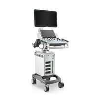
Diagnostic Ultrasound System
Brand: Mindray
|
Category: Medical Equipment
|
Size: 6 MB
Table of Contents
-
Warranty12
-
Exemptions12
-
Conventions15
-
Latex Alert25
-
Intended Use27
-
Imaging Mode28
-
Power Supply28
-
Options30
-
I/O Panel36
-
Symbols43
-
Power Supply46
-
Power ON/OFF47
-
Standby49
-
Basic Screen62
-
Touchscreen67
-
Pause Exam77
-
End Exam78
-
Imaging Mode79
-
Color M Mode105
-
Tdi108
-
Overview114
-
Note before Use118
-
Static 3D120
-
Ilive130
-
Smart 3D133
-
Iscape Viewing140
-
Cine Review142
-
Elastography142
-
Enter/Exit143
-
Cine Review144
-
Contrast Imaging144
-
Image Display149
-
Spot149
-
Pan150
-
Cine Review151
-
Image Compare154
-
Cine Compare154
-
Frame Compare154
-
Cine Saving154
-
Cine Memory155
-
Cine Settings156
-
Ecg157
-
ECG Review159
-
Measurement161
-
Basic Operations161
-
Adding Comments169
-
Moving Comments170
-
Comment Setting172
-
Body Marks172
-
Memory Media176
-
Thumbnails178
-
Ivision180
-
Istorage187
-
Print187
-
Setting187
-
Image Print187
-
Report Print188
-
Administration190
-
Access Setting190
-
System Login190
-
Modify Password192
-
Dicom193
-
DICOM Preset193
-
Network Preset193
-
Service Preset195
-
DICOM Services200
-
DICOM Storage200
-
DICOM Print202
-
DICOM Worklist203
-
Mpps204
-
Query/Retrieve205
-
Setup209
-
System Preset210
-
Region211
-
General212
-
Image Preset213
-
Application214
-
OB Preset214
-
Admin215
-
Exam Preset216
-
Measure Preset216
-
Print Preset217
-
Network Preset218
-
Local TCP/IP218
-
Istorage220
-
Maintenance221
-
Other Settings222
-
Probe223
-
Biopsy Guide233
-
Disposal253
-
Middle Line253
-
Acoustic Output255
-
MI/TI Display257
-
Acoustic Output258
-
Troubleshooting275
Advertisement
Mindray DC-40S Operator's Manual (291 pages)
Diagnostic Ultrasound System
Brand: Mindray
|
Category: Medical Equipment
|
Size: 7 MB
Table of Contents
-
Warranty12
-
Conventions15
-
-
Intended Use27
-
I/O Panel35
-
Symbols42
-
-
-
Power Supply44
-
Power ON/OFF45
-
-
Imaging Mode77
-
Color M Mode103
-
Tdi106
-
Overview112
-
Note before Use116
-
Static 3D118
-
Ilive128
-
Smart 3D131
-
Elastography140
-
Enter/Exit141
-
Cine Review142
-
-
-
Image Display143
-
-
Cine Review145
-
Image Compare148
-
Cine Compare148
-
Frame Compare148
-
-
Cine Saving148
-
Cine Memory149
-
Cine Settings150
-
-
7 Ecg
151 -
8 Measurement
155 -
-
Body Marks166
-
-
Istorage181
-
Print181
-
Setting181
-
Image Print181
-
Report Print182
-
-
Administration184
-
Access Setting184
-
System Login184
-
Modify Password186
-
-
11 Dicom
187-
DICOM Preset187
-
Network Preset187
-
Service Preset189
-
-
DICOM Services194
-
DICOM Storage194
-
DICOM Print196
-
DICOM Worklist197
-
Mpps198
-
Query/Retrieve199
-
-
-
12 Setup
203-
System Preset203
-
Region205
-
General206
-
Image Preset207
-
Application208
-
OB Preset208
-
Admin209
-
-
Exam Preset210
-
Measure Preset210
-
Print Preset211
-
Network Preset212
-
Local TCP/IP212
-
Istorage214
-
-
Maintenance215
-
Other Settings216
-
-
-
-
Probe217
-
Biopsy Guide227
-
Middle Line245
-
-
-
Acoustic Output250