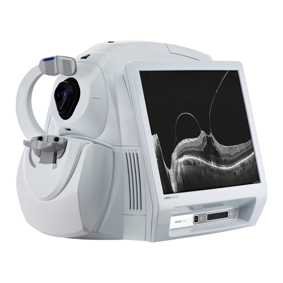
Sign In
Upload
Manuals
Brands
Zeiss Manuals
Medical Equipment
CIRRUS HD-OCT 500
Zeiss CIRRUS HD-OCT 500 Manuals
Manuals and User Guides for Zeiss CIRRUS HD-OCT 500. We have
2
Zeiss CIRRUS HD-OCT 500 manuals available for free PDF download: User Manual, Technical Specifications
Zeiss CIRRUS HD-OCT 500 User Manual (352 pages)
Brand:
Zeiss
| Category:
Diagnostic Equipment
| Size: 52.95 MB
Table of Contents
Table of Contents
3
Safety and Certifications
15
Symbols and Labels
15
Warning and Caution Definitions
16
Warnings
16
Cautions
17
Protection of Patient Health Information
18
Safety
18
CIRRUS HD-OCT User Manual 2660021169012 Rev. a
19
Record and Data Safety
23
Networking Guidelines
24
Electromagnetic Compatibility (EMC)
24
Rohs Compliance
26
Introduction
27
Intended Use
27
Indications for Use
27
Usage
27
Intended Operator Profile
28
Demographic
28
Occupational Skills
28
Job Requirements
28
Data Analyst Profile
29
Demographic
29
Occupational Skills
29
Job Requirements
29
Subject Profile
29
Installation and Setup
29
User Documentation
30
Accessing PDF Versions
30
Organization
30
Instruction Conventions
31
System Overview
33
Hardware
33
Power up
34
Power down
34
Power down through Hardware
34
Power down through Software
34
Software
35
Overview
35
Licensed Applications
35
Basic Screens
35
Toolbar Options
37
Navigation Bar
41
Status Area
41
Components of Status
42
System Administration
45
Institution Setup
45
Station Setup
46
Staff Accounts
47
Register New Staff
47
Edit Staff Records
48
Delete Staff Records
48
Category Registration and Maintenance
48
Category Registration
48
Edit Categories
49
Delete Categories
49
Archive Setup and Selection
50
Set up a CIRRUS HD-OCT Archive
50
Change the Current Archive
52
Set Preferences
52
Archive/Synchronize
52
Archive Alerts
52
Normative Data Settings
53
DICOM Archive
53
Display Options
54
Ipv4 / Ipv6
55
Preventive Maintenance Service Notifier
55
User Login/Logout
56
User Login
56
User Logout
57
Automatic Logout
57
Logout Locks the System
57
Clinical Workflow
59
The Importance of Good Clinical Workflow
59
Patient Good Practices
59
Specific to Iris Imaging
59
Specific to Fundus Imaging
59
Daily Tasks
59
Start of Day
59
End of Day
60
Archive Saved Exams
60
Shut down the System
60
Patient Records
60
Add New Patients
60
Find Existing Patients
62
Advanced Search
63
Add or Remove Categories for Existing Patient Records
65
View Today's Patients
66
How Today's Patients List Is Populated
67
Patient ID Search Conflicts
67
Patient Preparation
68
Optional Dilation of Patient's Eye(S) (Posterior Segment)
68
Position the Patient on the Chinrest
68
Optional Eyelid Elevation
69
Precautions
69
Patient Instructions
70
Of Special Note for Anterior Scans
70
Acquiring Scans
73
Overview of Scan Types
73
Posterior Segment Scans
74
CIRRUS OCT Angiography
74
Angiography Scan
74
Montage Angio Scan
74
ONH Angiography Scan
74
Macula
75
Macular Cube 200X200
75
Macular Cube 512X128
75
Optic Nerve
75
Optic Disc Cube 200X200
75
Integration
76
Macular and Optic Disc Cube Scans
76
Visualization
76
Macular or Optic Disc Cube Scans
76
Raster Scans
76
Guidelines for Raster Scans
77
Anterior Segment Scans
78
Scan Preparation
78
Instrument Imaging Process
78
Aligning Scans Corrected for Beam Scanning Geometry and Corneal Refraction
79
Center Corneal Scans on the Corneal Vertex
79
Scan Acquisition Controls
79
Scan Pattern Adjustments
79
Anterior Chamber and Cornea External Lenses
79
Attaching an External Lens
80
Anterior Chamber Scan (Chamber View™)
80
Guidelines for Anterior Chamber Scan Acquisition
81
Anterior Segment Cube Scan
82
Guidelines for Anterior Segment Cube 512X128 Scan Acquisition
83
HD Angle Scan
84
Guidelines for HD Angle Scan Acquisition
85
HD Cornea Scan
86
Guidelines for HD Cornea Scan Acquisition
86
Wide Angle-To-Angle Scan
87
Guidelines for Wide Angle-To-Angle Acquisition
88
Pachymetry
88
Guidelines for Pachymetry Scan Acquisition
89
Anterior Segment 5-Line Raster Scan
90
Guidelines for Anterior Segment 5 Line Raster Scan Cornea Acquisition
91
Acquire Screen and Controls
91
Acquire Screen Toolbar
92
Available Scans List
92
Scan Organizer
93
Iris Viewport
94
Scan Position Montage
94
6X6 MM Montage Angio Scan
94
8X8 MM Montage Angio Scan
95
Fundus Viewport
96
Mouse Controls
99
Live Scan Viewports
99
General Acquire Procedure
100
To Begin Scan Acquisition
100
Set up for Maximum Image Quality
103
B-Scans
103
Fundus Image
103
Iris Image
103
For All Scans
104
Tracking and Repeat Scans
104
Repeat Scans
104
Fasttrac
105
Troubleshooting Fasttrac
108
Scan Quality Check
111
Overview
112
Quality Check Tools
113
Criteria for Image Acceptance
115
Fundus
115
Oct
115
Check for Saccades and Banding
115
Advanced RPE Analysis Acceptance Criteria
116
CIRRUS OCT Angiography Acceptance Criteria
117
Signal Quality
117
Decorrelation Tails
118
Segmentation Errors
119
Anterior Segment Acceptance Criteria
121
Anterior Segment Cube Scan
121
General
121
Pachymetry Scan
121
Analysis
123
Overview
123
Image Menu Options
124
Normative Database Comparisons
125
Adjusting Slab Locations
125
Preset Slab Views
126
Create Global Custom
126
Create Scan Custom
126
Standard Presets
126
Posterior Segment
128
Macular Thickness Analysis
128
Fovea Location
128
ETDRS Position
129
ILM-RPE Layers
130
Slab Overlay ILM-RPE Thickness Map
130
Additional Features
132
Macular Change Analysis
133
Manual Selection
134
Macular Registration
135
Synchronized Data Review
138
XML Export
138
Macular Thickness ou Analysis
138
Advanced RPE Analysis
139
RPE Elevation Considerations
140
Sub-RPE Considerations
141
Ganglion Cell ou Analysis
144
En Face Analysis
147
En Face Analysis Preset Slabs
147
ONH and RNFL ou Analysis
150
RNFL Thickness Maps and Deviation from Normal Maps
151
Changing RNFL Calculation Circle Placement
152
TSNIT Thickness Profiles
152
RNFL Data Table
153
Parameter Summary Graphs
154
Summary Charts
155
RNFL & ONH Guided Progression Analysis
156
Ganglion Cell Guided Progression Analysis
160
Anterior Segment
163
Anterior Chamber Depth and Lens Vault
163
Angle-To-Angle Distance
165
Using Anterior Chamber Analysis
165
Using Wide Angle-To-Angle Analysis
165
Corneal Thickness
165
In Pachymetry Analysis
166
View Dropdown
166
In Anterior Chamber Analysis
171
In Anterior Segment Cube Analysis
171
In HD Cornea Analysis
171
In HD Images Analysis
172
Epithelial Thickness
173
In Pachymetry Analysis
173
Central Corneal Thickness (CCT) Measurement
174
Angle Measurements
175
In HD Angle Analysis
175
In Anterior Chamber Analysis
176
In Wide Angle-To-Angle Analysis
177
Specialized and Integrative Visualization Tools
177
High Definition Images Analysis
177
Change the Displayed Scan Line
179
Image Display Options Buttons
179
Single Eye Summary
180
The Panomap
181
3D Analysis
182
View Settings
183
Clip Selector
184
Show Settings
184
Animation
185
Animation Editor
185
Clip Surfaces
185
Greyscale Mode
185
Light Direction Adjust
185
Lighting
185
Movie Recording
186
Autorecord Keyframes
187
Take Snapshot
187
Mouse Rotates/Mouse Translates
188
Straighten Volume Data
188
Width, Height, and A-Scan Adjustments
188
Zoom
188
Reset
189
Transparent Surfaces
189
Advanced Visualization Analysis
190
Fundus Image Overlay Options
191
Retinal Layers Automatically Detected and Displayed
191
Slice and Slab Options
192
Function Buttons in Advanced Visualization
193
CIRRUS OCT Angiography
195
Overview
195
CIRRUS OCT Angiography, Montage Angio, and ONH Angiography Analysis
195
CIRRUS OCT Angiography Presets
198
Angioplex Metrix
201
B-Scan Settings
201
Current View References
202
Overlays
202
Vessel Density and Capillary Perfusion
205
Foveal Avascular Zone (FAZ)
206
Vessel Density Vs. Capillary Perfusion
206
Angioplex Metrix Measurements
210
Overlay Options
211
ETDRS Options
212
Angiography and ONH Angiography Change Analysis
213
Previous Scan
214
Options Bar
215
Toggle Icon
215
Angioplex Metrix in Change Analysis
216
ONH Overlay and Angio Metrix Bar
216
Selected Scan
216
ONH RNFL Thickness
217
OCT Angiography Change - Manual Selection
217
Angioplex Preset Algorithms
218
ONH Angiography Change Analysis Image Registration
221
10 Reports and Printing
223
Standard Print Options
223
The Report Preview Screen
224
Analysis Related Report Options
226
Macula Thickness Report Options
226
The Macula Multi-Slice Option
227
Macula Radial Report Option
228
Advanced Visualization Report Options
228
ONH and RNFL Thickness Report Options
230
Normative Data Details Report
231
High Definition Images Report (Raster Scan Options)
232
Guided Progression Report Options
233
Latest Scan
233
Parameters Summary Table Report
233
Summary
233
Normative Data Details Reports
237
Macula Thickness Normative Data Details Report
237
11 Data Management
239
FORUM/DICOM or Native Environment
239
Importing and Exporting Data
239
Patient Privacy
239
Data Integrity of Imported Records
240
Import/Export Native Mode
240
FORUM/DICOM Mode
243
Transferring Images in OPT IOD and OP IOD Formats
243
Data Queries and Search Options
245
XML Export
247
Batch XML Export
247
XML Export from Analysis Screens
247
Values Exported: Macular Thickness Analysis
248
Values Exported: ONH and RNFL ou Analysis
249
Export Values: Macular Change Analysis
250
Values Exported: Guided Progression Analysis
251
Values Exported: Ganglion Cell ou Analysis
252
Values Exported: Advanced RPE Analysis
253
Advanced Export
254
Dat Files of ILM-RNFL Thickness Maps
254
Txt Profiles of Neuro-Retinal Rim Thickness and RNFL Thickness
254
Move Scan Data (Native Mode Only)
254
Log Files
255
12 Routine Maintenance
257
Troubleshooting Power Problems
257
If Not Using the Optional Power Table
257
If Using the Optional Power Table
257
Handling Error Messages
258
Product Labels and Serial Number Location
258
Routine Cleaning
258
Forehead and Chinrests
259
External Surfaces of the Instrument
259
Imaging Aperture Lens and External Lenses
259
LCD (Monitor) Screen
259
Top Fan Filter
259
User Replacement Accessories
261
Performance Verification Check
262
Install the Verification Test Tool
262
Run the Check
263
13 Specifications
269
HD-OCT Imaging
269
HD-OCT Imaging for Posterior Segment Scans
269
HD-OCT Imaging for Anterior Segment Scans
269
Fundus Imaging
270
Iris Imaging
270
Electrical, Physical and Environmental
271
Maintenance
271
Measurement Units
271
14 Legal Notices
273
Software Copyright
273
Software License Agreement
273
Acknowledgment
274
A Normative Data Results
275
Overview
275
RNFL and Macula Normative Databases: Diversified
275
Inclusion and Exclusion Criteria
276
Exclusion Criteria
276
Inclusion Criteria
276
Data Collection
277
Scan Selection Criteria
277
Database Population
277
Data Analysis
278
Age Coefficient - RNFL Thickness
279
Description of Macular Scan Parameters Used in CIRRUS HD-OCT
280
Age Coefficient - Macula Thickness
282
Conclusion
283
Optic Nerve Head Normative Database: Diversified
283
Methods
284
Data Collection
284
Scan Selection Criteria
284
Data Analysis
284
Results
285
Factors that Affect CIRRUS ONH Normative Ranges
285
2Age
286
Optic Disc Area
286
Ethnicity
286
Calculation of Normal Limits
286
Presentation of Normative Limits
287
Conclusion
287
Report
288
RNFL and ONH Normative Databases
289
Distribution of Normals
290
Interactivity
293
Ganglion Cell Normative Database: Diversified
293
Methods
294
Data Collection
294
Scan Selection Criteria
294
Data Analysis
295
Results
295
Factors that Affect CIRRUS Ganglion Cell Analysis Normative Ranges
295
Age
296
Ethnicity
296
Axial Length and Refractive Error
297
Data Analysis
297
Conclusion
298
Asian Normative Database
298
Overview
298
Introduction
298
Inclusion and Exclusion Criteria
299
Exclusion Criteria
299
Inclusion Criteria
299
Data Collection
300
Scan Selection Criteria
300
The Asian Dataset
301
Normative Database Analyses
301
Description of RNFL Parameters
302
Age Coefficient - RNFL Thickness
302
Age Coefficient - Macula Thickness
303
Normal Values for Ganglion Cell Analyses
304
Normal Values for Asian ONH Measurements
305
Factors that Affect ONH Normative Ranges
306
Age
306
Ethnicity
306
Optic Disc Area
306
Calculation of Normal Limits
307
Conclusion
307
Methods
309
Purpose
309
Study 1: Retinal Segmentation and Analysis
309
Results and Discussion
310
Repeatability
314
Conclusion
316
References
316
Phase 1: Inter-Device Variability
317
Phase 2: Inter-Operator Variability
317
Study 2: Repeatability and Reproducibility of Illumination Area Measurements under the Retinal Pigment Epithelium
317
Phase 1: Inter-Device Variability
320
Phase 2: Inter-Operator Variability
320
Study 3: Repeatability and Reproducibility of Macular Retinal Pigment Epithelium Elevation Measurements
320
Study 4: Repeatability and Reproducibility of GCA and ONH Parameters
323
Benchtop Scanning Accuracy, Repeatability and Reproducibility
327
CIRRUS HD-OCT Repeatability in Measuring Central Corneal Thickness
327
Study 5: Anterior Segment Accuracy, Repeatability and Reproducibility
327
Performance of CIRRUS HD-OCT RNFL Analysis
328
References
330
Study 6: CIRRUS OCT Angiography
330
Reproducibility of Anterior Scan Measurements
331
100 C CIRRUS HD-OCT Repeatability and
331
Study 1: Performance of Pachymetry and Anterior Chamber Scan
331
Subjects, Including Repeatability, Reproducibility, and Comparison to Visante
331
Purpose
331
Data Collection
331
Inclusion Criteria
331
Corneal Pathology Group
331
Normal Cornea Group
331
Post-LASIK Group
332
Exclusion Criteria
332
Corneal Pathology Group
332
Normal Cornea Group
332
Data Analysis
332
Advertisement
Zeiss CIRRUS HD-OCT 500 Technical Specifications (2 pages)
Brand:
Zeiss
| Category:
Medical Equipment
| Size: 0.32 MB
Advertisement
Related Products
Zeiss CIRRUS HD-OCT 5000
Zeiss Acuitus 5010
Zeiss Acuitus 5015
Zeiss MultiSEM 505 Series
Zeiss MultiSEM 506 Series
Zeiss Axiocam 503 color
Zeiss Axiocam 503 mono
Zeiss Axiocam 506 color
Zeiss Axiocam 506 mono
Zeiss Conquest 50 T
Zeiss Categories
Microscope
Medical Equipment
Binoculars
Laboratory Equipment
Measuring Instruments
More Zeiss Manuals
Login
Sign In
OR
Sign in with Facebook
Sign in with Google
Upload manual
Upload from disk
Upload from URL