Table of Contents
Advertisement
Advertisement
Table of Contents

Summary of Contents for Planmeca Emerald
- Page 1 Planmeca Emerald User Manual...
-
Page 3: Table Of Contents
Session Usage ............................20 Autoclave Indicator ..........................20 Color Balancing ............................21 Positioning the Scanner for the First Scan ................21 Basic Scanning Steps ....................... 22 Model Scanning ........................23 © 2017 November Planmeca All rights reserved. Software Version 5.9.2 15698200.B... - Page 4 Selection Area Tool ........................43 Edit Pre-Op Tool ........................43 Drawing Pontic Margins ......................44 Exporting Data ..................45 Exporting a CAD/CAM Case ..................... 45 Exporting 3D Models ....................... 45 3D Model Export .............................. 45 DDX Export ..........................45 Planmeca Emerald User Manual...
- Page 5 Approvals (All Systems) ......................48 Guidance and Manufacturer’s Declaration - Electromagnetic Emissions ......49 Guidance and Manufacturer’s Declaration - Electromagnetic Immunity ......49 Recommended Separation Distances ...................... 51 Emerald Optical Specifications ........................52 Labels ............................53 Symbols ................................53 Product Identification Labels ........................54 External Components and Connectors ....................
-
Page 6: Introduction
Caution: US Federal law restricts the Planmeca Emerald to sale by or on the order of a dentist. The Planmeca Fit System requires Planmeca Romexis software revision 3.4.0.R or later. -
Page 7: Scanner
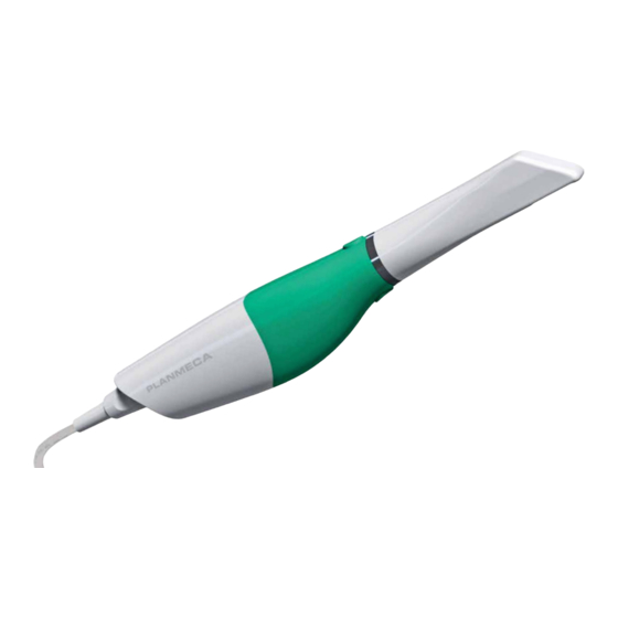
Scanner Accessories The Emerald system has a set of removable components. • Scanning Tip • Scanning Cable • Cradle • Color Balancer • Laptop • Mouse Connecting the Scanning Cable To connect the scanner cable, align the notch on the cable to the small notch on the back of the scanner. -
Page 8: Cradle
Squeeze the grey trigger and pull to separate the cradle from the base. The trigger is used every time that you want to insert or remove the scanner holder. Insert the cradle into the chair adapter. Insert the holder into a holder in your operatory equipment. Introduction Planmeca Emerald User Manual... -
Page 9: Relocating The Laptop And/Or Scanner
System software and hardware upgrades are initiated through Planmeca only. No software or hardware should be added or deleted to/from the Planmeca systems without prior approval of Planmeca. Doing so may result in damage to the system and will void the product warranty. -
Page 10: Cleaning The Scanner Tip
If there is any debris that cannot be removed or, if there is visible deterioration such as cracks or discoloration, the tip should be properly disposed of and replaced. Introduction Planmeca Emerald User Manual... -
Page 11: Storage
Individually pouch each scanner tip using an autoclave pouch (ex. HSD # 112-4854 or a comparable product). Autoclave baskets are not indicated for this cleaning procedure. Place one to three pouches per tray or cassette. DO NOT stack tips on or around other metal instruments. Select the Wrapped, Wrapped Instruments, or Pouch cycle on the autoclave with a minimum cycle of 132° C (269°... -
Page 12: Cleaning The System
For intraoral scanning systems only. Protect the keyboard with a disposable barrier. Cleaning Cycle: Before and after each use, clean all areas of the Planmeca System. Warning: Before and after each use, follow these instructions to disinfect the Planmeca System. -
Page 13: Moving/Viewing The 3D Model
Moving/Viewing the 3D Model Use the mouse to zoom in or out, move, and rotate the composite model. Rotating the Model Click and hold down the right mouse button. The pointer changes to Drag the mouse horizontally, vertically, or diagonally to rotate the image. Drag in small increments for more control. -
Page 14: The Settings Screens
This screen should be used only under the supervision of a customer service representative. These settings are pre- configured and should not be changed. Workbook Exercises (For Classic systems) Mill Notification Settings (For Classic systems) Milling Settings (For mill systems) Auto or Occlusal POI (For mill systems) Introduction Planmeca Emerald User Manual... -
Page 15: Additional Assistance
Additional Assistance Go to http://e4d.com for videos and downloadable documentation. If you have questions, please contact Customer Support at: Toll Free 800-537-6070 E-mail customersupport@e4d.com 972-479-1106 Hours of Operation 7 am – 6 pm CT (Monday - Friday) Web site www.e4d.com D4D Technologies LLC dba E4D Technologies Mailing Address 650 International Pkwy... -
Page 16: Scanning Safety
Planmeca could void the user’s authority to operate the equipment and/or void the warranty. Do not install or operate the Planmeca products in an environment where an explosion hazard exists, e.g., high oxygen area. Comply with all applicable regulations when disposing of waste materials from the Planmeca products. -
Page 17: Patients
Patients Managing Patients in Planmeca Romexis Creating new patients In Patient Management, click the Add Patient button. The Add Patient screen opens. Enter the necessary information and add a face photo if desired. The obligatory fields are in bold text. See the Romexis User Manual for more information. -
Page 18: Selecting And Opening Patients
• Start a new restoration - See below for more information • Open an existing restoration - See below for more information • Import 3D models - See Planmeca Romexis User’s manual for more information • Export 3D models - See Planmeca Romexis User’s manual for more information Deleting Files To delete an image (stl file) from the patient’s case files right-click on the file and select Inactivate STL. -
Page 19: Setup Screen
Setup Screen Use the Setup screen to set the restoration type, occlusal data type, material, and tooth library. If you open an existing restoration, many of these settings may already be selected. For diagnostic cases, orthodontic aligners, or when sending a large restorative case (without drawing margins) to a laboratory, select Full Arch as the restoration type and click Scan to proceed. - Page 20 Changing the Tooth Selection If the wrong tooth was highlighted for the restoration, right-click the tooth and click Deselect. Click on the correct restoration site. Patients Planmeca Emerald User Manual...
-
Page 21: Scanning
Scanning Warning The scanner is a high precision Class 2 laser scanning instrument. Always store the scanner in its cradle when not in use. To prevent damage or misalignment, do not drop or strike the scanner. Follow all stated precautions when using the scanner. -
Page 22: Scanner Tips
Session Usage is displayed above the scanner status icon and shows how long you have been scanning on this particular case. This number refreshes each time the Scan screen is opened or when you click the Refresh icon next to it. Autoclave Indicator The autoclave symbol indicates that an autoclave tip is in use. Scanning Planmeca Emerald User Manual... -
Page 23: Color Balancing
Color Balancing The color symbol displays when a color tip is in use. Balance the color weekly or as needed. This is an optional step to optimize the color represented on screen. This does not affect the stone model nor the amount of data collected by the scanner. -
Page 24: Basic Scanning Steps
Make adjustments as needed. Click the bottom scanner button or use the mouse to select the next scan type. Repeat the steps above. Click the Margin screen or click the Next button when finished with scanning. Scanning Planmeca Emerald User Manual... -
Page 25: Model Scanning
Model Scanning The default setting for the scanner is for intraoral scanning. Click the sun icon next to the scanner icon to switch to a dimmer setting (sunset icon) when needed for models or anytime the live view is too bright. Default setting - sun icon represents Activate the dimmer setting for external brighter laser for intraoral use. -
Page 26: Saving A Live Scan Image
Click Refresh. Click Photos to view the captured pictures. • Planmeca Romexis 4.5 and prior a. Open the This PC or My Computer in Windows Explorer. b. In the address bar at the top, type %temp% and hit Enter. -
Page 27: Active Filtering
There are two methods to activate this tool. The first way (for both scanners) is to click the trashcan icon just above the scanner in the lower left of PlanCAD. The second is to depress and release the bottom button of the Planmeca Emerald™... -
Page 28: Generate Model
Click Data Density View if it is not already activated. The model refreshes with the dark blue and purple areas indicating the least data. Rotate the model to analyze it. Dark areas on your restoration site and interproximal contact areas should be rescanned. Scanning Planmeca Emerald User Manual... -
Page 29: Reset Model
Look for colored areas on the prepared tooth, especially on the margin. The adjacent teeth should have good data on the interproximal contact area, occlusal surfaces, and of the lingual and buccal contours. Data below the height of contour is not as crucial on the adjacent teeth. If areas lack detail, take additional scans. -
Page 30: Time Saver
Scan Buccal 2 and refine if needed. Delete the less desirable buccal alignment before proceeding to the Margin tab. Basic Buccal Bite Workflow: If needed: • Scan Buccal 1 • Scan Buccal 2 • Refine • Refine • Verify alignment • Delete one of the two bite scans Scanning Planmeca Emerald User Manual... -
Page 31: Prep Clearance And Contact Strength
Prep Clearance and Contact Strength Prep Clearance displays the distance to the opposing dentition just by moving over the prep with your mouse. While in the Bite Align tool, rotate the model to view the prep. Use the mouse pointer to hover over the area. A distance indicator (in mm) appears as you move around the model. -
Page 32: Bite Registration Scanning
Click OK to use the Time Saver. If you do not wish to use the Time Saver option, the bite registration and adjacent teeth can be scanned on their own. The following instructions assume the use of the Time Saver option. Scanning Planmeca Emerald User Manual... -
Page 33: Selecting The Bite Registration
A copy of the preparation model is created in the bite registration model color. Click the Eraser Tool. Erase the preparation and the marginal ridges of the adjacent teeth. Click the Eraser Tool to deactivate it. Activate the scanner and scan begin the scans with the occlusal of one of the adjacent teeth. Once you have established where you are, you can begin scanning the bite registration data. -
Page 34: Model Alignment
The models will snap into place or will return to their original positions. In Buccal/Opposing cases, the opposing model appears after the prep and buccal bite are aligned. Click and drag the opposing model to match the buccal bite model. Scanning Planmeca Emerald User Manual... -
Page 35: Rotating The Models
When the models are aligned, you can rotate to view the amount of contact from above. The amount of contact is shown as a color map. To access the menu options at the top or to return to scanning, deactivate the selected Alignment icon. You cannot proceed if the Alignment icon is active (orange). -
Page 36: Scanning Impressions
Click the bottom button on the scanner, click the Generate Model button, or press M on the keyboard. Scanning Impressions Remove the excess impression material so that the scanner can get closer for scanning. Any impression material can be used. The system does not require a particular color or type of material. Scanning Planmeca Emerald User Manual... -
Page 37: Positioning The Scanner
Positioning the Scanner When scanning the impression, ensure the tip of the scanner is pointing towards the distal for the initial scans so that the orientation of the model is correct. Due to the nature of impressions, the normal positioning of the scanner may not be able to capture all of the walls of the impression. -
Page 38: Full Arch Scanning
• Models must be manually aligned. Use the method on the following pages when using Full Arch scans for the fabrication of appliances. Failure to capture using this method may result in loss of accuracy. Scanning Planmeca Emerald User Manual... - Page 39 15698200.B Scanning...
-
Page 40: Margin Tab
The Orientation tool is automatically activated when you first access the Margin tab. For Scan Only cases, this step can be skipped. If you are going to design the case, see the Planmeca PlanCAD manual for more information. Margin Tool Clicking the Margin Tool activates the margin editing mode in which various methods are available to create and edit the margin. -
Page 41: Margin Aids
Margin Aids View ICE Preparation For intraoral cases only. Use View ICE Preparation to toggle between ICE view and stone view. Show Features Click Show Features to highlight high contour areas in green. This can be helpful in finding the margin edge on supragingival preps, inlays, and onlays. -
Page 42: Lasso Tool
To delete the margin and start over, click the Paint, Trace, or Lasso button. If Lasso is having trouble finding the margin, you can change the ICE Margin Mode to Texture Only. See below. Margin Tab Planmeca Emerald User Manual... -
Page 43: Margin Tab Settings
Margin Tab Settings ICE Margin Mode For intraoral cases only. ICE Margin Mode determines which view the system uses to create the margin curve when using the Lasso tool. Click Settings. Click ICE Margin Mode. The default setting, Normal, means that the system uses both the stone and ICE view to determine where the Lasso line should appear. -
Page 44: Add Segments Tool
Margin drawn - With ditching Any changes to the margin will require the ditching to be redone. If you are doing a multiple restoration case, finish all of the margin edits before using the Retract tool. Margin Tab Planmeca Emerald User Manual... -
Page 45: Multiple Restorations
Click Toggle Margin to view the ditched area without the margin. This is similar to what your STL recipient will see. Click Toggle Retraction to show/hide the virtual ditching. Multiple Restorations On multiple restoration cases, the tooth number is assigned to each preparation when the margin is drawn. Click the desired tooth number tab. -
Page 46: Drawing Pontic Margins
Click Trace and designate the position and extension of the base of the pontic on the gingival tissue to fit the appropriate contour. Do not go too far down the curve of the gingival tissue or you may not be able to fit the bridge in the block. Margin Tab Planmeca Emerald User Manual... -
Page 47: Exporting Data
DDX. Exporting a CAD/CAM Case To export a file to share with another Planmeca system, click File - Export - Export CAD/CAM Case. Select the destination folder and enter a file name. Click Save. Exporting 3D Models 3D Model Export To export 3D models in STL format click the case in the Patient’s Case Files list then 3D model export. - Page 48 If the case does not automatically upload, click on the file in Existing Files and click the Add Files button. A PlanScan panel is available in the Dentrix Chart. See Dentrix user manual for details and instructions. Exporting Data Planmeca Emerald User Manual...
-
Page 49: Planmeca Emerald Specifications
Planmeca Emerald Specifications Model Number: Planmeca Emerald P/N: 15610001 Electrical Ratings: 5Vdc, Storage conditions: -20°C to 60°C (-4°F to 140°F) Operating conditions: 15 °C to 28 °C (59 °F to 82 °F) 5%-95% non-condensing relative humidity Environmental pressure range 0.74 atm - 1.05atm (75.3 kPa - 106.6 kPa) Dimensions: Scanner with tip - 1.6 x 1.8 x 9.8 inches (41 x 45 x 249 mm) -
Page 50: Packaging And Environmental
This device complies with part 15 of the FCC Rules. Operation is subject to the following two conditions: (1) This device may not cause harmful interference, and (2) this device must accept any interference received, including interference that may cause undesired operation. Planmeca Emerald Specifications Planmeca Emerald User Manual... -
Page 51: Guidance And Manufacturer's Declaration - Electromagnetic Emissions
Guidance and Manufacturer’s Declaration - Electromagnetic Emissions The Emerald is intended for use in the electromagnetic environment specified below. The customer or the user of the Emerald should assure that it is used in such an environment. - Page 52 IEC 61000-4-8 in a typical commercial or hospital environment. Note: Ut is the a.c. mains voltage prior to application of the test level. Planmeca Emerald Specifications Planmeca Emerald User Manual...
-
Page 53: Recommended Separation Distances
RF transmitters, an electromagnetic site survey should be considered. If the measured field strength in the location in which the Emerald is used exceeds the applicable RF compliance level above, the Emerald should be observed to verify normal operation. If abnormal performance is observed, additional measures may be necessary, such as reorienting or relocating the Emerald. -
Page 54: Emerald Optical Specifications
The scanner’s laser projection system utilizes a divergent beam powered by a non-accessible laser source with a maximum power output of 200 mW. The scanner incorporates design features that prevent exposure to any hazardous levels of laser radiation in normal operation modes and in any reasonable fault conditions. Planmeca Emerald Specifications Planmeca Emerald User Manual... -
Page 55: Labels
E4D Technologies. European conformity General mandatory action General warning Laser warning Lot number Mandatory Safety ISO 7010-M002 Manufacturer Catalog number Serial number Standby IEC 60417-5010 Type B Applied Part IEC 60417-5840 15698200.B Planmeca Emerald Specifications... -
Page 56: Product Identification Labels
UL Medical Equipment Listing MEDICAL - GENERAL MEDICAL EQUIPMENT AS TO ELECTRICAL SHOCK, FIRE AND MECHANICAL HAZARDS ONLY IN ACCORDANCE WITH ANSI/AAMI ES60601-1 (2005) CAN/CSA C22.2 No. 60601-1:2008 EN 60601-1 (2006) IEC 60601-1-2 IEC 60825-1 30SD Planmeca Emerald Specifications Planmeca Emerald User Manual... -
Page 57: Scanning Troubleshooting/Repair
Scanning Troubleshooting/Repair If you have questions, please contact Customer Support at Toll Free 800-537-6070 E-mail customersupport@e4d.com 972-479-1106 7 am – 7 pm CT (Monday - Thursday) Hours of Operation 8 am – 5 pm CT (Friday) Web site www.e4d.com D4D Technologies LLC dba E4D Technologies Mailing Address 650 International Pkwy Richardson, TX 75081 USA...


Need help?
Do you have a question about the Emerald and is the answer not in the manual?
Questions and answers