Table of Contents
Advertisement
Advertisement
Chapters
Table of Contents

Related Manuals for Hologic ThinPrep


Summary of Contents for Hologic ThinPrep
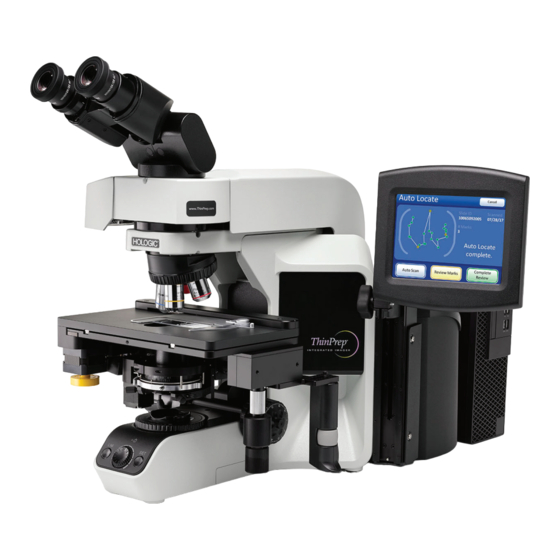
- Page 1 ThinPrep Integrated Imager ® Operator’s Manual...
- Page 2 ThinPrep ® Integrated Imager Operator’s Manual Hologic, Inc. 250 Campus Drive Marlborough, MA 01752 USA Tel: 1-800-442-9892 1-508-263-2900 Fax: 1-508-229-2795 Web: www.hologic.com For Use With Version 1.x.y Software MAN-05283-001...
- Page 3 Integrated Imager is a PC-based automated imaging and review system for use with ThinPrep cervical cytology sample slides. The ThinPrep Integrated Imager is intended to help a cytotechnologist or pathologist highlight areas of a slide for further manual review. The Product is not a replacement for manual review.
- Page 5 Operation Summary and Clinical Information The ThinPrep® Integrated Imager ...
- Page 6 The cytotechnologist determines specimen adequacy and the presence of infections during the review of the 22 fields of view presented by the ThinPrep Integrated Imager. Either of two methods can be used to determine specimen adequacy. The first method is to count cells and determine the average number of cells in the 22 fields of view presented by the Imager.
- Page 7 The ThinPrep Integrated Imager is only indicated for use with the ThinPrep Pap Test. The ThinPrep Integrated Imager is only indicated for the ThinPrep Pap Test slides prepared with the ® ® ThinPrep 2000 System and the ThinPrep 5000 processor.
- Page 8 Center Trial Evaluating the Primary Screening Capability of the ThinPrep Imaging System” was to show that routine screening of ThinPrep Pap Test slides using the ThinPrep Imaging System is equivalent to a manual review of ThinPrep slides for all categories used for cytologic diagnosis...
- Page 9 1.1% 0.7% 0.4% 0.6% HSIL+ prevalence 120,000 70,200 280,000 105,000 ThinPrep Pap Tests Per Year Number of Cytotechnologists Number of Cytotechnologists in Study Number of Cytopathologists Number of Cytopathologists in Study G.1.2 Descriptive Diagnosis Sensitivity and Specificity Estimates A panel of three independent cytopathologists adjudicated slides from all discordant (one-grade or higher cytologic difference) descriptive diagnosis cases (639), all concordant positive cases (355) and a random 5% subset of the 8550 negative concordant cases (428).
- Page 10 Table 2. Manual Review Versus Imager Review, Descriptive Diagnosis Summary Sensitivity Specificity Manual Imager Difference Manual Imager Difference Threshold (95% CI) (95% CI) (95% CI) (95% CI) (95% CI) (95% CI) 75.6% 82.0% +6.4% 97.6% 97.8% +0.2% ASCUS+ (72.2% to 78.8%) (78.8% to 84.8%) (2.6% to 10.0%) (97.2% to 97.9%)
- Page 11 ® The objective of the study entitled “Multi-Center Evaluation of the ThinPrep Integrated Imager” ® was to show that routine screening of ThinPrep Pap Test slides prepared on the ThinPrep 2000 ® System and the ThinPrep 5000 processor using the ThinPrep Integrated Imager is similar to the...
- Page 12 Main Eligibility Criteria Inclusion Criteria Study slides (two slides per case, one slide was prepared on the ThinPrep 2000 System and another slide was prepared on the ThinPrep 5000 processor) were produced, reviewed manually and adjudicated during the execution of a previous study .
- Page 13 (98.1% to 98.7%) (-0.2% to 0.3%) In addition, the data is presented below stratified by the type of processor used (ThinPrep 2000 System and ThinPrep 5000 processor). In all abnormal cases, the sensitivity for the Integrated Imager was higher than the ThinPrep Imaging System across all thresholds. There was a slight decrease in specificity for the Integrated Imager as compared to the ThinPrep Imaging System.
- Page 14 Table 6. ThinPrep Imaging System (TIS) Versus Integrated Imager (I2), Descriptive Diagnosis Summary (ThinPrep 2000 System-processed Slides Only) Sensitivity Specificity Difference Difference Threshold [# of reads] [# of reads] [# of reads] [# of reads] [# of reads] [# of reads]...
- Page 15 Table 8. “True Negative” (NILM) Contingency Table (for All Sites Combined) Overall Adjudicated NILM TIS vs. I2 UNSAT NILM ASCUS LSIL ASC-H AGUS HSIL Cancer UNSAT 3735 NILM ASCUS LSIL ASC-H AGUS HSIL Cancer Table 9. “True ASCUS” Contingency Table (for All Sites Combined) Overall Adjudicated ASCUS TIS vs.
- Page 16 Table 11. “True ASC-H” Contingency Table (for All Sites Combined) Overall Adjudicated ASC-H TIS vs. I2 UNSAT NILM ASCUS LSIL ASC-H AGUS HSIL Cancer UNSAT NILM ASCUS LSIL ASC-H AGUS HSIL Cancer Table 12. “True AGUS” Contingency Table (for All Sites Combined) Overall Adjudicated AGUS TIS vs.
- Page 17 7542, or 5.6%) than TIS Review (402 out of 7542, or 5.3%), however this was not statistically significant. Conclusion The sensitivity and specificity of Integrated Imager for review of ThinPrep 2000 slides and ThinPrep 5000 slides are similar to the sensitivity and specificity of the ThinPrep Imaging System.
- Page 18 (3) times on the same instrument with a washout period of a minimum of 14 days. The 260 slides used in this study were previously prepared from ThinPrep specimens and had an adjudicated cytology diagnosis.
- Page 19 Table 16. Summarized Results of Within-instrument Study Abnormal FOV Imager Abnormal Category+ Normal % zero Median 69.6% 11.0% 70.4% NILM 78.1% 4.3% 78.4% 75.9% 75.9% 13.3% 25.0% ASCUS 71.9% 71.9% 5.0% 28.1% 97.3% 93.2% 3.3% 2.8% LSIL 96.0% 94.0% 0.7% 4.0% 93.3% 86.7%...
- Page 20 Table 17. Summarized Results of Between-instrument Study Imager Abnormal Category+ Normal 90.0% NILM 88.1% 64.4% 64.4% ASCUS 71.7% 71.7% 95.0% 75.0% LSIL 96.9% 80.6% 87.7% 62.6% ASC-H 92.8% 63.6% 53.8% 37.6% AGUS 67.5% 57.3% 97.7% 54.7% HSIL 99.3% 64.7% 100% 63.2% CANCER 100%...
- Page 21 All slides were prepared from ThinPrep specimens of known cytology diagnoses, on a ThinPrep processor, and stained with ThinPrep Stain. Sets of 400 randomized slides per CT, each with approximately 10% abnormal diagnosis were provided in order to fully occupy a CT for three (3) full days of screening.
- Page 22 Table 19. Total Slides Reviewed by Review Type / CT (% Autoscan = #FOV+FMR / Total # Slides Reviewed over 3 Days) Sequential Review Batched Review CT #1 CT #2 CT #3 Overall CT #1 CT #2 CT #3 Overall Total # slides 1058 reviewed...
- Page 23 Table 21. PPA and NPA Results by Cytotechnologist Based on Adjudicated Results. (Positive Results Mean ASC-US+) Sequential Modality Batched Modality 100% CT #1 100% 100% CT #2 100% CT #3 Overall Workload is defined by CLIA as a maximum limit of 100 slides in no less than an 8-hour workday.
- Page 24 [(# slides FMR) (1) + (# slides FOV) (0.65) + (# slides FOV+FMR) (1.65)] = 100 slides ® Note: The ThinPrep Integrated Imager workload limit in an 8-hour workday includes all activities needed to process the cases, not exclusively time spent using the microscope: ...
- Page 25 24-hour period in no less than an 8-hour day. When conducting manual review, refer to the CLIA requirements for calculating workload limits. Manual review includes the following types of slides: o Slides reviewed on the ThinPrep Imaging System using the Autoscan feature o Slides reviewed without the ThinPrep Imaging System o Non-gynecologic slides.
- Page 35 Considering the technological similarity of the ThinPrep Imaging System and the comparative clinical study results, it is concluded that the ThinPrep Integrated Imager is similar to the ThinPrep ® Imaging System and may be used as replacement for manual review of ThinPrep...
- Page 36 Hologic, Inc. AW-17110-001 Rev. 001 4-2018 250 Campus Drive © 2018 Hologic, Inc. All rights reserved. Marlborough, MA 01752 USA 1-800-442-9892 www. hologic.com MAN-05359-001 -001 Rev. 001 page 32 of 32...
-
Page 38: Table Of Contents
T a b l e o f C o n t e n t s Contents Chapter One INTRODUCTION SECTION A: Overview ® SECTION B: The ThinPrep Imaging and Review Process SECTION C: Specimen Preparation SECTION D: Integrated Imager Technical Specifications SECTION E: Internal Quality Control 1.10 SECTION F: Integrated Imager Hazards 1.11... - Page 39 SECTION C: Using the Touch Screen and Review Controls SECTION D: Slide Imaging SECTION E: Slide Review 4.11 SECTION F: Review of Slides Not for Use with ® ThinPrep Imaging 4.21 Chapter Five MAINTENANCE SECTION A: General Cleaning SECTION B: Koehler Alignment...
-
Page 41: Chapter One
Integrated Imager is an automated cytology review microscope with ThinPrep slide imaging functionality. It is specifically designed to image and review ThinPrep Pap Test microscope slides on-demand. It also has the ability to act as a conventional microscope when not used in conjunction with ThinPrep imaging. -
Page 42: Section B: The Thinprep Imaging And Review Process
NTRODUCTION SECTION THE THINPREP IMAGING AND REVIEW PROCESS Imaging ® A prepared ThinPrep Pap Test microscope slide is loaded onto the stage of the device. A slide identification camera reads the slide label ID and compares it against slide IDs already in the computer database. - Page 43 Auto Locate and slide review 3a. Review CT controls stage Scan entire cell spot and (abnormal only) additional slide review 4. Remove Ready to remove slide Remove slide and place in tray Figure 1-2 Integrated Imager Workflow ® ThinPrep Integrated Imager Operator’s Manual...
- Page 44 Abnormal slides are reviewed by a cytopathologist for interpretation Normal slide and diagnosis. Figure 1-3 ThinPrep Imaging Process ® ThinPrep Integrated Imager Operator’s Manual...
-
Page 45: Section C: Specimen Preparation
Lubricants can adhere to the filter membrane and may cause poor cell transfer to the slide. Stain - do not substitute solutions for the ThinPrep Stain solutions. Follow the stain protocols exactly as they are written. Refer to the ThinPrep Stain User’s Manual. -
Page 46: Section D: Integrated Imager Technical Specifications
X,Y axis stage control knobs 40X objective (blue stripe) (stage control) 10. Microscope power switch 10X objective position sensor (on back left of microscope with Revolving nosepiece black side panel) Figure 1-4 Integrated Imager Components ® ThinPrep Integrated Imager Operator’s Manual... - Page 47 NTRODUCTION Dimensions 609 mm 24 in. 711 mm 28 in. 457 mm 18 in. Figure 1-5 Integrated Imager Dimensions ® ThinPrep Integrated Imager Operator’s Manual...
- Page 48 ThinPrep Microscope Slide for Use with the Imaging System The ThinPrep microscope slide is used by the ThinPrep Processor in preparing the patient slide. The slide utilizes fiducial marks, or fixed reference points, which are permanently printed features on the slide that are used to register the slide position on the stage.
- Page 49 The electromagnetic environment should be evaluated prior to operation of the equipment. Do not use this device in close proximity to sources of strong electromagnetic radiation (e.g., unshielded RF sources), as these may interfere with the proper operation. ® ThinPrep Integrated Imager Operator’s Manual...
-
Page 50: Section E: Internal Quality Control
The operator is alerted to any malfunction via a message on the user interface. If the system does not function or there are persistent errors, contact Hologic Technical Support(refer to Chapter 7, Service Information). -
Page 51: Section F: Integrated Imager Hazards
A CAUTION advises against actions or situations that could damage equipment, produce inaccurate data or invalidate a procedure, although personal injury is unlikely. • provides useful information within the context of the instructions being provided. Note 1.11 ® ThinPrep Integrated Imager Operator’s Manual... - Page 52 Section 5.1.1 Date of Indicates the date when the ISO 15223-1 Medical manufacture medical device was manufactured. devices—Symbols to be used with medical device labeling and information to be supplied, Section 5.1.3 1.12 ® ThinPrep Integrated Imager Operator’s Manual...
- Page 53 And this instrument uses the following markings: Waste Electrical and Electronic Equipment Do not dispose in municipal waste Contact Hologic for disposal of the instrument On (Power switch on the microscope) Off (Power switch on the microscope) Lamp intensity adjustment...
- Page 54 USB port USB port (5) Power Front and Rear of Computer (Note: The number and exact location of ports may be different, depending on the PC model you have.) Figure 1-7 Location of Labels 1.14 ® ThinPrep Integrated Imager Operator’s Manual...
-
Page 55: Section G: Disposal
NTRODUCTION Warnings Used in this Manual WARNING: Service Installation Only. This instrument is to be installed by trained Hologic personnel only. WARNING: Moving Parts. The instrument contains moving parts. Keep hands, loose clothing, jewelry, etc., clear. WARNING: Grounded Outlet. To ensure safe operation of the instruments, use a three-wire grounded outlet. - Page 56 NTRODUCTION This page intentionally left blank. 1.16 ® ThinPrep Integrated Imager Operator’s Manual...
-
Page 58: Chapter Two
PREPARATION PRIOR TO INSTALLATION Pre-Installation Site Assessment A pre-installation site assessment is performed by Hologic service personnel. Be sure to have prepared any and all site configuration requirements as instructed by the service personnel. The Integrated Imager will require two outlets to power the instrument. Make sure there is adequate electrical supply within 2 meters of the instrument. -
Page 59: Section D: Moving The Integrated Imager
The Integrated Imager is a precision instrument and should be handled with care. If the system must be moved, the controller and computer PC must be disconnected from one another, moved separately and reconnected at the new location. ® ThinPrep Integrated Imager Operator’s Manual... - Page 60 The computer may be set up to face either side, or with the use of the extension cable set, it Note: can be placed further away from the microscope and controller. Your final configuration may look slightly different than Figure 2-2. The cable connections to the computer ports remain the same. ® ThinPrep Integrated Imager Operator’s Manual...
-
Page 61: Section E: Connecting Integrated Imager Components
Figure 2-3 Moving the Integrated Imager SECTION CONNECTING INTEGRATED IMAGER COMPONENTS The Integrated Imager components must be fully assembled before turning on the power and using the instrument. Hologic service personnel will assemble the instrument: • Controller • Computer •... - Page 62 The light path and camera focus have been optimized by placement of spacers in the assembly of the optical components. Do not add or remove spacers or risers. If an optional telescoping head is being used, be sure to use the specific riser that Hologic supplies.
- Page 63 See Figure 2-5. Using the Allen screwdriver that comes with the Integrated Imager (see Figure 1-4), loosen but do not remove the Allen screw that holds the review control to the mounting bracket. ® ThinPrep Integrated Imager Operator’s Manual...
-
Page 64: Section F: Power On The Integrated Imager
2. Then power on the computer. Microscope power switch Computer power switch Figure 2-6 Power Switches The power switch for the Integrated Imager is located on the back left of the microscope. Press the switch to the on position. ® ThinPrep Integrated Imager Operator’s Manual... - Page 65 Figure 2-7 Integrated Imager Startup Screen The instrument is ready for use when the application main screen is displayed (Figure 2-8). Figure 2-8 Application Main Screen ® ThinPrep Integrated Imager Operator’s Manual...
-
Page 66: Section G: System Settings
The Integrated Imager may be stored in the location where it was installed. When it is not in use, the power should be turned off. Cover the instrument with the provided microscope dust cover. SECTION SYSTEM SHUTDOWN Normal Shutdown Shut Down button Figure 2-9 Shutdown ® ThinPrep Integrated Imager Operator’s Manual... - Page 67 Normal Shutdown. Remove any slides that may be on the stage. Completely remove power by unplugging the controller power cord and the computer cord from the power outlet. Cover the instrument with the provided dust cover. 2.10 ® ThinPrep Integrated Imager Operator’s Manual...
-
Page 69: Chapter Three
The slides are reviewed by a cytotechnologist. The instrument may also be used as a conventional microscope, for viewing slides not associated with the ThinPrep imaging process. The Integrated Imager enables the user to administer certain functions, such as user preferences, system settings and database backup. - Page 70 START........... . . 3.45 ® ThinPrep Integrated Imager Operator’s Manual...
-
Page 71: Section B: Startup
Service - This is a password-protected module for use by Hologic service personnel only. • Login - Enter a user ID to access the system for ThinPrep Imaging and Slide Review func- tions. Refer to “LOGIN” on page 3.30. •... -
Page 72: Section C: Administrative Options
Log In button to initiate a session with the Integrated Imager. Information associated with a User ID becomes part of the slide data record when a slide is imaged and when a slide is reviewed, using the Integrated Imager. ® ThinPrep Integrated Imager Operator’s Manual... - Page 73 “invalid ID” message will appear and a new ID number must be entered. Enter a 3-digit ID number (from 100 to 998) using the keypad. Figure 3-6 Add User Account Screen ® ThinPrep Integrated Imager Operator’s Manual...
- Page 74 User ID numbers must be between the range of 100 to 998. Numbers beginning with a zero Note: will cause errors. User ID 999 is reserved for Hologic service personnel. Do not use this ID. Note: Figure 3-7 Enter User Name Screen Press the letter buttons to enter a first name.
- Page 75 The three-digit User ID cannot be changed once it is created. It can only be retired. Note: A User Account cannot be edited or deleted once a slide data record is associated with it (by imaging or reviewing one or more slides). ® ThinPrep Integrated Imager Operator’s Manual...
-
Page 76: System Settings
The System Settings interface allows you to set or update Integrated Imager settings. The following parameters can be set: • Date • Time • Lab Name • Instrument Name • Label Format • Language ® ThinPrep Integrated Imager Operator’s Manual... - Page 77 Press the Save Changes button to return to the System Settings screen. See Figure 3-13. Figure 3-13 Edit Date Screen Depending on which language has been selected, the order of month and day on the display Note: may change to reflect customary usage. ® ThinPrep Integrated Imager Operator’s Manual...
- Page 78 Save Changes button to save and return to the System Settings screen. See Figure 3-15. Depending on which language has been selected, the clock on the display may change from Note: 12 hour to 24 hour, to reflect customary usage. Figure 3-15 Edit Time Screen 3.10 ® ThinPrep Integrated Imager Operator’s Manual...
- Page 79 Continue to save the entry and return to Sys- Continue to save the entry and return to Sys- tem Settings screen tem Settings screen Figure 3-17 Edit Lab Name Keyboard and Keypad Screens 3.11 ® ThinPrep Integrated Imager Operator’s Manual...
- Page 80 If a lab name is used, the name will appear on every report that is generated by the Integrated Note: Imager (usage history, system errors). It is not necessary to enable a lab name. 3.12 ® ThinPrep Integrated Imager Operator’s Manual...
- Page 81 Press the Switch Keys button to display a keypad screen to enter numbers. Switch between keyboard and keypad as often as desired before saving changes. Press the Continue button to save and return to the System Settings screen. Figure 3-20 Edit Instrument Name Screen 3.13 ® ThinPrep Integrated Imager Operator’s Manual...
- Page 82 1D barcode type(s) used in your facility Select 2D Barcode label format. See Table 3.1, “Slide Restrictions Based on Barcode Symbology Used,” on page 3.15 for more information. Figure 3-22 Edit Label Format Screens 3.14 ® ThinPrep Integrated Imager Operator’s Manual...
- Page 83 All printable ASCII 128 characters are supported. A minimum of 5 characters is required and a maximum of 8 characters will fit on a slide. 2-D datamatrix All printable ASCII 128 characters are supported. A maximum of 16 characters is supported. 3.15 ® ThinPrep Integrated Imager Operator’s Manual...
- Page 84 2-D barcode 1-Dimensional barcode examples example OCR format Figure 3-23 Examples of Barcodes on a ThinPrep Slide Language Figure 3-24 Language Settings Button Press the Language Settings button to change the language that is displayed on the user interface and on the reports.
-
Page 85: Reports And Logs
Usage History - Lists the number of slides imaged and reviewed on the Integrated Imager • Slide search - a specific slide ID or range of IDs and the associated slide data can be found in the database using this search. 3.17 ® ThinPrep Integrated Imager Operator’s Manual... - Page 86 Use the up/down arrows to scroll through the list using the touch screen. To download this report, place a USB key in the appropriate port of the computer and press the Save to USB button. 3.18 ® ThinPrep Integrated Imager Operator’s Manual...
- Page 87 Imager within a specific time period. Press the Usage History button. First you will select if the usage history report is for a one-week time period or for a one-day time period. See Figure 3-31. 3.19 ® ThinPrep Integrated Imager Operator’s Manual...
- Page 88 Press the Done button on the History screen to generate the report, displayed on the following page. The default view is the Usage Summary screen. It can be changed to the Usage Details screen. 3.20 ® ThinPrep Integrated Imager Operator’s Manual...
- Page 89 This summary may be saved to a USB key by pressing the Save to USB button. A detailed list of the slides reviewed is displayed by pressing the View Details button. Refer to the following section. 3.21 ® ThinPrep Integrated Imager Operator’s Manual...
- Page 90 Figure 3-34 Slide Search Report Button A specific slide number or range of slide numbers can be searched for in the database. After pressing the Slide Search button, a keypad will display. See Figure 3-35. 3.22 ® ThinPrep Integrated Imager Operator’s Manual...
- Page 91 Slide data Done button, to Save to USB return to Reports and Logs screen Figure 3-36 Slide Search Report Screen The slide IDs are listed with any available data for that ID: 3.23 ® ThinPrep Integrated Imager Operator’s Manual...
-
Page 92: Database Backup
From the Administrative Options screen, touch the Database Backup button to display the backup screen. Select the media type, CD or USB. Press Cancel to cancel the backup and return to the Administrative Options screen. Figure 3-38 Database Backup, Select Backup Type 3.24 ® ThinPrep Integrated Imager Operator’s Manual... - Page 93 The CD ROM must be blank, or the system will reject it. You cannot accumulate backups onto Note: a single disk. The USB storage device, however, does not need to be blank. The USB storage device only needs adequate space to store the database backup. 3.25 ® ThinPrep Integrated Imager Operator’s Manual...
-
Page 94: Password Settings
The Integrated Imager can use another USB storage device to save reports. Refer to “SAVE TO USB” on page 3.42. Refer to the Chapter 6, Troubleshooting, if any other messages display during backup. (Backup to CD shown) Figure 3-41 Database Backup Password Settings Figure 3-42 Password Settings Button 3.26 ® ThinPrep Integrated Imager Operator’s Manual... - Page 95 The word may be up to 20 alpha characters long and it is case-sensitive. Press the Continue button and the display returns to the Password Settings screen. The password is visible in the password field. 3.27 ® ThinPrep Integrated Imager Operator’s Manual...
- Page 96 Once Admin Options screen is exited, the system displays a keyboard and prompts for a password to access that screen again. See Figure 3-46. If the password is lost or forgotten, contact Hologic Technical Support (Chapter 7, Service Information). Figure 3-46 Password Required 3.28...
- Page 97 Figure 3-47 Disable the Password The password is removed. If an admin password is used later, the password has to be set up Note: again, as described in “To Set a Password” on page 3.27. 3.29 ® ThinPrep Integrated Imager Operator’s Manual...
-
Page 98: Section D: Login
ID with that number, or if that number has been retired. See “Add account” on page 3.5 for creating a user ID. See “User Preferences” on page 3.32 for selecting user preferences. 3.30 ® ThinPrep Integrated Imager Operator’s Manual... -
Page 99: Section E: Main Menu
Logout - to end the session with the Integrated Imager, press the Logout button. The system will return to the Startup screen. The instrument may be powered off or a user may log in to begin a new session. 3.31 ® ThinPrep Integrated Imager Operator’s Manual... -
Page 100: Section F: User Preferences
Direction Up-Down or Direction Left-Right. (Figure 3-51.) To view the selection through the eyepieces, ensure the 10X objective is in position, load a slide in the slide holder for reference, and press the Preview button. 3.32 ® ThinPrep Integrated Imager Operator’s Manual... - Page 101 Type The Auto Scan function presents the entire cell spot in a defined path at 10X magnification. Three types of scan motion are selectable: • Automatic Start/Stop • Semiautomatic Start/Stop • Manual 3.33 ® ThinPrep Integrated Imager Operator’s Manual...
- Page 102 Upon resuming, the area of review will return to the part of the cell spot where you left off and continue to present the rest of the cell spot. The display on the touch screen is shown below. Press the Cancel Scan button on the touch screen to stop the preview. 3.34 ® ThinPrep Integrated Imager Operator’s Manual...
- Page 103 From the User Preferences screen, press Save Changes to retain your preference now, or continue to set your next preference. Auto Scan - Semiautomatic Start/Stop Stage speed Slower Faster Done. Save setting adjustments and return to User Preferences screen. Figure 3-55 Select Semiautomatic Start/Stop Scan 3.35 ® ThinPrep Integrated Imager Operator’s Manual...
- Page 104 Figure 3-56 Semiautomatic Scan Mode Preview Continue to adjust and preview the stage speed until it is satisfactory. Press the Done button to save the settings and return to the User Preferences screen. 3.36 ® ThinPrep Integrated Imager Operator’s Manual...
-
Page 105: Scan Direction
Both axis stage control knobs will be activated and the user can move about the cell spot. The Auto Scan must be resumed from pause in order to complete the scan. Note: To resume Auto Scan: 3.37 ® ThinPrep Integrated Imager Operator’s Manual... -
Page 106: Speed
The Auto Locate Speed setting adjusts how quickly the stage moves from field to field during presentation of the 22 fields of view. The stage moves to each field of view and stops until the user advances using the Next function. 3.38 ® ThinPrep Integrated Imager Operator’s Manual... - Page 107 To view the selection through the eyepieces, load a slide in the slide holder for reference, and press the Preview button. Observe the speed at which the stage advances. The preview displayed on the touch screen is shown below. Figure 3-60 Auto Locate Speed Preview Screen 3.39 ® ThinPrep Integrated Imager Operator’s Manual...
-
Page 108: Sound
The volume of the beep may be adjusted quieter or louder by repeatedly pressing the -5 or +5 buttons to decrease or increase the volume. (Figure 3-61.) Test it by pressing the Preview button to hear the beep. To disable the audible beep, adjust it to its lowest setting. 3.40 ® ThinPrep Integrated Imager Operator’s Manual... -
Page 109: Mark Indicator
Therefore, if an audible beep is desired to indicate end of Auto Locate and end of Auto Scan, it will also be heard for mark/unmark. If the audible beep is turned too low to hear, then it will not be heard for Auto Locate, Auto Scan and mark/unmark. 3.41 ® ThinPrep Integrated Imager Operator’s Manual... -
Page 110: Section G: Save To Usb
USB key, if desired. These are: • User Accounts • System Events • Usage Summary • Slide Search The Integrated Imager has two USB ports available for use. Either may be used. (See Figure 3-66.) 3.42 ® ThinPrep Integrated Imager Operator’s Manual... - Page 111 Reports are automatically named by the convention of “Report type - Instrument Name - Year Month Day Hour Minute Second. XML.” This is illustrated below. A style sheet file IIReports.xsl” is also written to the USB key. This is needed to view the reports on a browser. 3.43 ® ThinPrep Integrated Imager Operator’s Manual...
- Page 112 Figure 3-69 Example of a Usage Summary Report The Integrated Imager can use a USB storage device to save the database backup. Refer to “Database Backup” on page 3.24. 3.44 ® ThinPrep Integrated Imager Operator’s Manual...
-
Page 113: Section H: Start
NTERFACE SECTION START (Begin using the Integrated Imager) Press the Start button to begin imaging and review of a slide. Refer to Chapter 4, Operation for instructions on operating the Integrated Imager. 3.45 ® ThinPrep Integrated Imager Operator’s Manual... - Page 114 NTERFACE This page intentionally left blank. 3.46 ® ThinPrep Integrated Imager Operator’s Manual...
-
Page 116: Chapter Four
ThinPrep imaging process. Slide Preparation Proper slide preparation is critical to successful imaging of the ThinPrep Pap Test microscope slide. Prior to being imaged on the Integrated Imager, the slide must be: ®... - Page 117 Serial number of the Integrated Imager that reviewed the slide • Operator ID for each review of the slide (including subsequent reviews) • Status whether Auto Scan was completed for each review • Electronic mark coordinates ® ThinPrep Integrated Imager Operator’s Manual...
-
Page 118: Section B: Materials Required Prior To Operation
1. Run Shut Down Next Slide 2. Power off Microscope Figure 4-1 Typical Slide Review Process SECTION MATERIALS REQUIRED PRIOR TO OPERATION ® Prepared ThinPrep Pap Test microscope slides Integrated Imager Marking pen for slide marking ® ThinPrep Integrated Imager Operator’s Manual... -
Page 119: Section C: Using The Touch Screen And Review Controls
A slide can only be imaged once on an Integrated Imager. • The mounting medium must be thoroughly dry. ® • The label must be appropriate for the ThinPrep Integrated Imager (refer to page 3.14). • Always keep the instrument in proper Koehler alignment (refer to page 5.2). •... - Page 120 The review control has a scroll wheel that acts like the scroll wheel found on a computer mouse. It enables the operator to execute the main review functions (Next, Previous, Mark) without having to take their eyes away from the binoculars. ® ThinPrep Integrated Imager Operator’s Manual...
- Page 121 Previous used to adjust user preference settings Mark used to electronically mark or unmark areas for review or dotting ® ThinPrep Integrated Imager Operator’s Manual...
-
Page 122: Section D: Slide Imaging
Load a slide into the slide holder on the stage. (See Figure 4-6.) With your right thumb against the slide clip button, open the slide clip. With your left hand, load the slide onto the stage with the label ® ThinPrep Integrated Imager Operator’s Manual... - Page 123 Figure 4-7 Reading Slide ID If the slide ID is already in the database, a message will state so. Review is available as an option or the slide review may be cancelled. See Figure 4-8. ® ThinPrep Integrated Imager Operator’s Manual...
- Page 124 Do not bump the instrument while imaging a slide. Cancel button, to stop imaging and return to the main screen Slide ID and date scanned Status message Figure 4-9 Imaging Slide Screen ® ThinPrep Integrated Imager Operator’s Manual...
- Page 125 Imager performs functional checks prior to completion. Figure 4-10 Imaging Slide in Process Do not remove the slide from the stage during imaging. To cancel imaging, press the Cancel button. Figure 4-11 Imaging Complete 4.10 ® ThinPrep Integrated Imager Operator’s Manual...
-
Page 126: Section E: Slide Review
Mark using the review control or the touch screen To advance to the next location, the 10X objective must be in the engaged position. Press Next using the review control or the touch screen. 4.11 ® ThinPrep Integrated Imager Operator’s Manual... - Page 127 When the review control or touch screen is pressed to make a mark, the indicator will blink or beep once. If it is pressed again to unmark the area, the indicator blinks or beeps twice, to differentiate. 4.12 ® ThinPrep Integrated Imager Operator’s Manual...
- Page 128 After presenting the 22 fields of interest in the Auto Locate mode, the stage positions the cell spot at the 6:00 position (on the stage) and stops. (On the user interface, the path through the fields of view is removed.) See Figure 4-14. 4.13 ® ThinPrep Integrated Imager Operator’s Manual...
- Page 129 Cancel button to cancel the review (No slide review data will be written to the data- base.) 1. Nayar R, Wilbur DC. (eds). The Bethesda System for Reporting Cervical Cytology: Definitions, Criteria, and Explanatory Notes. 3rd ed. Cham, Switzerland: Springer: 2015 4.14 ® ThinPrep Integrated Imager Operator’s Manual...
- Page 130 Previous button, to go back one field of view Mark button, to make addi- tional marks or remove exist- Next button, to ing marks advance to next field of view Figure 4-16 Review Marks Screen 4.15 ® ThinPrep Integrated Imager Operator’s Manual...
- Page 131 For all modes, changing the objective will pause the scan. The scan cannot con- tinue until the 10X objective is in position. Electronic marks may be added, removed or left as is. 4.16 ® ThinPrep Integrated Imager Operator’s Manual...
- Page 132 ID and writes slide data to the database. See page 4.18. Figure 4-18 Auto Scan Complete The operator may: • press Review Marks to see the electronically marked locations again • manually mark the slide 4.17 ® ThinPrep Integrated Imager Operator’s Manual...
- Page 133 If the slide has not been imaged, the Integrated Imager will automatically image it. (Refer to “Imag- ing” on page 4.1.) If the slide has been imaged already, the Review Slide button is displayed. (See Figure 4.8.) 4.18 ® ThinPrep Integrated Imager Operator’s Manual...
- Page 134 Auto Locate pres- Auto Locate Com- ents the 22 fields plete screen of view including any electronic marks that were made previously. Figure 4-21 Auto Locate During Subsequent Review 4.19 ® ThinPrep Integrated Imager Operator’s Manual...
- Page 135 (Electronic marks from previous reviews can- not be deleted.) The Cancel Scan button will cancel the scan and transition to the Auto Locate Complete screen. See Figure 4-23. 4.20 ® ThinPrep Integrated Imager Operator’s Manual...
-
Page 136: Thinprep Imaging
Coordinates of any electronic marks that were added during the review SECTION REVIEW OF SLIDES NOT FOR USE WITH THINPREP IMAGING If the Integrated Imager is used to look at non-Imager slides, the power must be on in order for the controller to power the illumination, stage and X,Y axis stage controls. - Page 137 PERATION This page intentionally left blank. 4.22 ® ThinPrep Integrated Imager Operator’s Manual...
-
Page 139: Chapter Five
Either remove it before air dusting, or hold it down with a finger while cleaning. Figure 5-1 Functional Check Features To Be Kept Clean Do not detach or remove any covers or panels on the microscope, controller or computer. Note: ® ThinPrep Integrated Imager Operator’s Manual... -
Page 140: Section B: Koehler Alignment
7. Open the collector aperture until it just disappears from view. 8. Adjust the condenser aperture to achieve desired contrast by moving the condenser diaphragm adjustment lever left or right to close or open the aperture. ® ThinPrep Integrated Imager Operator’s Manual... -
Page 142: Chapter Six
Once the OK button has been pressed, slide imaging and review may be performed. However, if the problem is not resolved by the next backup time, the system will lock and will require technical ser- vice intervention. ® ThinPrep Integrated Imager Operator’s Manual... -
Page 143: Section B: User-Initiated Database Backup Failed
Problem with the computer. Contact Hologic Technical Support. Failed to write data to the media Check that the disk is CD ROM and not DVD. Confirm it is blank and seated correctly in the drive. Otherwise contact Hologic Technical Support. Load a writable CD into the drive The CD drive is empty or the system does not recognize the disk. -
Page 144: Section C: Invalid Slide Id
The label format is not compatible with the system. • The label is damaged, illegible or missing. • Mechanical failure of the slide ID reader device After attempting to scan the slide ID and failing, a message is displayed: ® ThinPrep Integrated Imager Operator’s Manual... - Page 145 Continue to review the slide as usual. At the end of slide review, when the system would normally scan the ID to confirm identity of the slide, a message prompts the user to confirm the slide ID. ® ThinPrep Integrated Imager Operator’s Manual...
-
Page 146: Section E: Slide Id Mismatch While Completing The Review
This may be caused by: • Removal of the slide from the stage during review • Malfunction of the slide ID reader ® ThinPrep Integrated Imager Operator’s Manual... -
Page 147: Section F: Error Handling
Proper slide preparation is critical to the success of imaging by the Integrated Imager. If your Note: ® laboratory does any of the ThinPrep slide preparation processes, please consult the appro- priate user documentation that came with the equipment. Recoverable Errors Recoverable errors are system errors that the Integrated Imager can recover from with user interven- tion. - Page 148 OK button. The user interface transitions to a restricted version of the main screen, with only the Restart, Shut Down and Service buttons enabled. See Figure 6-7. Figure 6-7 System Disabled Screen ® ThinPrep Integrated Imager Operator’s Manual...
- Page 149 The error should be cleared when the system next boots up. If it persists or the instrument cannot successfully boot up, contact Technical Support. The Service button is available for Hologic service personnel to access the service mode, if a field ser- vice call is necessary.
- Page 150 Slide cannot be processed Recoverable Press OK. Attempt to reimage slide. 11307 Slide cannot be processed Recoverable Press OK. Attempt to reimage slide. 11308 Slide cannot be processed Recoverable Press OK. Attempt to reimage slide. ® ThinPrep Integrated Imager Operator’s Manual...
- Page 151 Press OK. Restart or shut down instrument and restart. 12202 Invalid database operation Non-recoverable Press OK. Restart or shut down instrument and restart. 12203 Database null reference Non-recoverable Press OK. Restart or shut down instrument and restart. 6.10 ® ThinPrep Integrated Imager Operator’s Manual...
- Page 152 Error Type Action Number 12500 Slide record has invalid data Recoverable Press OK. Slide can only be manually reviewed. 12501 Slide record has invalid data Recoverable Press OK. Slide can only be manually reviewed. 6.11 ® ThinPrep Integrated Imager Operator’s Manual...
- Page 153 ROUBLESHOOTING This page intentionally left blank. 6.12 ® ThinPrep Integrated Imager Operator’s Manual...
-
Page 155: Chapter Seven
Orders can also be faxed to the attention of Customer Service at 508-229-2795. Warranty A copy of Hologic’s limited warranty and other terms and conditions of sale may be obtained by contacting Customer Service at the numbers listed above. Technical Support ®... - Page 156 ERVICE NFORMATION This page intentionally left blank. ® ThinPrep Integrated Imager Operator’s Manual...
-
Page 158: Chapter Eight
Orders can also be faxed to the attention of Customer Service at 508-229-2795. Warranty A copy of Hologic’s limited warranty and other terms and conditions of sale may be obtained by contacting Customer Service at the numbers listed above. Protocol for Returned Goods ®... - Page 159 Optional objective ASY-03287 * If the telescoping head is installed, it must be configured with ONE Hologic riser. The telescoping head must not be used with the Olympus riser. ** The standard tilting binocular head is limited to accommodating only ONE Olympus riser.
- Page 161 Auto Scan direction 3.32 Auto Scan overlap 3.32 Auto Scan preference settings 3.32 Auto Scan type 3.32 automated database backup automatic start/stop Auto Scan 3.34 backup failed barcode format 3.14 beep volume 3.40 Index.1 ® ThinPrep Integrated Imager Operator’s Manual...
- Page 162 3.20 database backup, automated database backup, user-initiated 3.24 date, set dimensions dust cover error messages error, non-recoverable error, recoverable extended shutdown 2.10 eyepieces 1.6, 2.5, fiducial mark field of view 1.4, 4.11 Index.2 ® ThinPrep Integrated Imager Operator’s Manual...
- Page 163 Imaging process 1.2, imaging the slide Installation instrument name 3.13 Koehler alignment lab name 3.11 label format 3.14 labels, location on instrument 1.14 language, select 3.16 light intensity adjustment knob login 3.30 Index.3 ® ThinPrep Integrated Imager Operator’s Manual...
- Page 164 PreservCyt Solution 1.15 materials required microscope microscope slide Next function non-recoverable errors normal shutdown nosepiece object of interest objective objectives, 4X, 10X, 40X 1.6, 2.5, Operator’s Manual ordering information overlap, Auto Scan 3.32 Index.4 ® ThinPrep Integrated Imager Operator’s Manual...
- Page 165 1.6, review control, adjust review marks 4.15 Review process safety standards save to USB 3.42 screwdriver (on board) 1.6, semiautomatic start/stop Auto Scan 3.35 set date set time 3.10 shutdown slide data record Index.5 ® ThinPrep Integrated Imager Operator’s Manual...
- Page 166 4.2, 4.19 system disabled screen system errors 3.18 system settings system software version Technical Support temperature range time, set 3.10 touch screen 2.5, Troubleshooting usage details 3.22 usage history 3.19 usage summary 3.21 Index.6 ® ThinPrep Integrated Imager Operator’s Manual...
- Page 167 NDEX USB ports 3.43 USB storage device 3.25 user accounts User Interface user preferences 3.32 voltage volume (sound) 3.40 Warnings 1.11 weekly history report 3.20 weight 1.8, X,Y axis stage control knob tension Index.7 ® ThinPrep Integrated Imager Operator’s Manual...
- Page 168 NDEX This page intentionally left blank. Index.8 ® ThinPrep Integrated Imager Operator’s Manual...
- Page 169 Integrated Imager operator’s manual for full operating instructions, Next (roll back) including any warnings, contraindications and safety information. (roll forward) Hologic, Inc. • 250 Campus Drive • Marlborough, MA 01752 USA Review Control with Scroll Wheel Touch Screen (example) 1-800-442-9892 • 1-508-263-2900 • www.hologic.com AW-18070-001 Rev. 001...
-
Page 170: Start
Typical Slide Review Process Software Menu Overview Power On Power On 1. Microscope 2. Computer User Login Manual Admin Login Service Review Options Load Slide Optionally Password Password Scan Slide ID Protected Protected User User Accounts Slide ID in Database Start Preferences Slide ID not in database... - Page 172 Hologic, Inc. 250 Campus Drive Marlborough, MA 01752 USA +1-508-263-2900 www.hologic.com MAN-05283-001 Rev. 002...
Need help?
Do you have a question about the ThinPrep and is the answer not in the manual?
Questions and answers