Table of Contents
Advertisement
Quick Links
Advertisement
Table of Contents

Related Manuals for OPTO-EDU A33.1502


Summary of Contents for OPTO-EDU A33.1502
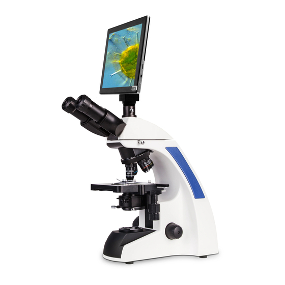
- Page 1 A33.1502 9.7" LCD Digital Microscope, 5.0M, Android Pad Instruction Manual To ensure the safety and obtain satisfactory performance, please study this instruction manual thoroughly before start to use your instrument. www.optoedu.com Page 1 of 15 sale@cnoec.com...
-
Page 2: Working Environment
Caution Attention A33.1502 Biological Microscope can only be used inside the room. Place microscope on a stable plane and keep it in balance. Power line must be retained in an easily accessible place to prevent any unnecessary accident. Working Environment As microscope is a precision instrument, improper using will make it unworkable or reduce its precision. -
Page 3: Technical Parameter
IV. Technical Parameter A33.1502 9.7" LCD Digital Biological Microscope Trinocular Head, 30° Inclined, Interpupillary Distance 48-75mm, Both Eyepiece Tube Head Diopter Adjustable Eyepiece WF10x/20mm, Diopter Adjustable, Dia. 30mm High Contrast Chromatic Free Objective 4x, 10x, 40x(S), 100x(Oil, S) Coaxial Coarse & Fine Focusing, Fine Division 0.002mm, Coarse Stroke 37.7mm/Rotation,... -
Page 4: Preparation Before Use
V. Preparation Before Use Assemble the Binocular Head 1.Loosen the fixing screw on the top of the microscope body. 2.Put the binocular head on the body properly, then tighten the fixing screw. Insert the Eyepiece 1.Standard gemel binocular head. Special-processed ring is fixed in the observation tube to tighten the eyepiece. 2.Insert the eyepiece properly. -
Page 5: Oil Immersion
6.Periodical inspection To maintain the performance of the microscope, periodical inspection is recommended. Changing of Bulb A33.1502 Series biological microscope is configured with LED illumination. There is no need to change bulb if there is no certain situation. www.optoedu.com Page 5 of 15 sale@cnoec.com... - Page 6 LCD Digital Camera (A59.3520) Real-Time Measurement Software for Android www.optoedu.com Page 6 of 15 sale@cnoec.com...
-
Page 7: Camera User Interface
1. Introduction The APP installed in Android Pad A59.3520 can adjust image parameter, measure the objects, capture image & video from the camera. It also provided simple image processing feature for particles analysis. 2. Camera User Interface 1. Preview Image – show live video of the camera. 2. - Page 8 4. Adjust image parameter of camera When the color of image is not very good, adjust the effect of image “Effect” panel. Exposure: Auto mode: Brightness of the image will be automatically adjusted, you can also adjust the target brightness. Manual mode: Manually adjust exposure time and gain.
- Page 9 Enter calibration mode Follow the Tips: 1. Drag the yellow ruler, let endpoints of the ruler close to physical ruler’s scale. We use the 0.01mm physical ruler, each big grid is 10μm, we pick two grid, that’s 20μm. www.optoedu.com Page 9 of 15 sale@cnoec.com...
- Page 10 2. Input the name of the calibration and the physical length of the ruler. We input 40x for the name, that means the magnification of objective is 40X, Then input the physical length of the ruler, that is 20μm. 3. Click “Calculate” to calculate the calibration value for current objective and preview size, and save to list.
- Page 11 TwoCircles Measure distance of two circle. Perpendicular Measure length of perpendicular Concentric Measure radius of two circles. Text Draw text annotation on the image. Annotation Change stroke width and color of rulers, and the size and color Option of the text. export Export the image with measurement rulers.
-
Page 12: Image Analysis
Use the line ruler to measure the physical ruler. We measured 1.5 big grid, the line ruler show 14.910μm, that represent the result is right. 6. Image Analysis Image is an application which used to process microscopic images on Android devices. It depends on computer vision library Open CV. - Page 13 6.1.2 Convert the image into gray scale Choose “Grayscale” function from left sidebar, click “Apply” button to confirm conversion. 6.1.3 Adjust contrast and brightness Adjust contrast and brightness of the gray scale image, increase the difference between the objects and background 6.1.4 Binarization With the binarization tool, you can change the minimum and maximum value of threshold.
- Page 14 Note: Try to make the red area do not overlap each other. 6.1.5 Particle analysis After apply binarization, open particle analysis tool: First of all, because some noise or dirty point was not clear by binarization. You need set the range of particle’s size.
- Page 15 Click “Analyze Particles” button to start analysis, the result will show blow. Click “CSV” button to export to data to report file, the default path is: /mnt/sdcard/Image/Reports www.optoedu.com Page 15 of 15 sale@cnoec.com...
Need help?
Do you have a question about the A33.1502 and is the answer not in the manual?
Questions and answers