
Related Manuals for Olympus EVIS EXERA TJF-160VR


Summary of Contents for Olympus EVIS EXERA TJF-160VR
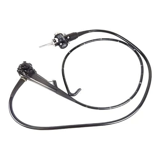
- Page 1 INSTRUCTIONS EVIS EXERA DUODENOVIDEOSCOPE OLYMPUS TJF TYPE 160VR Refer to the endoscope’s companion manual, the “OLYMPUS TJF TYPE 160VR REPROCESSING MANUAL” for reprocessing information.
-
Page 3: Table Of Contents
Contents Contents Symbols..................Important Information — Please Read Before Use....Intended use .................... Applicability of endoscopy and endoscopic treatment ......Instruction manual..................User qualifications ..................Instrument compatibility ................Reprocessing before the first use/reprocessing and storage after use..Spare equipment ..................Repair and modification ................ - Page 4 Contents Chapter 5 Troubleshooting ............Troubleshooting guide ..............Withdrawal of the endoscope with an abnormality......Returning the endoscope for repair..........Appendix..................System chart .................... EMC information..................EVIS EXERA TJF TYPE 160VR OPERATION MANUAL...
-
Page 5: Symbols
Symbols Symbols The meaning(s) of the symbol(s) shown on the package with the components, the back cover of this instruction manual and/or this instrument are as follows: Refer to instructions. Endoscope TYPE BF applied part Manufacturer Authorized representative in the European Community EVIS EXERA TJF TYPE 160VR OPERATION MANUAL... -
Page 6: Important Information - Please Read Before Use
Important Information — Please Read Before Use Intended use This instrument has been designed to be used with an Olympus video system center, light source, documentation equipment, video monitor, endo-therapy accessories (such as a biopsy forceps) and other ancillary equipment for endoscopy and endoscopic surgery within the duodenum. -
Page 7: Instruction Manual
Keep this and all related instruction manuals in a safe, accessible location. If you have any questions or comments about any information in this manual, please contact Olympus. User qualifications The operator of this instrument must be a physician or medical personnel under the supervision of a physician and must have received sufficient training in clinical endoscopic technique. -
Page 8: Reprocessing Before The First Use/Reprocessing And Storage After Use
Repair and modification This instrument does not contain any user-serviceable parts. Do not disassemble, modify or attempt to repair it; patient or operator injury and/or equipment damage can result. This instrument is to be repaired by Olympus technicians only. Signal words... -
Page 9: Warnings And Cautions
Important Information — Please Read Before Use Warnings and cautions Follow the warnings and cautions given below when handling this instrument. This information is to be supplemented by the warnings and cautions given in each chapter. • After using this instrument, reprocess and store it according to the instructions given in the endoscope’s companion reprocessing manual. - Page 10 Important Information — Please Read Before Use • Never insert or withdraw the endoscope’s insertion tube with excessive force. Otherwise, patient injury could result. • Do not pull the universal cord during an examination. The endoscope connector will be pulled out from the output socket of the light source and the endoscopic image will not be visible.
-
Page 11: Examples Of Inappropriate Handling
CV-160. Although the memory chip is durable, damage will prevent data from being backed up on it. When data are lost or damaged, contact Olympus. • Electromagnetic interference may occur on this instrument... -
Page 12: Chapter 1 Checking The Package Contents
Match all items in the package with the components shown below. Inspect each item for damage. If the instrument is damaged, a component is missing or you have any questions, do not use the instrument; immediately contact Olympus. This instrument was not disinfected or sterilized before shipment. - Page 13 Chapter 1 Checking the Package Contents Endoscope Channel cleaning brush Water-resistant cap (MH-553) (BW-20T) Injection tube (MH-946) Channel-opening cleaning AW channel cleaning brush (MH-507) adapter (MH-948) Suction valve Air/water valve Biopsy valve (MH-443, 2 pcs) (MH-438, 2 pcs) (MB-358, 10 pcs) Channel plug (MH-944) Mouthpiece Washing tube...
-
Page 14: Chapter 2 Instrument Nomenclature And Specifications
Chapter 2 Instrument Nomenclature and Specifications Chapter 2 Instrument Nomenclature and Specifications Nomenclature Universal cord 1. Suction connector 5. Electrical connector 2. S-cord connector mount Air pipe 3. Air supply connector 3. Water supply connector Light guide Product name and serial number Electrical contacts 4. - Page 15 Chapter 2 Instrument Nomenclature and Specifications 8. Suction valve (MH-443) 9. Air/water valve (MH-438) 7. UP/DOWN angulation lock 6. UP/DOWN angulation control knob 19. Elevator control lever 18. RIGHT/LEFT angulation control knob Control Suction cylinder section 17. RIGHT/LEFT angulation lock Air/water cylinder Grip section Guidewire fixing...
-
Page 16: Endoscope Functions
This connector connects the endoscope to the suction tube of the suction pump. 2. S-cord connector mount This mount connects the endoscope with the Olympus electrosurgical unit via the S-cord. The S-cord conducts leakage current from the endoscope to the electrosurgical unit. To connect the S-cord, refer to the instruction manual for the electrosurgical unit. - Page 17 Chapter 2 Instrument Nomenclature and Specifications 11. Insertion tube limit mark This mark shows the maximum point to which the endoscope may be inserted into the patient’s body. 12. Forceps elevator The elevator moves endo-therapy accessories when the elevator control lever is operated.
-
Page 18: Specifications
Chapter 2 Instrument Nomenclature and Specifications Specifications Environment Operating Ambient temperature 10 – 40°C (50 – 104°F) environment Relative humidity 30 – 85% Atmospheric pressure 700 – 1060 hPa (0.7 – 1.1 kgf/cm (10.2 – 15.4 psia) Transportation and Ambient temperature –47 to 70°C (–52.6 to 158°F) storage Relative humidity... - Page 19 Chapter 2 Instrument Nomenclature and Specifications Specifications Endoscope functions Model TJF-160VR Optical system Field of view 100° Direction of view Backward Sideviewing 5° Depth of field 5 – 60 mm Insertion tube Distal end outer ø 13.5 mm diameter Distal end enlarged 1.
- Page 20 Chapter 2 Instrument Nomenclature and Specifications Air flow rate 25 cm Note: Standard when CLV-160 (high air pressure) is used. Bending section Angulation range UP 120°, DOWN 90° RIGHT 110°, LEFT 90° Total length 1550 mm Medical Device This device complies with the Directive requirements of Directive 93/42/EEC concerning medical devices.
-
Page 21: Chapter 3 Preparation And Inspection
If the irregularities are suspected after inspection, follow the instructions given in Chapter 5, “Troubleshooting”. If this instrument malfunctions, do not use it. Return it to Olympus for repair as described in Section 5.3, “Returning the endoscope for repair”. -
Page 22: Preparation Of The Equipment
Chapter 3 Preparation and Inspection Preparation of the equipment Prepare the equipment shown in Figure 3.1 (for compatibility, see the “System chart” in the Appendix) and personal protective equipment, such as eye wear, face mask, moisture-resistant clothing and chemical-resistant gloves, before each use. -
Page 23: Inspection Of The Endoscope
Chapter 3 Preparation and Inspection Inspection of the endoscope Clean and disinfect or sterilize the endoscope as described in the “REPROCESSING MANUAL” whose cover lists the model of your endoscope. Then remove the water-resistant cap from the endoscope connector. Inspection of the endoscope Inspect the control section and the endoscope connector for excessive scratching, deformation, loose parts or other irregularities. - Page 24 Chapter 3 Preparation and Inspection Using both hands, bend the insertion tube of the endoscope into a semicircle. Then, moving your hands as shown by the arrows, confirm that the entire insertion tube can be smoothly bent to form a semicircle and that the insertion tube is sufficiently pliable (see Figure 3.3).
- Page 25 Chapter 3 Preparation and Inspection Inspection of the bending mechanisms Perform the following inspections while the bending section is straight. If the movement of the UP/DOWN angulation lock, RIGHT/LEFT angulation lock and their angulation control knobs are loose and/or not smooth, or the bending section does not angulate smoothly, the bending mechanism may be abnormal.
- Page 26 Chapter 3 Preparation and Inspection Inspection of the UP/DOWN angulation mechanism Move the UP/DOWN angulation lock all the way in the opposite direction of the “F ” mark. Then turn the UP/DOWN angulation control knob in the “ U” or the “D ”...
-
Page 27: Preparation And Inspection Of Accessories
Chapter 3 Preparation and Inspection Move the elevator control lever slowly all the way in the opposite direction of the “ U” direction. Confirm that the lever can be operated smoothly and that the forceps elevator is lowered smoothly (see Figure 3.5). Elevator control lever Forceps elevator... - Page 28 Chapter 3 Preparation and Inspection Confirm that the holes of the valves are not blocked (see Figures 3.6 and 3.7). Confirm that the valves are not deformed or cracked (see Figures 3.6 and 3.7). Check for excessive scratching or tears in the air/water valve’s seals (see Figure 3.6).
- Page 29 Chapter 3 Preparation and Inspection Inspection of the biopsy valve The biopsy valve is a consumable item that should be inspected before each use. Replace it with a new one if irregularities are observed by following inspection. An irregular, abnormal or damaged valve can reduce the efficacy of the endoscope’s suction system, and may leak or spray patient debris or fluids, posing an infection-control risk.
- Page 30 Chapter 3 Preparation and Inspection Inspection of the distal cover • The distal cover has not been sterilized prior to shipping. Using a distal cover that has not been disinfected or sterilized may result in patient infection. • Should the slightest irregularity be suspected when inspecting the distal cover, do not use it.
- Page 31 Chapter 3 Preparation and Inspection Inspection of the mouthpiece Do not use a mouthpiece that is damaged, deformed or reveals other irregularities. Doing so may cause patient injury and/or equipment damage. Placing the mouthpiece in the patient’s mouth before the procedure prevents the patient from biting and/or damaging the endoscope’s insertion tube.
-
Page 32: Attaching Accessories To The Endoscope
Chapter 3 Preparation and Inspection Attaching accessories to the endoscope The air/water valve and the suction valve do not require lubrication. Lubricants can cause swelling of the valves’ seals, which will impair valve function. Attaching the suction valve Align the two metal ridges on the underside of the suction valve with the two holes in the suction cylinder. - Page 33 Chapter 3 Preparation and Inspection Attaching the air/water valve Attach the air/water valve to the air/water cylinder of the endoscope (see Figure 3.13). Confirm that the valve fits properly without any bulging of the skirt. Air/water valve Suction valve Skirt Suction cylinder Air/water cylinder Figure 3.13...
- Page 34 Chapter 3 Preparation and Inspection Attaching the biopsy valve If a biopsy valve is not properly connected to the instrument channel port, it can reduce the efficacy of the endoscope’s suction system and may cause patient debris to leak or spray from the endoscope.
- Page 35 Chapter 3 Preparation and Inspection Attaching the distal cover • Never use the endoscope unless the distal cover is properly attached to the distal end. If the distal cover is not attached correctly, it may slip off or fall off the distal end during the examination.
- Page 36 Chapter 3 Preparation and Inspection Keep the bending section straight and move the elevator control lever to set the forceps elevator beside the side wall area of the distal end as shown in Figure 3.15. White ring Forceps elevator Hook Distal end Bending section Side wall area...
- Page 37 Chapter 3 Preparation and Inspection Push the distal cover straight onto the distal end of the endoscope until the bottom end of the distal cover contacts the end part of the white ring. Hold the bending section lightly close to the distal end and press the distal cover about 1 mm onto the distal end.
- Page 38 Chapter 3 Preparation and Inspection Confirm that there are no gaps between the distal end of the endoscope and the distal cover at the two positions indicated by the arrows in Figure 3.19. White ring Figure 3.19 Confirm that the part of the distal cover indicated by an arrow and the optic surface of the endoscope are aligned as shown in Figure 3.20.
- Page 39 Chapter 3 Preparation and Inspection Hold the bottom end of the distal cover and turn it to adjust the indication mark to the straight position as shown in Figure 3.21. Indication mark Figure 3.21 Confirm that the bottom end of the distal cover does not spread as shown by the arrows in Figure 3.22, and that the white ring of the distal end is not covered by the distal cover as shown in Figure 3.22.
- Page 40 Chapter 3 Preparation and Inspection Exchange the distal cover when it is rolled up. Figure 3.23 Pull the distal cover gently and confirm that the distal cover and the distal end of the endoscope do not separate (see Figure 3.24). Twist the distal cover gently in both directions and confirm that the distal cover and the distal end of endoscope do not separate (see Figure 3.24).
-
Page 41: Inspection And Connection Of Ancillary Equipment
Chapter 3 Preparation and Inspection Inspection and connection of ancillary equipment Inspection of ancillary equipment • Attach the water container to the specified receptacle on the trolley or the light source. If the water container is attached anywhere else, water may drip from the water container’s water supply tube, and equipment malfunction can result. - Page 42 Chapter 3 Preparation and Inspection If any ancillary equipment is ON, turn it OFF. Insert the endoscope connector completely into the scope socket (output socket when using the CLV-U20/U40) of the light source. Connect the water container’s connection adapter to the air supply connector and water supply connector (see Figure 3.25).
-
Page 43: Inspection Of The Endoscopic System
Chapter 3 Preparation and Inspection Turn the connector of the videoscope cable clockwise until it stops (see Figure 3.26). Confirm that the mark on the videoscope cable is aligned with mark 2 on the endoscope connector. Connect the suction tube from the suction pump to the suction connector on the endoscope connector (see Figure 3.27). - Page 44 If this fails to stop air bubbles from being emitted, do not use the endoscope, as there may be a malfunction. Contact Olympus. EVIS EXERA TJF TYPE 160VR OPERATION MANUAL...
- Page 45 Chapter 3 Preparation and Inspection When the distal end of the insertion tube is immersed less than 10 cm below the surface of the sterile water, a small amount of air bubbles may be emitted from the air/water nozzle even when the air/water valve is not operated. This does not indicate a malfunction.
- Page 46 If the reattached or replaced suction valve fails to operate smoothly, the endoscope may be malfunctioning; stop using it and contact Olympus. • If the biopsy valve leaks, replace it with a new one. A leaking biopsy valve can reduce the efficacy of the endoscope’s...
- Page 47 Chapter 3 Preparation and Inspection Inspection of the instrument channel and forceps elevator Keep your eyes away from the distal end when inserting endo-therapy accessories. Extending the endo-therapy accessory from the distal end could cause eye injury. Confirm that the forceps elevator is lowered, then insert the endo-therapy accessory through the biopsy valve.
-
Page 48: Chapter 4 Operation
Chapter 4 Operation Chapter 4 Operation The operator of this instrument must be a physician or medical personnel under the supervision of a physician and must have received sufficient training in clinical endoscopic technique. This manual, therefore, does not explain or discuss clinical endoscopic procedures. - Page 49 • If the forceps elevator cannot be lowered while using an endo-therapy accessory, stop the procedure immediately and contact Olympus on keeping the condition. • If the distal cover should fall off the distal end during the examination, or seems to fall off, immediately stop the examination, and slowly withdraw the endoscope from the patient.
-
Page 50: Insertion
Chapter 4 Operation Set the brightness of the light source to the minimum necessary to perform the procedure safely. If the endoscope is used for a prolonged period at or near maximum light intensity, vapor like smoke may be observed in the endoscopic image. - Page 51 Chapter 4 Operation Insertion of the endoscope • Keep the elevator control lever moved all the way in the opposite direction of the “ U” direction while inserting or withdrawing the endoscope into or from the patient. If the elevator control lever is moved all the way in the “ U”...
- Page 52 Chapter 4 Operation Move the elevator control lever in the opposite direction of the “ U” direction until it stops. If necessary, apply a medical-grade, water-soluble lubricant to the insertion tube. Place the mouthpiece between the patient’s teeth or gums, with the outer flange on the outside of the patient’s mouth.
- Page 53 Chapter 4 Operation Air/water feeding and suction • Before using a syringe to inject liquid through the biopsy valve, detach the valve’s cap from the main body. Then insert the syringe straight into the valve and inject the liquid. If the cap is not detached and/or the syringe is not inserted straight, the biopsy valve could be damaged, which could reduce the efficacy of the endoscope’s suction system, and...
- Page 54 Chapter 4 Operation Cover the air/water valve’s hole to feed air from the air/water nozzle at the distal end (see Figure 4.3). Depress the air/water valve to feed water onto the objective lens (see Figure 4.3). Suction valve Air/water valve Figure 4.3 EVIS EXERA TJF TYPE 160VR OPERATION MANUAL...
- Page 55 Chapter 4 Operation Suction • Avoid aspirating solid matter or thick fluids; channel or valve clogging can occur. If the suction valve clogs and suction cannot be stopped, disconnect the suction tube from the suction connector on the endoscope connector. Turn the suction pump OFF, detach the suction valve and remove solid matter or thick fluids.
-
Page 56: Using Endo-Therapy Accessories
Chapter 4 Operation Using endo-therapy accessories For more information on combining the endoscope with particular endo-therapy accessories, refer to the “System chart” in the Appendix and the instruction manuals of the accessories. Refer to the accessories’ instruction manuals for operating instructions. •... - Page 57 Chapter 4 Operation • Do not insert or withdraw an endo-therapy accessory by force when the forceps elevator is raised to its maximum height. The instrument channel and/or the endo-therapy accessory may be damaged and patient injury, bleeding and/or perforation can result. If the endo-therapy accessory cannot be inserted or withdrawn, move the elevator control lever in the opposite direction of “...
- Page 58 Chapter 4 Operation • Do not let the endo-therapy accessory ‘hang down’ from the biopsy valve. Doing so can create a space between the accessory and the valve’s slit or hole and/or damage the valve, which can reduce the efficacy of the endoscope’s suction system, and may leak or spray patient debris or fluids, posing an infection-control risk.
- Page 59 Chapter 4 Operation Manipulate the elevator control lever to adjust the height of the elevator. Operation of endo-therapy accessories Operate the endo-therapy accessory according to the directions given in its instruction manual. Withdrawal of endo-therapy accessories • Patient debris might spray when the endo-therapy accessories are withdrawn from the biopsy valve.
- Page 60 Chapter 4 Operation • Do not use a guidewire when its outer surface is damaged, ripped or torn. Leakage current can be discharged from damaged parts of the guidewire, which could cause burns to the patient, operator and/or assistant, damage the endoscope, equipment and/or endo-therapy accessory.
- Page 61 Chapter 4 Operation • Do not withdraw the endoscope if the guidewire is stuck in the guidewire-locking groove at the distal end. Patient injury, bleeding and/or perforation can result. In this case, insert a wire guided type endo-therapy accessory over the guidewire from its proximal end, while observing the endoscopic images to confirm that the guidewire does not penetrate patient tissue.
- Page 62 Chapter 4 Operation When the assistant function of the guidewire fixation does not work effectively, using a guidewire with a length of less than 4.5 m may make it difficult to exchange the wire guided type endo-therapy accessories. Prepare the guidewire with a length of 4.5 m or more.
- Page 63 Chapter 4 Operation Withdraw the endo-therapy accessory slowly while holding the elevator control lever stationary so that the elevator and the guidewire do not move forward to the “ U” direction. Observe the endoscopic and X-ray images while withdrawing the accessory. The assistant function of the guidewire fixation may not function effectively due to the position of the distal end of the endoscope and the papilla, because the guidewire comes off...
- Page 64 Chapter 4 Operation Insertion of wire guided type endo-therapy accessories Move the elevator control lever all the way in the “ U” direction slowly until it stops while only the guidewire is extended from the endoscope’s distal end. Hold the elevator control lever stationary that it can no longer move forward in the “...
-
Page 65: Withdrawal Of The Endoscope
Chapter 4 Operation • If the distal cover should fall off or slip off the distal end during the examination, immediately stop the examination, and slowly withdraw the endoscope from the patient. If the distal cover falls off or slips off the distal end, do not perform high frequency cauterization treatment. -
Page 66: Transportation Of The Endoscope
Chapter 4 Operation Transportation of the endoscope Transporting within the hospital When carrying the endoscope by hand, loop the universal cord, hold the endoscope connector together with the control section in one hand and hold the distal end of the insertion tube securely, but gently without squeezing, in the other hand (see Figure 4.6). -
Page 67: Chapter 5 Troubleshooting
Troubles or failures due to other causes than those listed below should be serviced. As repair performed by persons who are not qualified by Olympus could cause patient or user injury and/or equipment damage, be sure to contact Olympus for repair following the instructions given in Section 5.3, “Returning the endoscope for repair”. - Page 68 Chapter 5 Troubleshooting Endoscope functions Angulation Irregularity Possible cause Solution description Resistance is The angulation lock(s) is Rotate angulation lock(s) in the encountered when (are) engaged. “F ” direction. rotating angulation control knob(s). Air/water feeding Irregularity Possible cause Solution description No air feeding.
- Page 69 Chapter 5 Troubleshooting Suction Irregularity Possible cause Solution description The suction is absent or The biopsy valve is not Attach it correctly. insufficient. attached properly. The biopsy valve is Replace it with a new one. damaged. The suction pump is not Adjust the suction pump’s setting set properly.
- Page 70 Chapter 5 Troubleshooting Endo-therapy accessories Irregularity Possible cause Solution description Endo-therapy accessory An incompatible Refer to the “System chart” in the does not pass through endo-therapy accessory Appendix and select a compatible the instrument channel is being used. endo-therapy accessory. Confirm smoothly.
-
Page 71: Withdrawal Of The Endoscope With An Abnormality
If the endoscope or endo-therapy accessory cannot be withdrawn from the patient smoothly, do not attempt to forcibly withdraw it. If any irregularities are suspected, immediately contact Olympus. Forcibly withdrawing the endoscope or endo-therapy accessory may cause patient injury, bleeding and/or perforation. - Page 72 Chapter 5 Troubleshooting When using an endo-therapy accessory, close the tip of the endo-therapy accessory and/or retract into its sheath. Withdraw the endo-therapy accessory slowly while lowering the forceps elevator gradually. Aspirate accumulated air, blood, mucus or other debris by depressing the suction valve.
-
Page 73: Returning The Endoscope For Repair
Olympus. Before returning the endoscope for repair, contact Olympus. With the endoscope, include a description of the malfunction or damage and the name and telephone number of the individual at your location who is most familiar with the problem. - Page 74 Chapter 5 Troubleshooting EVIS EXERA TJF TYPE 160VR OPERATION MANUAL...
-
Page 75: Appendix
New products released after the introduction of this instrument may also be compatible for use in combination with this instrument. For further details, contact Olympus. If combinations of equipment other than those shown below are used, the full responsibility is assumed by the medical treatment facility. - Page 76 Appendix Suction pumps KV-4/5 SSU-2 Videoscope cable EXERA Videoscope cable 100 (MAJ-843) (MH-976) EVIS EXERA video system EVIS video system center center (CV-160) (CV-100/140) Mouthpiece (MB-142) Distal cover (MAJ-311) EVIS EXERA light source EVIS universal light source (CLV-160) (CLV-U20/U40) Water container (MH-884/MAJ-901) EVIS EXERA TJF TYPE 160VR OPERATION MANUAL...
- Page 77 Appendix Biopsy valve (MB-358) Endo-therapy accessories Electrosurgical units See next page. Electrosurgical accessories UES-20/30 See next page. PSD-20/30/60 Cleaning and disinfection equipment Channel-opening cleaning brush (MH-507) Ultrasonic cleaner Injection tube Washing tube (MH-974) (KS-2, ENDOSONIC) (MH-946) ∗1 Endoscope washer (EW-30) ∗1 Channel cleaning Endoscope reprocessor...
- Page 78 Appendix Endo-therapy accessories BIOPSY FORCEPS Alligator type with Fenestrated type Rat tooth Alligator type rat tooth Endoscope TJF-160VR FB-19N-1/26N-1 FB-39Q-1/40Q-1 FB-45Q-1 FB-46Q-1 GRASPING FORCEPS Disposable cytology brush Rat tooth Basket type Flower basket type Endoscope FG-18Q-1/22Q-1/ TJF-160VR BC-23Q/24Q FG-14P-1 FG-301Q 23Q-1 DISPOSABLE GRASPING FORCEPS Flower basket type...
- Page 79 Appendix DISPOSABLE MECHANICAL LITHOTRIPTOR MECHANICAL CANNULA LITHOTRIPTOR Basket type Slide type Slide type Standard type Endoscope PR-104Q-1/ BML-201Q/202Q/ TJF-160VR BML-1Q-1/2Q-1 BML-3Q-1/4Q-1 ∗1, ∗2 203Q/204Q 106Q-1/304Q CANNULA Indwelling type Metal-tip with stylet Metal-tip type Hard Endoscope ∗1 ∗1 TJF-160VR PR-7Q-1 PR-108Q-1 PR-5Z-1 PR-11Q-1/128Q-1 CANNULA...
- Page 80 Appendix DISPOSABLE CANNULA Stiff type Slitted Short tapered Long tapered Endoscope ∗1 PR-214Q /218Q/ ∗1 PR-227Q ∗1 ∗1, ∗2 ∗2 TJF-160VR PR-217Q 225Q /414Q PR-220Q/420Q ∗1, ∗2 427Q ∗2 418Q DISPOSABLE CANNULA DISPOSABLE WASHING PIPE BENDING Ball tip Metal tip Spray type CANNULA Endoscope...
- Page 81 Appendix NASAL BILIARY DRAINAGE TUBE (7 Fr., 5 Fr.) BALLOON CATHETER α type Reverse α type Pigtail type Endoscope ∗1 ∗1 B7-2Q /2LA ∗1 ∗1 ∗1 ∗1 ∗1 ∗1 TJF-160VR B5-2Q/2LA PBD-21Z /25Z PBD-22Z /26Z PBD-23Z /27Z ∗1 B-230Q-A/B MEASURING DEVICE GUIDE CATHETER Straight type...
- Page 82 Appendix Electrosurgical accessories ELECTROSURGICAL SNARE DISPOSABLE HOT BIOPSY FORCEPS Crescent Hexagonal Endoscope ∗2 TJF-160VR SD-7P-1 SD-8P-1 FD-5U PAPILLOTOMY PAPILLOTOMY KNIFE KNIFE WITH SIDE HOLE Pull type Push type Push-pull type Pull type Endoscope KD-4Q-1/5Q-1/16Q- KD-6Q-1/28Q-1 TJF-160VR KD-27Q-1 KD-7Q-1/8Q-1/9Q-1 1 to 26Q-1/30Q-1 /29Q-1 PAPILLOTOMY KNIFE (WIRE...
- Page 83 Appendix DISPOSABLE PAPILLOTOMY TRIPLE LUMEN DISPOSABLE TRIPLE LUMEN KNIFE (WIRE SPHINCTEROTOME SPHINCTEROTOME GUIDED TYPE) Pull type Pull type Pull type Pull type (clever cut) (clever cut) Endoscope ∗1 ∗1 ∗1 ∗1 TJF-160VR KD-211Q KD-301Q /321Q KD-401Q /421Q KD-411Q /431Q PRECUTTING KNIFE SINGLE USE TRIPLE LUMEN Needle type...
-
Page 84: Emc Information
Appendix EMC information This model is intended for use in the electromagnetic environments specified below. The user and the medical staff should ensure that it is used only in these environments. Magnetic emission compliance information and recommended electromagnetic environments Emission standard Compliance Guidance RF emissions... - Page 85 Appendix Electromagnetic immunity compliance information and recommended electromagnetic environments IEC 60601-1-2 Immunity test Compliance level Guidance test level Electrostatic Contact: Same as left Floors should by be made of wood, concrete, ±2, ±4, ±6 kV discharge (ESD) or ceramic tile that hardly produces static. If IEC 61000-4-2 floors are covered with synthetic material that Air:...
- Page 86 Appendix Cautions and recommended electromagnetic environment regarding portable and mobile RF communications equipment such as cellular phones IEC 60601-1-2 Compliance Immunity test Guidance test level level Formula for recommended separation distance =3 according to the compliance level) Conducted RF 3 Vrms 3 V (V ------ - IEC 61000-4-6...
- Page 87 Appendix Recommended separation distance between portable and mobile RF communications equipment and this instrument Separation distance according to frequency of transmitter (m) (calculated as V =3 and E Rated maximum output 150 kHz – 80 MHz 80 MHz – 800 MHz 800 MHz –...
- Page 89 ©2004 OLYMPUS MEDICAL SYSTEMS CORP. All rights reserved. No part of this publication may be reproduced or distributed without the express written permission of OLYMPUS MEDICAL SYSTEMS CORP. OLYMPUS is a registered trademark of OLYMPUS CORPORATION. Trademarks, product names, logos, or trade names used in this document are generally registered trademarks or trademarks of each company.
- Page 90 Manufactured by 2951 Ishikawa-cho, Hachioji-shi, Tokyo 192-8507, Japan Fax: (042)646-2429 Telephone: (042)642-2111 Distributed by 3500 Corporate Parkway, P.O. Box 610 Center Valley, PA 18034-0610, U.S.A. Fax: (484)896-7128 Telephone: (484)896-5000 One Corporate Drive, Orangeburg, N.Y. 10962, U.S.A. Fax: (845)398-9444 Telephone: (845)398-9400 5301 Blue Lagoon Drive, Suite 290 Miami, FL 33126-2097, U.S.A.
Need help?
Do you have a question about the EVIS EXERA TJF-160VR and is the answer not in the manual?
Questions and answers