Table of Contents
Advertisement
Advertisement
Table of Contents

Related Manuals for Atmos i View PRO Series


Summary of Contents for Atmos i View PRO Series
- Page 1 English ATMOS i View PRO ® GA1GB.120102.0 2018-04 Index: 17...
-
Page 2: Table Of Contents
Cleaning and care ..........22 (not with an integrated HD camera) ....11 General information on cleaning 3.2.6 Rear view of the control device of the ATMOS ® and disinfection ...........22 View 31 PRO with an integrated HD camera ..12 Cleaning the mechanical microscope surface ..22 Integration possibilities........12... -
Page 3: Introduction
View PRO and are therefore a must besides regular cleaning. ® Repair work and safety inspections may be carried out only by expert personnel authorised by ATMOS. By applying only original spare parts you will have the guarantee that operational safety, readiness for work and the value of your ATMOS i View PRO will be preserved. -
Page 4: Intended Use
Application sites are clinics, practices and operating rooms for ENT doctors and phoniatrists as well as temporal bone laboratories. The examination with the microscope may only be executed by medically trained persons. The microscope may only be used in closed rooms, on firm ground, wall-mounted, ceiling-mounted or mounted to ATMOS ENT units. -
Page 5: Function
ATMOS i View PRO is a guarantor for best image quality. ® In connection with the mechanical support arm and the numerous connection possibilities to units and stands the ATMOS i View ®... -
Page 6: Explanation Of Pictures And Symbols
24.07.13 O.Eirich 09.01.14 C.Reinhardt Label transport position Mikolp 09.01.14 C.Reinhardt Blatt 9576/13 22.11.13 ATMOS Medizin T echnik GmbH & Co. KG 060.0604.0 Ludwig - Kegel - Str. 16 (sheet) 79853 Lenzkirch / Germany 9463/13 15.07.13 Bl.1/1 Tel: +49 7653 689 -0 Fax: +49 7653 689 -190 atmos@atmosmed.de... -
Page 7: Scope Of Supply
- at an air pressure of 700...1060 hPa padded and offers sufficient protection. If damage occurs during transport: • Document and report the transport damage. • Send the device to ATMOS (Chapter „6.2 Sending in the device“ on page 24). -
Page 8: For Your Safety
60601-1-2 / EN 60601-1-2 „Electromagnetic Compatibility - installed earth conductor. Medical Electrical Devices“. • Power cables, accessories and access cables need to be • ATMOS is not liable for personal injury and damage to checked for defects prior to setting up the ATMOS i View ®... -
Page 9: Setting Up And Starting Up
3.0 Setting up and starting up 3.1 Overview ATMOS i View 21 PRO ATMOS i View 31 PRO ® ® Examination microscope with an integrated, Examination microscope with an integrated, Description fanless, high transmission, high performance fanless, high transmission, high performance... -
Page 10: Setting Up
3.2 Assembly Please make sure that the static conditions stated by ATMOS MedizinTechnik are met (for details see the separately enclosed document „Static requirements for installing the ATMOS i View“). The fulfilment of these requirements must be confirmed by an authorized expert. -
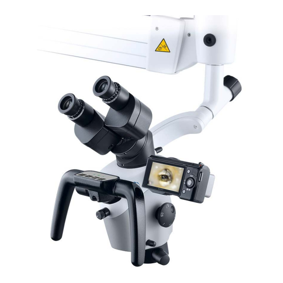
Page 11: Operating Elements At The Microscope
With an integrated HD camera: (start / stop) Adjusting the light mode of the camera 3.2.4 Rear view of the control device of the ATMOS i View 21 PRO ® Output of the power supply Connection for potential... -
Page 12: Rear View Of The Control Device Of The Atmos ® I
3.0 Setting up and starting up 3.2.6 Rear view of the control device of the ATMOS i View 31 PRO with an integrated HD camera ® HD video input. Can HD video input from HD video outlet from the Connection to the foot... -
Page 13: Starting Up
• Consider, when setting up the microscope, that the elastic force of the arm – without microscope head – is exceedingly strong. Operate the break of the height adjustment carefully. • To activate the ATMOS i View PRO please use the power switch on the front side of the control device. -
Page 14: Starting Up At A Glance
3.0 Setting up and starting up 3.6 Starting up at a glance Adjust microscope to initial position on the microscope suspension by use of the fixing wheel. Adjust microscope horizontally and vertically. Adjust all the clamps on the carrier and float arm to secure the movability of the arm in compliance with the requirements. -
Page 15: Operation
Automatic light switching: Once the arm is in the upper position the LED light switches off automatically. 4.3 Hand grips When purchasing the ATMOS i View PRO you may choose ® between two versions of handles. -
Page 16: Adjusting The Interocular Distance
4.0 Operation 4.4 Adjusting the interocular distance The interocular distance is adjustable between 50-75 mm. • Swivel the microscope into the work space. • Look through the eye lenses and push or pull the eye lens tubes together or apart with both hands. The interocular distance is perfectly adjusted when you look through with both eyes and a circular field is visible! 4.5 Adjusting the eye pieces... -
Page 17: Exchanging The Lenses
Tighten the three grub screws. Grub screw 4.9 Adjustment of the 5-fold magnification changer The 5-fold magnification changer from ATMOS enables free range zoom between 0.5x up to 2.0x. • Select the desired zoom factor by selecting one of the lateral rotating knobs. -
Page 18: Focussing
4.0 Operation 4.10 Focussing • Adjust the zoom to maximum (2.0) on the magnification unit. • Approach the object with the microscope until the image is sharp. • If the zoom level is changed the pre-adjusted degree of sharpness is still maintained. 4.10.1 Fine-focussing The optional fine focussing allows for sensitive and precise focussing in a 17 mm range. -
Page 19: Shadowless Illumination
4.0 Operation 4.14 Shadowless illumination The option shadowless illumination prevents instruments from causing shadows in the field of view. This option cannot be retro- fitted. • For the shadowless illumination no operating steps are required. 4.15 Microscope zoom and object field size Lens f in mm Factor display on the magnification unit Eyepieces with... -
Page 20: Image And Video Recording
Benennung (designation) Tasterfolie Erstellt 27.03.3013 C.Reinhardt Bearb. Gepr. Blatt Zeichnungs / ATMOS Medizin T echnik GmbH & Co. KG 060.0621.0 Ludwig - Kegel - Str. 12, 14-16, 18 (sheet) Artikel-Nr. 79853 Lenzkirch / Germany (part no.) Tel: +49 7653 689 -0... -
Page 21: Endoscope Adapter
ATMOS Cam or other external endoscope or digital camera (third party products). The ATMOS Cam can be easily and swiftly attached to the endoscope adapter by means of a special clip seal. Other endoscope cameras which provide a standardised connection interface can also be adapted without any trouble. -
Page 22: Cleaning And Care
Residues can be removed with a mixture made from equal parts of ethyl alcohol and distilled water to which a drop of standard washing-up liquid is added. If fluids penetrated the ATMOS i View PRO it needs to be sent in and may only be used after the check up of an ATMOS ® authorised person. -
Page 23: Fogging Of Optical Surfaces
5.3.3 Fogging of optical surfaces To prevent the eyepiece optics from fogging, we recommend using an anti-fogging agent. Note: Anti-fogging agents used by opticians for eyeglass lenses are also suitable for the ATMOS i View PRO. ® • Please observe the instructions supplied with each anti-fogging agent. -
Page 24: Maintenance And Service
• Place used accessories with the product. • ATMOS recommends: Work should be carried out by an • Fill in the form QD 434 „Delivery complaint / return shipment“ authorized ATMOS service partner. This ensures that repairs and the respective decontamination certificate. -
Page 25: Troubleshooting
View PRO is hot ® Switch off and let cool down for 2-3 hours ATMOS i View PRO is ® Please contact the ATMOS service. overheated Switch on the ATMOS i View PRO at the ® No function whatsoever... -
Page 26: Accessories And Options
538.9200.0 45° adaption for binocular tubes 606.1106.0 Binocular rotary disk with detent 538.3300.0 Cable (only for the ATMOS i View 31) ® S-VHS cable professional, 5 m (not with an integrated HD camera) 008.0882.0 Cable HDMI type A/C, L = 5 m (only with an integrated HD camera) 538.1902.0 Cable HDMI extension, L = 5 m (only with an integrated HD cam- 008.0909.0... -
Page 27: Technical Data
9.0 Technical data Voltage 100-240 V~ ± 10 %; 50/60 Hz Power consumption max. 45 VA Fuses 2 x T 3.15 A / 250 V Operation time Continuous operation Light intensity F 200 min. 120 kLux F 250 min. 80 kLux F 300 min. -
Page 28: Disposal
Register) is this type of device excluded from the ElektroG regulations. In order to guarantee a proper disposal of your old device, please either pass on your old device to your specialised dealer or send it directly to ATMOS MedizinTechnik for a professional disposal. -
Page 29: Notes On Emc
• The use of other accessories, other converters and cables than stated may lead to an increased emission or a reduced interference immunity of the equipment or system. 11.1 Guidelines and Manufacturer´s Declaration - Emissions The ATMOS i View PRO is intended for use in the electromagnetic environment specified below. The customer or user of the ®... - Page 30 ® for 0.5 Cycle for 0.5 Cycle IEC 61000-4-11 function during interruptions of the energy supply, it is recommended to supply the ATMOS ® i View PRO from an uninterruptible power supply 40 % UT 40 % UT or a battery.
- Page 31 ATMOS i View PRO is to be observed to verify the intended use. If abnormal performance characteristics are noted, additional ® measures might be necessary, e. g. a changed arrangement or another location for the ATMOS i View PRO. ®...
-
Page 32: Medizintechnik Gmbh & Co. Kg Ludwig-Kegel-Straße
ATMOS MedizinTechnik GmbH & Co. KG Ludwig-Kegel-Straße 16 79853 Lenzkirch / Germany Phone: +49 7653 689-0 atmos@atmosmed.de www.atmosmed.com...
Need help?
Do you have a question about the i View PRO Series and is the answer not in the manual?
Questions and answers