
Related Manuals for STEINDORFF CX50 Series


Summary of Contents for STEINDORFF CX50 Series
- Page 1 STEINDORFF ® INSTRUCTION MANUAL 360° Rotatable Head Nosepiece Coaxial Coarse Achromatic color-coded DIN Objectives Fine Focusing Double layer Machanical Stage Iris Diaphragm LED Illuminator Rubber Feet Light Intensity Control...
- Page 2 CONTENTS A.Illustration of Microscope…………………………….……..………COVER B.Unpacking and Preparation for Use…..………….…...…….....…… 1 C.Features / Definitions…………………………..……..…….....…….. 2 D.Operating Procedure…….……………………………......… 4 E.Maintenance and Care …….………………………………..…..…..6 F.Electrical Maintenance ………..………….……....…....7 G.Troubleshooting….…………….……………......…...…...9 H.Optional Digital Cameras…………………..……...………...….…...10 B. UNPACKING & PREPARATION Your The microscope is supplied with an expanded two-part Styrofoam/polystyrene case.
- Page 3 C. FEATURES AND DEFINITIONS EYEPEICE: 10X wide field eyepiece with built in calibrated pointer for measurement. HEAD: monocular head: 360° rotatable monocular head. Binocular head: Dual view head allows 2 people to observe the microscope simultaneously. OBJECTIVES: The lower functional component of the optical system. Avail- able in 4X, 10X and 40XR, 100XR.
- Page 4 crisp, cool-to-touch white light that lasts up to 60x longer than traditional tungsten bulbs. STAGE: The stage of the microscope is equipped with built-in double layer mechanical stage. SAFETY RACK STOP: Controls the maximum upward movement of the stage. SPECIFICATIONS (With 10x Eyepiece) Objective Total Magnification Field of View...
- Page 5 4. Place a low powered objective into position. 5. Place the micro-slide specimen to be observed under the spring stage clips. If using a mechanical stage, pull back the lever on the left side of the stage, insert the slide, then bring the crescent shaped holder into contact with the slide.
- Page 6 II. Microscope equipped with an 1.25 Abbe-condens- er with or without the 100XR oil immersion objective. 1. Follow steps 1 through 6 and then: 2. FOCUSING THE CONDENSER. The sub-stage Abbe condenser is mounted beneath the stage in a rack & pinion focusing mount.
- Page 7 E. MAINTENANCE & CARE Cleaning of the optical surfaces Never take objectives or eyepieces apart. They should be cleaned on the instrument since they are not easily removed. To clean lens surfaces, first remove dust using a soft brush or blow off with a small syringe. Use a cotton-tip applicator and a small amount of xylene.
- Page 8 F. ELECTRICAL MAINTANENCE WARNING DISCONNECT POWER SUPPLY CORD FROM WALL RECEPTACLE BEFORE REPLACING LAMP OR FUSE Charging/Re-charging NiMH rechargeable batteries. (Rechargeable models only) Your microscope is equipped with rechargeable Nickel Metal Hydride (NiMH) batteries. Batteries should be charged prior to first use. Fully charge batteries, approximately 8 hours, by plugging the supplied charger into a standard 110v outlet, and attaching to the power inlet on the microscope.
- Page 9 Rechargeable Battery Replacement (Rechargeable models only) 1. Lay the microscope on its side on a flat surface, and un- screw the four rubber feet 2. Remove the base and lay on a flat surface 3. Unscrew the two screws on both ends of the rectangular plastic box 4.
- Page 10 G. TROUBLE SHOOTING Objectives If you are using any type of liquid on the slides and then view through the microscope, make sure that you clean the ob- jectives immediately after you are done. You can clean with paper towel and xylene. If you do not clean after contact with liquid the objective lens will become very dirty.
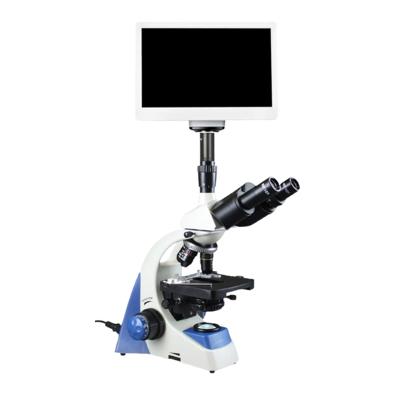
- Page 11 H. OPTIONAL DIGITAL CAMERAS T hree digital cameras to choose from: DG 0.35 / DG 1.3 / DG 3.0 The digital cameras are specifically designed for use with any standard biological or stereo microscope. It acts as a converter by converting your analog microscope into a digital microscope enabling you to capture, analyze or share the digital images.
- Page 12 100 Lauman Lane, Suite A, Hicksville, NY 11801 Tel: (877) 877-7274 | Fax: (516) 801-2046 Email: Info@nyscopes.com www.microscopeinternational.com...
Need help?
Do you have a question about the CX50 Series and is the answer not in the manual?
Questions and answers