Table of Contents
Advertisement
Quick Links
®
STEINDORFF
Inverted Biological
Microscope
NYMCS-290
User Manual
This manual is written for Inverted Biological Microscope NYMCS-290. For safety and for exerting the best
performance, making you familiar with the instrument entirely, it is strongly recommended that you read this
manual carefully before using the microscope. For the further reference, please place this manual in a position
where nearby the worktable and fetched easily.
Advertisement
Table of Contents

Related Manuals for STEINDORFF NYMCS-290


Summary of Contents for STEINDORFF NYMCS-290
- Page 1 NYMCS-290 User Manual This manual is written for Inverted Biological Microscope NYMCS-290. For safety and for exerting the best performance, making you familiar with the instrument entirely, it is strongly recommended that you read this manual carefully before using the microscope. For the further reference, please place this manual in a position...
-
Page 2: User Notice
® STEINDORFF USER NOTICE Safety Note Do not keep the instrument in a direct sunlight, high temperature or humidity, dusty and easy shaking environment. Make sure the stage is plane, horizontal and stable enough. When moving the microscope, please hold up the instrument with one hand on the lower side of the eyepiece tube ①, and the other hand on the illumination bracket... - Page 3 ® STEINDORFF Do not disassemble any parts of the microscope. That will affect the function or decline the performance of the microscope. If haven’t mounted the objective, please cover the dust cap to prevent the dust and the splashed liquid of the tissue culture entering the inside.
-
Page 4: Component Names
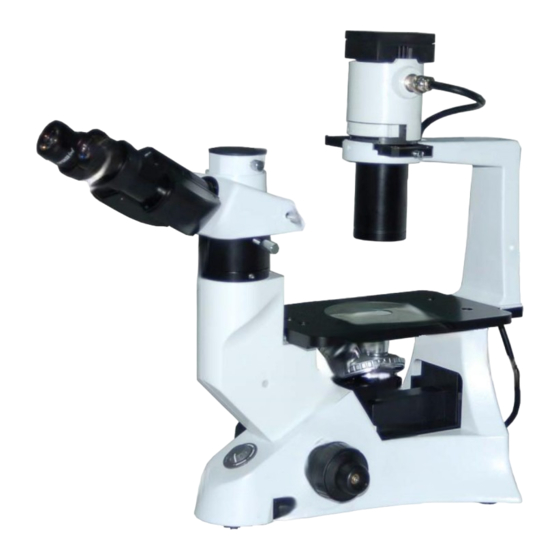
® STEINDORFF 1. COMPONENT NAMES Lamp house Objectives Phase contrast slider 10x (PH) 20x (PH) Illumination bracket Ocular (eyepiece) Condenser Trinocular viewing tube (Fixed) Stage inserted plate Objective turret (Nosepiece) Microscope base Stage (Fixed) (Fixed) Figure 1... -
Page 5: Installation
® STEINDORFF 2. INSTALLATION 2.1. Installing diagram The following figure shows the installation sequence of the components. Before installing, be sure every components is clean, do not score any parts or glass surface. Keep well with the supplied hexagon wrench. When changing the components, you will need it again. - Page 6 ® STEINDORFF 2.2. Installing steps 2.2.1. Installing and replacing the lamp (Figure 3) Please use the specified halogen Lamp 6V30W 1. Hold to the bulb ① after you wrap it with gauze or other protection materials, then depress the plugs ② into the jack ③...
- Page 7 ® STEINDORFF 2.2.4. Installing the objective (Figure 6 and 7) Turning the coarse fusing knob ① like the figure shows till the nosepiece get to its lowest position. For ensuring the safety of the instruction on transportation, the nosepiece is located in the lowest position and the tension adjustment collar ②...
- Page 8 ® STEINDORFF 2.2.5. Installing the stage lengthen splint and the mechanical ruler (Figure 8) Stage lengthen splint can be installed in either side of the stage to enlarge the work surface. But you can’t install the mechanical ruler together. Generally, the mechanical ruler will be installed in the right side for comfortable adjustment.
- Page 9 ® STEINDORFF 2.2.9. Connecting the power cord (Figure 12, 13 and 14) Do not bring the power cord to bear a powerful stress. When being bent or wrapping, the cable and wires will be broken easily. Turn the main switch ① on “O” (off) state before connecting the power cord.
-
Page 10: Operating The Adjustment
® STEINDORFF 3. OPERATING THE ADJUSTMENT 3.1. Microscope base 3.1.1. Turning on the lamp (Figure 15) Connect the power, turn on the main switch ① which on the bottom side of the base to “ - ” (on). 3.1.2. Adjusting the brightness (Figure 16) Turning the brightness adjustment knob clockwise, the voltage raise, and the brightness strengthen;... - Page 11 ® STEINDORFF 3.2. Stage 3.2.1. Setting the specimen (Figure 18 and 19) Set the specimen in the center of the stage, please. To obtain the best observe effect, please select the containers, such as culture dish and culture bottle, with the bottom thickness is 1.2mm, and the same thickness is also...
- Page 12 ® STEINDORFF 3.3. The viewing tube 3.3.1. Adjusting the diopter (Figure 20) 1. Look into the right ocular by your right eye, then revolving the coarse focus knob to focus on the specimen. 2. Then use your left eye to look into the left ocular. If the image is not sharp, just use the diopter adjustment ring ①...
- Page 13 ® STEINDORFF 3.3.3. Switching the light path (Figure 22) Pulling out the light path selector lever ① by your thumb, select the light path you needed. When in the binocular observation, pushing in the lever until you heard a “clicked”. While in video or photography, pulling out the lever until it reached the “clicked”...
- Page 14 ® STEINDORFF 3.4.2. Using the aperture diaphragm (Figure 24) When in the bright field observation, the aperture diaphragm control the numerical aperture of the illumination system. Only when the numerical aperture of the objective and the illumination system being matching, you can obtain the higher image resolution and contrast, and the increased depth of field, too.
-
Page 15: Phase Contrast Viewing
® STEINDORFF 4. PHASE CONTRAST VIEWING 4.1. Name of the components 4.1.1. Phase contrast objective (Figure 25) The optional magnification of the phase contrast is: 10X, 20X. If you want to know how to mount the phase contrast objective, please see 2.2.4. You ought to mount it on the turret. - Page 16 ® STEINDORFF 4.2. Installation and use 4.2.1. Installing the phase contrast slider (Figure 27) 1. Keep the slider ① face (the surface which had character) up towards, then inserted it into the illumination system from the right to the left like the figure showed.
- Page 17 ® STEINDORFF 6. The 10X and the 20X phase contrast objective use the same light ring on the phase contrast slider. So you need to check the coincidence of the light ring center and the phase contrast center when changing the objective.
- Page 18 ® STEINDORFF 5. MICROSCOPE PHOTOGRAPHY AND VIDEO 5.1. Microscope video 5.1.1. Selecting the light path (Figure 30) Just used in the trinocular observation 1. Pulling out the light path selector lever, until you heard the “clicked”. In the dark specimen observation, you can make the focus by both eyes at first, then change the light path 5.1.2.
- Page 19 ® STEINDORFF 5.2. Microscope photography 5.2.1. Selecting the light path Just used in the trinocular observation The operation diagram is shown in 5.1.1, and the details reference is in 3.3.3. 5.2.2. Installing the photography set (Figure 32) 1. Loosen the locking bolt ① on the trinocular viewing tube, and take out the dust cap ②.
- Page 20 ® STEINDORFF 5.2.3. Focus Do the binocular observation at 20% brightness, and focus primary. When in microscope photography, do use the camera viewfinder to focus the specimen. Please refer the user manual of the camera set to obtain the details.
-
Page 21: Technical Specifications
® STEINDORFF 6. TECHNICAL SPECIFICATIONS 6.1. Main specifications Optical System Infinite Optical System Compensation Free Trinocular Tube Inclined at 30; Viewing Tube Division ratio: 20% of Binocular Viewing and 80% of Video Viewing & Micrography Eyepiece Wide Field Eyepiece 10X, Linear Visual Field: 22 mm... -
Page 22: Troubleshooting
® STEINDORFF 7. TROUBLESHOOTING Under certain condition, some no-fault factors will bring a reversible influence to the instrument’s performance. If the problem is happened, please take proper measures according to the follow table. If you can’t solve the trouble by the supplied methods, please contact with the sales department of our company. - Page 23 ® STEINDORFF The nosepiece is not in the center of the light Insure the nosepiece is in the “clicked” position path Place the specimen on the stage The specimen don’t place properly 6. One side of the image correctly. is unfocused...
Need help?
Do you have a question about the NYMCS-290 and is the answer not in the manual?
Questions and answers