
Related Manuals for Zeiss 3000


Summary of Contents for Zeiss 3000
- Page 1 Stratus OCT Model 3000 Instrument and Stratus Review Software Version 7.0 User Manual...
- Page 2 © 2010 Carl Zeiss Meditec, Inc. All rights reserved. Trademarks Stratus OCT and GPA are either registered trademarks or trademarks of Carl Zeiss Meditec, Inc. in the United States and/or other countries. Windows is either a registered trademark or trademark of Microsoft Corporation in the United States and/or other countries.
-
Page 3: Table Of Contents
Contents Contents (1) Introduction ....... . . 1-1 •Intended Use/Indications For Use ......1-1 •Purpose Of This User Manual . - Page 4 Contents •Scan Protocol Descriptions, Options And Tips ....4-6 •Repeat Scan Function ........4-11 (5) Analyze Scans .
- Page 5 •Carl Zeiss Meditec, Inc. Electronic End User License Agreement . . 12-4 •Carl Zeiss Meditec, Inc., Limited Warranty for Software ..12-6 •Software Support Agreement ......12-8 (A) Networking Guidelines.
- Page 6 Contents •Notice ..........A-1 •Instrument Network Capabilities .
- Page 7 •Install and Configure the NAS Device ..... . .E-2 •Cleaning the NAS Device ....... . .E-6 (F) Printer Configuration Instructions .
- Page 8 viii Stratus OCT User Manual PN 2660021134133 A...
-
Page 9: Introduction
Introduction (1) Introduction The ZEISS Stratus OCT Model 3000 (Stratus OCT) enables examination of the posterior pole of the eye at an extremely fine spatial scale, without surgical biopsy or even any contact with the eye. Stratus OCT Review Software enables you to view, analyze and manage Stratus OCT data on a personal computer;... - Page 10 Introduction Part of the Body The Stratus OCT instrument is designed for in-vivo viewing and axial cross-sectional imaging and measurement of posterior ocular structures. In addition, the system physically interacts with the patient’s forehead and chin. Application The Stratus OCT instrument is designed for continuous use, although it is expected that most sites operate the instrument for 10 hours or less per day, indoors, within a medical office or hospital setting.
-
Page 11: Purpose Of This User Manual
Purpose Of This User Manual Carl Zeiss Meditec designed this User Manual to serve as a training, usage and reference guide. We assume that users are clinicians or technicians with professional training or experience in the use of retinal imaging equipment, and in diagnostic interpretation of the images generated. -
Page 12: What's New
Introduction What's New? This User Manual includes information on new and modified features of the Stratus OCT system. For customers who are upgrading from version 6.0 or earlier, the following describes differences and where information can be found in this user manual. If you are upgrading from version 6.0 Features that can be licensed: •... -
Page 13: Stratus Oct User Manual
Introduction If you are upgrading from version 4.0 Additional features that can be licensed: • Guided Progression Analysis (GPA™) Advanced Serial Analysis that helps the user interpret changes in RNFL measurements over time (see page 6-26). • Layer Editing tool gives the ability to manually adjust the retinal layer boundaries for macula or RNFL analysis (see page 6-8). -
Page 14: Stratus Oct System Described
Introduction Organization of The Manual This introductory chapter provides a system description, installation and safety information. Chapters through are organized according to the normal sequence of operation of the Stratus OCT, followed by data management and data transfer functions in Chapters through (9), as follows: Prepare To Scan, explained in Chapter (2). - Page 15 Introduction The Stratus OCT interferometer electronically detects, collects, processes and stores the echo delay patterns from the retina. With each scan pass, the Stratus OCT captures from 128 to 768 longitudinal (axial) range samples, i.e., A-scans. Each A-scan consists of 1024 data points over 2 mm of depth.
-
Page 16: Stratus Oct System Hardware
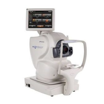
Figure 1-1 Stratus OCT System Hardware Software and Storage Media Carl Zeiss Meditec pre-installs all software necessary to operate the Stratus OCT. We recommend the use of a network solution for daily archive and backup, either a network file server or network attached storage device. You can purchase a network attached storage device designed for this purpose from Carl Zeiss Meditec. -
Page 17: Warning: User Changes To Software Or Hardware
Unauthorized modification also voids the instrument warranty. Approved Software Note: Carl Zeiss Meditec does not provide technical support for the use of third party software. Please refer to the Stratus OCT Technical Support section of our website (www.meditec.zeiss.com/stratus) for the current list of approved software, including updates and patches to the Windows®... -
Page 18: Instrument Installation
Introduction Instrument Installation Only an authorized Carl Zeiss Meditec service representative should install the Stratus OCT. We do not provide assembly and installation instructions. In consultation with the buyer, Carl Zeiss Meditec schedules a free on-site installation appointment to coincide with delivery. -
Page 19: Embedded Windows License
WARNING: To prevent electric shock, the instrument must be plugged into an earth grounded outlet. Do not remove or disable the ground pin. Only an authorized Carl Zeiss Meditec service representative may install the instrument. WARNING: Do not use the printer or the instrument with an extension cord or a power strip (multiple portable socket outlet). - Page 20 Disclaimer: Carl Zeiss Meditec does not offer advice or instruction in the diagnostic interpretation of OCT images. It is the clinician’s responsibility to make diagnostic interpretations of OCT scans.
-
Page 21: Electromagnetic Compatibility (Emc)
Introduction 1-13 Applicable Phototoxicity Statements (FDA CDRH Ophthalmoscope Guidance #71): Because prolonged intense light exposure can damage the retina, the use of the device for ocular examination should not be unnecessarily prolonged. While no acute optical radiation hazards have been identified for direct or indirect ophthalmoscopes, it is recommended that the exposure time for the patient’s eye be limited to the minimum time that is necessary for diagnosis. - Page 22 1-14 Introduction Guidance and manufacturer’s declaration - electromagnetic emissions The Stratus OCT is intended for use in the electromagnetic environment specified below. The customer or user of the Stratus OCT should assure that it is used in such an environment Emissions Test Compliance Electromagnetic environment -...
- Page 23 Introduction 1-15 Guidance and manufacturer’s declaration - electromagnetic immunity The Stratus OCT is intended for use in the electromagnetic environment specified below. The customer or user of the Stratus OCT should assure that it is used in such an environment Immunity Test IEC 60601 test level Compliance level...
-
Page 24: Accessory Equipment
1-16 Introduction Recommended separation distances between portable and mobile RF communications equipment and the Stratus OCT The Stratus OCT is intended for use in an electromagnetic environment in which radiated RF disturbances are controlled. The customer or the user of the Stratus OCT can help prevent electromagnetic interference by maintaining a minimum distance between portable and mobile RF communications equipment (transmitters) and the Stratus OCT as recommended below, according to the maximum output power of the communications equipment. -
Page 25: Symbols And Labels
Introduction 1-17 Symbols and Labels Caution, consult accompanying documents. Note: There are important operating and maintenance instructions found in the manual. Presence of electrical shock hazard. Note: Indicates risk of electrical shock due to the presence of uninsulated high voltage inside the instrument. Do not remove the instrument cover or parts. Fuse Type B applied parts Manufacturer... -
Page 26: Password Protection
Passwords are case-sensitive. Stratus OCT shipped with Version 6.0 or later Stratus OCT shipped before Version 6.0 User Name Zeiss Administrator Password November171846 tomography Workgroup Name... - Page 27 Figure 1-2 Users and Passwords Dialog Activated WARNING: Do not attempt to remove, edit the properties or edit the passwords of the “Administrator” or “Zeiss” or “Tech Support” accounts. Failure to observe this warning could disable access to the system.
- Page 28 1-20 Introduction 6. The Add New User dialog box now lets you select the level of access of the new user account. Select the Other radio button and then select Administrators from the drop down list, as in the example below. Figure 1-3 Select Other, Administrators 7.
-
Page 29: Instrument Disposition
STRATUS OCT Host desktop icon to launch the Stratus OCT software. Instrument Disposition When it comes time to upgrade the Stratus OCT, please contact Carl Zeiss Meditec to inquire about trade-in or upgrade values we may offer. Should you not wish to trade in the instrument, please dispose of it in accordance with local and national requirements. - Page 30 1-22 Introduction Stratus OCT User Manual PN 2660021134133 A...
-
Page 31: Prepare To Scan
Prepare To Scan (2) Prepare To Scan Chapter Overview This chapter explains in detail how to prepare for scanning with the ZEISS Stratus OCT. Along the way, it describes the Stratus OCT M , from which all functions INDOW originate. Scan preparation consists of the following: •... -
Page 32: Navigate To The Main Window
Prepare To Scan Windows Start menu at lower left. For later models with soft shutdown, this will power down the whole system. On earlier models, you must flip the computer power switch off to shut down the whole system. Soft Shutdown To prevent abrupt shutdowns of the system, later models of Stratus OCT power down only through the computer software. - Page 33 Prepare To Scan • The Patient and Scan Group Images Tabs (lower left) Figure 2-3 The Stratus OCT Main Window Each area has its own functionality, which the manual describes in the context of its use. Resize Functional Areas • You can resize any area by placing the pointer over its interior edge until the two-headed arrow appears.
-
Page 34: Prepare The Patient
Prepare To Scan Prepare The Patient Patient preparation includes dilating the patient (optional), selecting the fixation method, and preparing the patient for the exam experience. Note: The forehead and chin rests should be cleaned between each examination with an alcohol prep swab. Dilate the Patient (Optional) Dilation is an optional procedure to aid in the acquisition of an OCT image. -
Page 35: Enter Patient Data
Prepare To Scan • It is important to situate the target light level with the patient's fellow eye and as far as possible from the patient. The Patient Experience The patient experience with the Stratus OCT is normally brief and comfortable. An experienced operator can acquire several scans from each eye in the space of 5-7 minutes. - Page 36 Prepare To Scan Note: The DICOM workflow for Modality Worklist is available if DICOM1 Integration is licensed and activated. To display a list of scheduled patients, click on Search Worklist Patients. The M ODALITY dialog appears. ORKLIST Figure 2-4 Modality Worklist By default, the Date Range will be the current date.
- Page 37 Prepare To Scan Category Field If you have created patient categories (select Options > Register > Categories to create categories), you can use the drop down list to select a category of patients for display in the patient list. Today’s Patients Today’s Patients is the list that is shown when you click on Search Worklist Patients.
- Page 38 Prepare To Scan Advanced Search Stratus OCT provides an Advanced Search dialog to serach for patients using additional parameters such as patient category, diagnosis, scan type, insurance company, doctor, clinical trial ID, or subject reading ID, etc. Advanced Search is available from the Toobar. Figure 2-6 Advanced Search Dialog Using the available fields, enter or select search parameters and click Search.
- Page 39 Prepare To Scan The Patient Tab On the lower left, this tab shows the selected patient's information. Figure 2-7 The Patient Tab in Main Window Add/Edit Patient Records Click the Patient: Add or Edit buttons in the upper center of the M , next to the INDOW Search field, to access the P...
- Page 40 2-10 Prepare To Scan • To edit a patient record, select a patient from the list and click Edit. Edit the fields as desired and click OK when finished. • If desired, you can enter a variety of other patient information using the available tabs.
-
Page 41: Acquire Scans
Acquire Scans (3) Acquire Scans Chapter Overview This chapter explains in detail how to acquire Stratus OCT scans. It covers the following steps: • Adjust the Height for patient comfort (below). • Initiate a Scan (on page 3-3). • Position The Patient Module (on page 3-5). - Page 42 Acquire Scans • For finer adjustments, you can raise or lower the chin cup. Use the knobs below the chin cup on either side (see Figure 3-1 below). Eye Position Marker Chin Cup Chin Cup Height Adjustment Knob Figure 3-1 Height Adjustment With the patient seated, follow these steps to adjust the height: 1.
-
Page 43: Initiate A Scan
Acquire Scans Initiate a Scan To position the patient module for scanning, you must initiate a scan. First, select a scan acquisition protocol using the Scan Tab, below. Figure 3-2 The Scan Tab in Stratus OCT Main Window • If not in the M , click the Go to Scan Acquisition button to display it, as INDOW pictured at left. - Page 44 Acquire Scans double-click the protocol. This activates the S window, as pictured CQUISITION below. Figure 3-3 The Scan Acquisition Window The S window allows you to adjust video and scan parameters from a single CQUISITION interface, enabling the acquisition of high quality scans. The Repeat button to the left of the Scan button allows you to repeat any saved scan group using the same parameters.
-
Page 45: Position The Patient Module
Acquire Scans In preparation for the next step, the important features to note are the scan image (left side) and video image (upper right). The other functions available in this window will be detailed in the usual sequence of their application. Center Line Display Right-click anywhere in the scan image and a vertical line appears in the center of the scan Adjust Scan... -
Page 46: Optimize Scan Image
Acquire Scans ocular lens and positioning further. All the while, use the live video image to observe the position and focus with respect to the study eye. (While not usually necessary, adjustments to the video image are possible—see Video and Lamp Parameter Tab, page 3-18, for instructions.) 1. - Page 47 Acquire Scans scanned, and you see nothing but noise in the live scan image. The 2 mm axial “window” must be positioned to bracket the retina, making it visible in the scan image. • Once you bring the retina into range for the first scan, it is likely to be visible subsequently.
-
Page 48: Adjust Scan Placement
Acquire Scans • Once you have a retinal scan image on the screen, click the Optimize Polarization button on the Scan Parameter Tab (pictured above). • You can incrementally adjust the polarization using the Polarization arrow buttons. If you do, you may note that there are several local maxima in the 180-degree range of polarization. -
Page 49: Acquire Scans
Acquire Scans Move the Fixation LED Click and drag works on the fixation LED if it is sufficiently separated from the scan pattern, or if you select Move Fixation LED from the drop-down list as illustrated in Figure 3-5 below. After you save the first scan in a related series, you can no longer move the fixation LED (see Which Scan Parameters Can Be Adjusted When, page 4-4, for more... - Page 50 In most cases, the fovea and/or the optic disc are the primary points of clinical interest. Carl Zeiss Meditec designed most of its scan acquisition protocols for scans on one or the other location by default, although they may be used in other locations. Thus, the Stratus OCT places the fixation target either centrally (fovea mode) or approximately 15°...
- Page 51 Acquire Scans 3-11 • Blue patient module buttons: Again, press the left button once. Flash is used unless you uncheck the Freeze with Flash option in the Scan menu (click Scan > Freeze with Flash, as at left). 3. Save the scan. •...
- Page 52 3-12 Acquire Scans Note that media problems associated with many retinal diseases (vitreous opacities, hemorrhages, lens opacities, etc.) can make it impossible to achieve a signal strength of 5, even with careful scanning. Nevertheless, such scans can be useful for visual analysis of the retinal structure and the association between the retinal layers.
-
Page 53: Other Scan Adjustments
Acquire Scans 3-13 Scan Review Window When you click the Review button after freezing a scan, the S appears, EVIEW INDOW as pictured below. Figure 3-7 The Scan Review Window Along the bottom, it displays thumbnails of the last several images captured during scanning, up to eight total. - Page 54 3-14 Acquire Scans Adjust Scan Pattern Parameters The Scan Pattern Parameter area (pictured at left) is found in the upper left of the Scan Parameter Tab (see page 3-7). You can use it to adjust several parameters including scan size, angle or number of lines in the pattern. •...
- Page 55 Acquire Scans 3-15 Adjust Scan Variables From the Scan menu, select Scan Variables (click Scan > Scan Variables) to reveal the Scan Variables dialog box. The name of the currently selected scan protocol appears at the top. Figure 3-8 The Scan Variables Dialog Box Number of A-Scans By default, the number of A-scans is 512 for all protocols except the Fast Scan protocols (fixed at 768 A-scans total).
- Page 56 3-16 Acquire Scans Define Custom Scan This Scan menu option enables you to define your own scan acquisition protocol. Click Scan > Define Custom Scan (as pictured at left) to display the Define Custom Scan dialog box. Figure 3-9 The Define Custom Scan Dialog Box •...
- Page 57 Acquire Scans 3-17 OCT Image Tab Click the OCT Image Tab on the S to adjust OCT image noise CQUISITION INDOW and range values, or the number of A-scans for the scan protocol you have selected. Figure 3-10 The OCT Image Tab (Scan Acquisition Window) Noise and Range These two sliders are set by default to filter the low end (background noise) and high end (saturation signal) of the OCT interferometer signal.
- Page 58 3-18 Acquire Scans signal color). This makes the retinal image poorly defined or appearing as undifferentiated white. • The slider operates on a percentage scale. At zero, the weakest detectable signal is assigned the saturation color in the scan image, and the image is all white. At 100, only the strongest detectable signal is assigned the saturation color in the scan image, and very little retinal signal is strong enough to be depicted in the scan image.
- Page 59 Acquire Scans 3-19 Indicators Tab This tab provides patient and scan information. Figure 3-12 The Indicators Tab in Scan Acquisition Window Details Tab This tab specifies the x and y position of the scan pattern, the landmark and the fixation LED, in mm with respect to the center of the video image.
- Page 60 3-20 Acquire Scans Stratus OCT User Manual PN 2660021134133 A...
-
Page 61: Scan Acquisition Protocols
Scan Acquisition Protocols (4) Scan Acquisition Protocols Chapter Overview This chapter explains the applications and attributes of the scan acquisition protocols offered by the Stratus OCT. While the previous two chapters provide operating instructions, this chapter is designed to help the user select which protocol to use and know how to use it. - Page 62 Scan Acquisition Protocols Activity Radio Buttons The four Activity radio buttons above the Scan Tab enable you to display the protocol groups: those designed for examination for Glaucoma, or of the Retina (other retinal pathologies), All scan protocols and any Custom protocols you have created (see Define Custom Scan on page 3-16).
-
Page 63: General Tips
Image Processing Protocols (see page 6-37). Carl Zeiss Meditec designed the image processing protocols for use with any scan. However, we designed each quantitative analysis protocol for use with a certain scan type (line or circle) or scan pattern made on a certain retinal location (macula or disc). Several of the scan protocols are designed for use with a limited subset of analysis protocols. - Page 64 Scan Acquisition Protocols Analysis Protocol Designed for Scan Group(s) RNFL Thickness Average Circle, Proportional Circle, (Fast) RNFL Thickness (3.4), RNFL Thickness (2.27xdisc) Nerve Head Circle: 1 OS and/or 1 OD of same radius around disc Rings: 1 OS and/or 1 OD group around disc RNFL Thickness Map (Fast) RNFL Map,...
- Page 65 Scan Acquisition Protocols • For scans in a related series, only scan and fixation LED placement are adjustable after the first scan. Several scan protocols consist of a series of related scans you acquire one by one. To preserve the design integrity of these scan protocols, you cannot adjust the size, number of lines, number of A-scans or placement of the landmark after you save the first scan in the series.
-
Page 66: Scan Protocol Descriptions, Options And Tips
Scan Acquisition Protocols image appears most nearly horizontal on your monitor. Try to minimize this variation by adjusting the patient module laterally by small amounts. This adjusts where the scan beam enters the pupil and its angle of incidence on the retina. Scan Protocol Descriptions, Options And Tips There are two basic scan patterns: lines and circles. - Page 67 Scan Acquisition Protocols Fast Macular Thickness Map (Fast Mac) The Fast Macular Thickness Map (Fast Mac) protocol combines the six Macular Thickness Map scans into one scan (see page 4-7). This protocol acquires six 6 mm radial line scans in 1.92 seconds of scanning. You cannot alter the size or number of lines. This retina protocol is designed for use with the retinal thickness analyses.
- Page 68 Scan Acquisition Protocols repeated in several other protocols with variations in size and adjustability of parameters. This protocol provides maximum adjustability, and you can use it to Define Custom Scan (see page 3-16). The default pattern has 6 lines of 6 mm length. You can adjust the length of all the scan lines by adjusting the aiming circle size, or by adjusting the length of the first scan in the series.
- Page 69 Scan Acquisition Protocols glaucoma protocol provides a simple way to acquire scans to which you can apply RNFL thickness analyses. RNFL Thickness (2.27xdisc) Select RNFL Thickness (2.27xdisc) to acquire a single circle scan around the optic disc that is 2.27 times the radius of the aiming circle. The default pattern has an aiming circle of 1.5 mm radius.
- Page 70 4-10 Scan Acquisition Protocols Concentric 3 Rings The Concentric 3 Rings protocol consists of three equally spaced concentric circle scans with radii in the ratio of 1:2:3. Like other circle scan patterns, this protocol is designed for use around the optic disc to measure nerve fiber layer thickness. You can use it to build custom scans.
-
Page 71: Repeat Scan Function
Scan Acquisition Protocols 4-11 Repeat Scan Function The Repeat function allows you to repeat any saved scan group using the same set of parameters. Repeat parameters include scan size, angle and placement, fixation LED placement, and landmark placement. When you repeat a scan group, the parameters unique to each scan in the series is recalled and repeated in the same order. - Page 72 4-12 Scan Acquisition Protocols 7. In the video window you will see the prior and new fundus images toggle back and forth automatically. The prior scan will have the word PRIOR in the upper left corner. You can change the toggle speed in the Video and Lamp dialog. Move the slider in the Prior Fundus Toggle or select a number.
-
Page 73: Analyze Scans
(5) Analyze Scans Chapter Overview Carl Zeiss Meditec equips the Stratus OCT with software to assist you with analysis of scan images. This chapter explains how to analyze scans with the Stratus OCT. We designed the Stratus OCT software for analysis of the posterior segment of the eye. To analyze scans using the Stratus OCT software involves the following: •... - Page 74 Analyze Scans The Scan List The scan list shows all saved scan groups of the selected patient. Figure 5-1 The Scan List in the Main Window It is organized by scan Date, OD Scan Groups and OS Scan Groups. The latter two name the scan group by the scan acquisition protocol used (see Chapter (4)).
- Page 75 Analyze Scans Note: Some analyses assume or require a predetermined number of scans for analysis. Deselecting scans may prevent the analysis from working. Some analyses may work with less than the optimal number of scans, but yield output that relies more on interpolation and may be less accurate in interpolated areas.
-
Page 76: Selection Window
Analyze Scans • Scan selection is optional, and you can do the same by using the Scan Group Images Tab, as explained on page 5-2. If you try to run an incompatible analysis, a message will so inform you. Selection Window When you click the Scan Selection button at the bottom of the Analyze Tab, the S ELECTION appears. - Page 77 Analyze Scans • For the scan image, select Color or Grayscale. Use the sliders to adjust Level, Window and % Zoom. Click the Default button to reset the scan image to the default settings. • Level sets the reflectivity threshold that is filtered out as background noise, and apportions the color scale according to the relative reflectivity found in the scan image.
-
Page 78: Print Analysis Output
Analyze Scans Note: From applicable analysis output windows, Stratus gives you the ability to edit the retinal boundary layers automatically provided by the retinal and RNFL thickness algorithms. For more information, see Edit Layers on page 6-8. Print Analysis Output To print any analysis output screen, select Print from the File menu (click File >... -
Page 79: Export To Dicom
Analyze Scans 1. Click Options > Register > Clinical Site ID. The Clinical Site ID dialog box appears. Figure 5-5 The Clinical Site ID Dialog Box 2. To enter the clinic name, type up to 64 characters, including spaces, in the Clinical Site ID field. - Page 80 Analyze Scans 2. When the DICOM export is completed, you will see the following dialog confirming that the report was sent to the DICOM system: Figure 5-7 Report Confirmation 3. Click OK. Note: Before using the Export to DICOM function, make sure that the Clinical Site ID is configured on the instrument.
-
Page 81: Analysis Protocols
Analysis Protocols (6) Analysis Protocols Chapter Overview The Stratus OCT offers analysis protocols, divided between: • Analyses and Reports (page 6-13). • Image Processing Protocols (page 6-37). This chapter describes each analysis protocol and provides general and specific usage tips. See Chapter (5) Analyze Scans for detailed instructions to perform analyses of scan... - Page 82 Analysis Protocols Analyses and Reports The Stratus OCT offers analysis and report protocols, which you can group in the Glaucoma and Retina lists. Some Glaucoma protocols are repeated in the Retina protocol list, since they are useful for both applications. Glaucoma •...
-
Page 83: General Tips
Analysis Protocols Are Correlated with Scan Patterns When selecting a protocol, it is important to remember that the protocols are of two kinds: image processing protocols and quantitative analysis protocols. Carl Zeiss Meditec designed the image processing protocols for use with any scan. However, each quantitative analysis protocol is designed for use with a certain scan type (line or circle) or scan pattern made on a certain retinal location (macula or disc). - Page 84 Analysis Protocols Scan Direction Legend The analyses that display a processed scan image include a legend that indicates the path and direction of the currently displayed scan, as shown in the examples at left. The legend enables you to orient the scan image with respect to the scan path. The scan angle (in degrees, for line scans) is at lower right.
- Page 85 Analysis Protocols Scan Messages The scan messages “Scan too high,” “Scan too low” and “Scan missing data” are descriptive of what you see on screen. When the scan is too high or too low on the screen, the analysis algorithm frequently fails. When either message appears, the operator should repeat the scan, using the Z-Offset arrow buttons to raise or lower the scan image as necessary.
-
Page 86: Normative Data And Its Applicability
Normative Data and Its Applicability Carl Zeiss Meditec gathered data from several hundred normal eyes acquired at multiple sites to create normative databases for RNFL thickness and for macular thickness. The Stratus OCT normative database study was designed to obtain normative data for RNFL and macular thickness from healthy subjects ages 18 to 86. - Page 87 Analysis Protocols to each particular A-scan location in the graph, to the quadrant and clock hour averages in the circular graphs, and to the OD and OS columns of the data table. Among same-age individuals in the normal population, the percentiles apply to each particular RNFL thickness measurement as follows: •...
-
Page 88: Edit Layers
Note: Edit Layers is an optional feature that may not be activated on all instruments. If you do not have this feature and want to purchase it, contact Carl Zeiss Meditec. In the U.S., call 1-877-486-7473; outside the U.S., contact your local Carl Zeiss affiliate or distributor. - Page 89 Analysis Protocols Figure 6-1 The Layer Edit Dialog If the analysis includes multiple scan groups and/or scans from more than one eye, initially it displays the first scan in the first group for the right eye. The currently selected scan is open for editing in the large pane at the top;...
- Page 90 6-10 Analysis Protocols field is empty and white; click in it to select the layers you want to use as the basis for the edits you are about to make. The Undo button becomes available after you begin editing layers and remains available until you save the current layer edits. Adjust the Edit Layer Line Colors Click Options >...
- Page 91 Analysis Protocols 6-11 Note: You can also hold down the Alt key, click and drag the cursor (Alt-click) repeatedly; each click creates an anchor at the clicked point, from which the cursor will trace a straight line until you click again. This method enables you to trace the desired boundary in straight line segments.
- Page 92 6-12 Analysis Protocols 1. Click either of these links and a selection dialog called Edit History Data for the Selected Exams will appear, as below. Figure 6-4 Edit History Data for the Selected Exams Dialog The dialog displays one row for each scan series (scan group) in the current analysis. You can change the layer edits used for the analysis for each series independently.
-
Page 93: Analyses And Reports
Analysis Protocols 6-13 • Click Cancel to discard your selection of saved layer edits without applying them. A dialog will prompt you to confirm your choice. Analyses and Reports These protocols yield quantitative and visual information that facilitates the detection and management of retinal diseases. - Page 94 6-14 Analysis Protocols Output Display Figure 6-6 Retinal Thickness/Volume Analysis Output • The output display has the same layout for both thickness and volume analysis. The upper map always presents retinal thickness using a color code. The color scale appears to the right. The lower map shows either average retinal thickness (in micrometers) or volume (in mm ) in each area.
- Page 95 Analysis Protocols 6-15 Output Display The output display includes all the elements of the Retinal Thickness/Volume analysis (see above), with a different arrangement to accommodate the table. Figure 6-7 Retinal Thickness/Volume Tabular Analysis Output When applied to a Fast Macular Thickness Map (Fast Mac) scan, the macula normative data color code applies to the OD and OS columns of the data table, and to the lower circular maps.
- Page 96 6-16 Analysis Protocols Note: Clinicians must exercise judgment in the interpretation of the macula normative data. For any particular measurement, note that 2 out of 20 normal eyes (10%) will fall either above or below green. Note: Subject ethnicity was self-reported by the patients in the population comprising the normative database but was NOT used as a variable in constructing the normative database for macular thickness.
- Page 97 Analysis Protocols 6-17 • A key of the map circle diameters appears at upper right. The default diameters are 1, 3 and 6 mm. Click the 3.45 mm radio button above to change to diameters of 1, 2.22 and 3.45 mm. RNFL Thickness Application: Select RNFL Thickness to obtain graphs of retinal nerve fiber layer thickness along circle scans made around the optic disc (peripapillary region).
- Page 98 6-18 Analysis Protocols • On the right appear circular diagrams showing quadrant and clock-hour RNFL thickness averages. Overall RNFL Average thickness appears at lower right. • Click the Caliper ON checkbox to measure distances between the cross-shaped calipers that appear. Caliper Length appears at lower right. Export Results The Export button enables you to export the results of the RNFL Thickness analysis.
- Page 99 Analysis Protocols 6-19 appropriate number of scans selected, as described in RNFL Normative Database page 6-6. Output Display Figure 6-11 RNFL Thickness Average Analysis Output The graphs indicate the nasal, superior, temporal and inferior quadrants. When applied to the appropriate kinds of scans (see RNFL Normative Database on page 6-6), the OD and OS graphs include a white-green-yellow-red color code based on the age-matched...
- Page 100 6-20 Analysis Protocols Note: Subject ethnicity was self-reported by the patients in the population comprising the normative database but was NOT used as a variable in constructing the normative database for RNFL thickness. Normative data will not appear for patients under 18 years of age.
- Page 101 Analysis Protocols 6-21 Output Display Figure 6-12 RNFL Thickness Map Analysis Output • The upper map presents RNFL thickness using a color code. The color scale appears at right. The lower map shows average RNFL thickness in the inner and outer areas of eight map sectors, in micrometers.
- Page 102 6-22 Analysis Protocols Output Display Figure 6-13 RNFL Thickness Change Analysis Output • The graphs show RNFL thickness change between examinations (vertical axis) versus A-scan location (horizontal axis). The graphs indicate the temporal, superior, nasal and inferior quadrants. • To the right of the graphs, for each eye, circular diagrams show quadrant and clock-hour RNFL average thickness changes between examinations.
- Page 103 Analysis Protocols 6-23 Output Display Figure 6-14 RNFL Thickness Serial Analysis Output • The legend at bottom distinguishes the lines by exam date. Optic Nerve Head Application: Select Optic Nerve Head to access a multi-featured interactive analysis of the optic nerve head. You can apply this analysis to one ) Optic Disc scan group at a time.
- Page 104 6-24 Analysis Protocols Method of Optic Nerve Head Analysis For each scan in the group, the Optic Nerve Head analysis detects the anterior surface of the RNFL and the RPE. It detects the RNFL surface by searching each A-scan from anterior to posterior until it finds reflectivity above a threshold value.
- Page 105 This is the most important interactive functionality because it determines all the measurements. Carl Zeiss Meditec designed this feature in case the user believes, based on visual analysis of the scan image, that the disc reference points can be placed more accurately.
- Page 106 Note: GPA Advanced Serial Analysis is an optional feature that may not be activated on all instruments. If you do not have this feature and want to purchase it, contact Carl Zeiss Meditec. In the U.S., call 1-877-486-7473; outside the U.S., contact your local Carl Zeiss affiliate or distributor.
- Page 107 Analysis Protocols 6-27 regression analysis and reports whether or not any observed RNFL loss over the included visits is statistically significant with 95% or higher confidence. The analysis can be applied to one or both eyes and can include up to 8 exams (from 8 visits) for each eye—up to 16 total exams per analysis.
- Page 108 6-28 Analysis Protocols Application: Select GPA Advanced Serial Analysis to do a statistical analysis of RNFL thickness over time. You can apply this analysis simultaneously to up to eight OD and/or eight OS scan groups of equal radius and same scan type. All scans you choose must be from either RNFL Thickness (3.4) Fast RNFL Thickness...
- Page 109 Analysis Protocols 6-29 population, the percentiles apply to each particular RNFL thickness measurement as follows: • The thinnest 1% of measurements fall in the red area. Measurements in red are considered outside normal limits (red < 1%, outside normal limits). •...
- Page 110 6-30 Analysis Protocols • Rate of change (slope): The reported rate of change is the best unbiased estimate of the slope, or rate of change, based on the given points. Stratus reports the rate of change in units of micrometers per year (μ/yr). •...
- Page 111 Analysis Protocols 6-31 Output Display Figure 6-16 Retinal Thickness Analysis Output • The output graph shows retinal thickness (black line, in micrometers) on the vertical axis versus A-scan location on the horizontal axis. The scale is linear. If you analyzed more than one scan, use the scroll bar on the left to see results for the other scans.
- Page 112 6-32 Analysis Protocols • The thickest 5% of measurements fall in the light yellow area or above (95% < light yellow 99%, suspect above normal) • 90% of measurements fall in the green area (5% green 95%). •...
- Page 113 Analysis Protocols 6-33 Note: It is the clinician’s responsibility to associate patient information with exported files. Retinal Map Application: Select Retinal Map to obtain two maps of retinal thickness in a circular area centered on the macula. One map shows retinal thickness using a color code, and the other shows average retinal thickness (in micrometers) in nine map sectors.
- Page 114 6-34 Analysis Protocols • Below the right map a pair of radio buttons appear, to adjust the diameter of the maps displayed. The default map circle diameters are 1, 3 and 6 mm. Click the 3.45 mm radio button to change to circle diameters of 1, 2.22 and 3.45 mm. •...
- Page 115 Note: Multi-Slice Report is an optional feature that may not be activated on all instruments. If you do not have this feature and want to purchase it, contact Carl Zeiss Meditec. In the U.S., call 1-877-486-7473; outside the U.S., contact your local Carl Zeiss affiliate or distributor.
- Page 116 6-36 Analysis Protocols To generate a report: 1. Select a patient and highlight the exams that you want to include in your report, You can select up to two time periods. Figure 6-20 Select Multiple Scans for Multi-Slice Report 2. Select Multi-Slice Report and then click on the Scan Selection button. The following window appears.
-
Page 117: Image Processing Protocols
Analysis Protocols 6-37 3. Thumbnails of the scans appear along the left side of the screen, with checked boxes indicating they are selected. Un-check the box to the right of each scan you do not want to include in your report. 4. - Page 118 6-38 Analysis Protocols Format of Display Six of seven image processing protocols share a common format of display, as seen below. The exception is the Scan Profile protocol. Figure 6-22 Common Output Window for Image Processing The top left quadrant shows the processed OCT Image. The bottom left shows the raw Scanned Image.
- Page 119 Analysis Protocols 6-39 Normalize Select Normalize to eliminate background noise and to use the whole color scale in the processed scan image. This function normalizes scan images with respect to noise and signal strength. In other words, when you apply this function to scan images made with different noise or signal strength, the resulting images appear equally “bright,”...
- Page 120 6-40 Analysis Protocols average value weighted by location. The advantage of Median Smoothing is that it removes noise while preserving small details in the data. Scan Profile Select Scan Profile to display an interactive profile of all signal values for any single scan group, as pictured below.
- Page 121 Analysis Protocols 6-41 Each A-scan consists of 1024 data points on the longitudinal axis (from 1 to 1024 anterior to posterior) over 2 mm of depth. There are two vertical cursors. You can drag the red cursor 1 or green cursor 2 along the 1024 data points. At lower right, the signal value at the indicated data point appears in decibels.
- Page 122 6-42 Analysis Protocols Stratus OCT User Manual PN 2660021134133 A...
-
Page 123: Data Management
Data Management (7) Data Management Chapter Overview This chapter explains how to perform data management functions. The topics covered in the current chapter include: • Find and Select Records, page • Print Patient Data, page • Print Clinical Notes, page •... -
Page 124: Print Patient Data
Data Management Advanced Search Stratus OCT provides an Advanced Search dialog to search for patient records using additional parameters such as patient category, diagnosis, scan type, insurance company, doctor, clinical trial ID or subject reading ID, etc. You can access Advanced Search from the Toolbar. - Page 125 Data Management To create a new patient record in the database, follow these steps: 1. From the M , click the Add button. The P appears, INDOW ATIENT ECORD INDOW Figure 7-1 The Patient Record Window 2. Only the fields in red are required to save a record. These are Last Name, First Name and Date of Birth.
-
Page 126: Edit Patient Records
Data Management Edit Patient Records WARNING: Every patient record has an identifier that consists of the Name (Last, First, Middle), Date of Birth, Gender and Patient ID in combination. It is possible to unintentionally merge patient records by editing a patient record so that it has the exact same identifying information as another record. - Page 127 Data Management 1. In the Patient Menu, select Merge Patients. The Merge Patient dialog appears. Figure 7-2 Merge Patients dialog 2. You can search for a patient using the last name, patient ID, or category. Use Ctrl and left-click to select two patients, then click on the Merge Patients button. The Patient Merge dialog appears.
-
Page 128: Create And Edit Visit Records
Data Management Note: Stratus OCT will not permit you to merge patient records with identical visit dates because it assumes that patients can visit only once per day. You cannot edit visit dates in Stratus OCT. Create and Edit Visit Records In the V , you can edit visit notes or create new visit records. -
Page 129: Edit Exam Record Notes
Data Management • Or: Select Visit History from the Patient menu (click Patient > Visit History). The appears listing all visits for that patient. To edit one, select it ISIT ISTORY INDOW and click the Edit Current Visit Record button. 3. -
Page 130: Edit Clinical Notes
Data Management Edit Clinical Notes While clinical notes may be of a general nature regarding a patient, in Stratus OCT, clinical notes pertain to specific visits and exams. You must edit (or add) clinical notes in either the or the V . - Page 131 Data Management Create, Edit and Delete Categories Create Only the Default category is pre-installed. To create new categories, follow these steps: 1. Click Options > Register > Categories. The R ECORD ATEGORY ETTINGS INDOW appears. Figure 7-7 The Record Category Settings Window 2.
- Page 132 7-10 Data Management • If before saving you decide not to create a new category, click the Undo Changes button or just close the window. You must confirm your choice in the C ONFIRM dialog box. Edit To edit a category, click the category you wish to edit and then click the Edit Selected Patient Record Category button.
-
Page 133: Create And Assign Patient Diagnoses
Data Management 7-11 2. At the top in the Select Operation area, select the Add Patients to Selected Categories radio button. 3. In the Select Patients area, select the patient or patients to whom you will apply categories. You can either click the checkbox next to the name, or you can select all patients at once by clicking the Select All checkbox above the list. - Page 134 7-12 Data Management Create, Edit and Delete Diagnoses To create diagnoses for use in Stratus OCT, follow these steps: 1. In the M , select Options > Register > Diagnosis. The D INDOW IAGNOSIS opens. EGISTRATION INDOW Figure 7-9 The Diagnosis Registration Window 2.
- Page 135 Data Management 7-13 description will read “Glaucoma,” as if the record were created on importing instrument B. To delete a diagnosis and remove references to it in all records, click the diagnosis you wish to delete and then click the Delete Selected Diagnosis Forms button. You must confirm your choice in the C dialog box.
-
Page 136: Register, Edit And Assign Medical Staff
7-14 Data Management 4. Click the OK button to accept the diagnosis changes and close the window, or click the Apply button to apply the changes without closing the window. Click the Cancel button to discard the changes and close the window. Register, Edit and Assign Medical Staff Stratus OCT enables you to create and edit medical staff records and assign staff to patient records. -
Page 137: Register, Edit And Assign Insurance Companies
Data Management 7-15 • To edit a staff record, select it from the Available Staff Records list, and then click the Edit Selected Staff Record button. 3. In either case, this activates the fields for editing. Edit the staff record fields as desired and then click the Save Staff Record button. - Page 138 7-16 Data Management Register (Create), Edit and Delete Insurance Company Records To register (create) and/or edit insurance company records, follow these steps: 1. From the M , select Options > Register > Insurance Company. The INDOW appears: NSURANCE OMPANY EGISTRATION INDOW Figure 7-12 The Insurance Company Registration Window 2.
-
Page 139: Delete Patient, Visit And Exam Records
Data Management 7-17 Delete Patient, Visit and Exam Records Delete Patient Records Stratus OCT permits you to delete a patient record and all its associated visit and exam records at once. You must delete one patient record at a time. To do so, follow these steps: 1. - Page 140 7-18 Data Management • Click OK to confirm deletion or Cancel to cancel deletion. In the M , you can verify that the deleted exams no longer appear in the list. INDOW Note: If you delete all the exam records for a visit using this method, the visit record also is deleted automatically.
-
Page 141: Site Id And Logo
Data Management 7-19 Site ID and Logo Note: You must create a Clinical Site ID to successfully export images and data, because exported data must be associated with the source clinic. You can create a clinical site ID and designate a graphic logo. The logo graphic must be in bitmap format and have a “.bmp”... -
Page 142: Defragment Database
7-20 Data Management Defragment Database To maintain or restore peak performance of the Stratus OCT database, we recommend that you defragment the database after each 1,000 patients, or when you notice that the patient list is loading more slowly. Note: Depending on the database size, defragmentation may take several minutes. -
Page 143: Archive, Retrieve & Backup
Archive, Retrieve & Backup (8) Archive, Retrieve & Backup Chapter Overview The Stratus OCT offers several functions that together enable you to preserve your valuable patient data and maintain database performance. Archive and backup are the core functions required to preserve and recover your data in case of computer malfunction. These functions can be set to occur automatically, and you can also archive manually. -
Page 144: Data Maintenance Requirements
Archive, Retrieve & Backup Data Maintenance Requirements Archive and Backup Two actions must occur to protect your data from permanent loss: Archive Backup Index Database Scan Data To enable excellent database performance along with long-term data protection, the patient database separates data-intensive scan data from the index database. The complete index database always remains on the Stratus OCT computer hard drive (unless you delete individual patient data). -
Page 145: Recommendations For Automated Data Maintenance
Archive, Retrieve & Backup Recommendations For Automated Data Maintenance To maximize data security and long-term database performance with minimal effort, we recommend that you automate data maintenance. To do this, use the Data Management Tab of the Preferences dialog, as described next. WARNING: It is the user’s responsibility to protect their exam data from loss. -
Page 146: Archive Registration And Management
Archive, Retrieve & Backup Note: The Stratus OCT hard drive can hold several thousand scans before it will prompt you to clear scan data. Scan data clearance is for long-term database maintenance. To preserve it and ensure fast access to it, the Stratus OCT tries to retain exam data, and clears only enough exams to forestall the reappearance of the yellow or red indicator for several days or weeks, depending on the acquisition rate of new exams. -
Page 147: Care Of Dvd Archives
Archive, Retrieve & Backup is a process called synchronization. (See Access Archived Scans on page instructions to do this.) First you must register the network archive(s) of the other instruments. To do so follow these steps: 1. From the M , select Options >... -
Page 148: Format Dvd-Ram Disks
Format (UDF1.5). Carl Zeiss Meditec provides unformatted DVD-RAM cartridges that must be formatted correctly to work in your DVD-RAM drive. Brand: Carl Zeiss Meditec has qualified the following brands to work with the Stratus OCT: • Verbatim 4.7 GB DVD-RAM Type 2 •... - Page 149 Archive, Retrieve & Backup 3. Click Format DVD Ram Disk. • The utility first checks the installed DVD for the presence of an OCT archive database. If one is present, the following dialog appears. Figure 8-5 Cannot format an archive disk You cannot format an archive disk.
-
Page 150: Dicom Archive And Retrieve Workflow
Archive, Retrieve & Backup 5. Click on the down-arrow next to the Format Type field and select Universal Disk Format (UDF1.5). Figure 8-9 Select the UDF1.5 Format Type 6. Click Start. Formatting will begin. The progress bar across the bottom will indicate formatting progress. - Page 151 Archive, Retrieve & Backup begin the process. Confirm archive process by Clicking Yes on the popup dialog, as shown below. Note: If you do not establish an exam date interval to archive, DICOM Archive will attempt to archive every exam it can find on the instrument which has not been archived.
- Page 152 8-10 Archive, Retrieve & Backup 1. Click on DICOM Retrieve in the Data pull down menu on the M , as INDOW shown at left. The Q dialog is displayed, similar to the one shown UERY ETRIEVE below. Worklist query parameters Patient list Figure 8-13 Query Retrieve Form Dialog...
-
Page 153: Retrieve Scan Data
Archive, Retrieve & Backup 8-11 3. If you want to view details of a particular patient, click on the desired patient, and then click on the Details button. The P screen is displayed: ATIENT ETAILS Figure 8-14 Patient Details Screen 4. - Page 154 8-12 Archive, Retrieve & Backup Registration and Retrieval When you retrieve from a DVD archive volume, it is registered automatically on the Stratus OCT. In contrast, to retrieve from a network archive volume, it must first be registered. If it is not yet registered, see the instructions to Register Existing Network Archive(s) page 8-4.
- Page 155 Archive, Retrieve & Backup 8-13 3. To select a different source archive, click the Select Source Archive button, or click File > Select Source Archive. The Archive Selection dialog appears. Figure 8-16 The Archive Selection Dialog 4. The Archive Selection dialog shows the name, location and description of all registered network archives.
-
Page 156: Clear Archived Exams
8-14 Archive, Retrieve & Backup 7. The Retrieve Options dialog gives you the option to remove references to the source archive media, which will enable you to re-archive the exams to a new or different archive volume. To enable re-archiving, select the Remove reference to the archive media checkbox. - Page 157 Archive, Retrieve & Backup 8-15 WARNING: We recommend that you do not clear scan data unless it has been archived. If you attempt to clear un-archived scan data, the Stratus OCT will prompt you twice to confirm your choice before permanently deleting the scan data.
- Page 158 8-16 Archive, Retrieve & Backup Stratus OCT User Manual PN 2660021134133 A...
-
Page 159: Export And Import Scan Data
Export and Import Scan Data (9) Export and Import Scan Data Chapter Overview The Stratus OCT offers the capability of exporting data for viewing and analysis on another Stratus OCT instrument. This chapter explains how to export and import Stratus OCT data, but first it details the technical measures employed to ensure patient privacy and data integrity. -
Page 160: Oct Image Export
Note: OCT Image Export is an optional feature that may not be activated on all instruments. If you do not have this feature and want to purchase it, contact Carl Zeiss Meditec. In the U.S., call 1-877-486-7473; outside the U.S., contact your local Carl Zeiss affiliate or distributor. - Page 161 Export and Import Scan Data Image Export, and it is available both in the M and through the E INDOW XPORT INDOW Note: When you start OCT Image Export through the E , you have the XPORT INDOW flexibility to select specific exams for image export. You must export all scan images for the selected patient when you start OCT Image Export from the M INDOW ...
- Page 162 Export and Import Scan Data 2. Select the patients and exams whose scan images you wish to export. When you select a patient, all their visits and exams appear and are selected by default. Deselect those you do not wish to export. You can select exams from multiple patients. •...
- Page 163 Export and Import Scan Data 3. Click the OCT Image Export button on the toolbar, or select OCT Image Export from the File menu. The OCT Image Export Options dialog appears. Figure 9-3 OCT Image Export Options Dialog 4. Select the desired options: •...
-
Page 164: Database Export
Export and Import Scan Data 5. When finished making your selections, click OK to start export. A Progress dialog appears and shows export progress. A Notice dialog tells you when export is finished. Figure 9-4 Export Progress and Completion Notice From the Main Window Follow these steps to use OCT Image Export from the M INDOW... - Page 165 Export and Import Scan Data 2. To a network destination. The export options dialog enables you to select a network destination. Users are responsible for setting up and maintaining their own networks, and for selecting the desired network destination when exporting. 3.
- Page 166 Export and Import Scan Data 2. From the M , select Export from the Data menu (click Data > Export) to INDOW reach the E . The Patients list displays every name in the patient XPORT INDOW database. Each name has an empty checkbox by it. Figure 9-5 The Export Window 3.
- Page 167 Export and Import Scan Data 4. When you have finished making your selections, select Database Export from the File menu (click File > Database Export). The Export Options dialog box appears: Figure 9-6 The Export Options Dialog 5. The dialog prompts you to Enter Media Label Name. The left field, which is grayed out and unalterable, contains the instrument serial number followed by a dash and an archive number generated automatically, starting with “0001”...
-
Page 168: Import Data
9-10 Export and Import Scan Data physician. Instead, anonymous patient records include a unique Patient Export ID and the originating clinic’s Site ID. You will then have the further option of obscuring the date of birth: either fully, partially or not at all. 8. - Page 169 Export and Import Scan Data 9-11 database has been copied there) or from any location in a network path through a mapped drive. Click Browse to find and select the desired import path, or type in the desired import path. When finished, click OK. •...
- Page 170 9-12 Export and Import Scan Data number of exams in the export database. A message notifies you when the operation is complete. Figure 9-9 Window Showing Import Progress and Finished Notice • At any time, you can click the Cancel button to abort import. The import process will stop after the next whole patient record has been imported.
-
Page 171: (10) Routine Maintenance
(10) Routine Maintenance Chapter Overview Carl Zeiss Meditec designed the Stratus OCT instrument to require very little user maintenance besides cleaning. This chapter covers the handling of error messages, defragmentation of the hard drive, routine cleaning and reporting signs of wear. -
Page 172: Routine Cleaning
In the U.S., call 1-800-341-6968. Outside the U.S., contact your local Carl Zeiss affiliate or distributor. Only Carl Zeiss service engineers are authorized to disassemble the instrument and replace any parts. -
Page 173: List Of User Replacement Accessories
Cable BNC to RT-ANGL-BNC 3 Ft. (for OCT3 P5 PC) 2660021123281 Printer, HP 6940 2660021125246 Monitor 20” Black To order: In the U.S., call 1-800-341-6968. Outside the U.S., contact your local Carl Zeiss affiliate or distributor. Stratus OCT User Manual PN 2660021134133 A... - Page 174 10-4 Routine Maintenance Stratus OCT User Manual PN 2660021134133 A...
-
Page 175: (11) Specifications
Specifications 11-1 (11) Specifications Tomographic Imaging • Purpose: Cross sectional imaging of fundus • Signal Type: Optical scattering from tissue • Signal source: Super Luminescent Diode (SLD), 820 nm • Optical Power: 750 microwatts at cornea. SLD current will shut off if a safety circuit is activated (upon scanner failure) •... -
Page 176: Fuse Ratings
11-2 Specifications 220/240V~ systems:(± 10%), 50/60Hz, 3A • Power consumption: 700VA Fuse Ratings • Main fuses: (under power input of table and back of computer): Fuse ratings Table Computer Quantity ~ systems 100/120V 6.3A 6.3A 2 each ~ systems 220/240V 3.15A 6.3A 2 each... -
Page 177: System Weight
WARNING: Do not reconfigure system components on the table, nor add non-system devices or components to the table, nor replace original system components with substitutes not approved by Carl Zeiss Meditec. Such actions could result in failure of the table height adjustment mechanism, instability of the table, tipping and damage to the instrument, and injury to operator and patient. - Page 178 11-4 Specifications Stratus OCT User Manual PN 2660021134133 A...
-
Page 179: (12) Legal Notices
You bear the entire risk as to the quality and performance of the software. ZEISS does not warrant that the software will meet Your requirements, that the operation of the software will be uninterrupted or error-free, or that all software errors will be corrected. - Page 180 ZEISS. Should such software prove defective following its purchase, You (and not ZEISS) assume the entire cost of all necessary service, repair, or correction. ZEISS has no liability or responsibility to any person or entity with respect to any claim, loss, liability, or damage caused or alleged to be caused directly or indirectly by any software supplied with the Stratus OCT or by ZEISS.
- Page 181 Software License Agreement This Software license agreement (“License”) is a legal contract between the Purchaser (“You”, “Your” “Licensee”) and ZEISS governing Your use of the Software. Opening the sealed package indicates Your acceptance of the terms and conditions of this License. If You have any questions concerning this License, contact Carl Zeiss Meditec, Attention Customer Service, 5160 Hacienda Drive, Dublin, CA 94568.
-
Page 182: Stratus Review Software Legal Notices
Meditec, Inc., Attention Customer Service, 5160 Hacienda Drive, Dublin, CA 94568. License Agreement This is a license agreement and not an agreement for sale. ZEISS continues to own the copy of the Software and the physical media contained in this package and any other copy that you are authorized to make pursuant to this Agreement. - Page 183 Software even if ZEISS has been advised of the possibility of such damages. For further warranty information, please contact the Carl Zeiss Meditec, Inc., Customer Service Department.
-
Page 184: Carl Zeiss Meditec, Inc., Limited Warranty For Software
This Agreement may only be modified in writing signed by an authorized officer of Carl Zeiss Meditec, Inc. This is the entire agreement between ZEISS and You relating to the Software and the... - Page 185 You (“Warranty Term”). Scope of Limited Warranty for Software: ZEISS warrants that (i) it has the right to license or sublicense the Software to You for the purposes and subject to the terms and conditions set forth in ZEISS’...
-
Page 186: Software Support Agreement
BY ANY ZEISS REPRESENTATIVE SHALL NOT MODIFY OR EXPAND THESE WARRANTIES. This Warranty May Be Transferred if: before You make the proposed transfer, You provide ZEISS with a written request and You receive written confirmation from ZEISS that it will accept the transfer. -
Page 187: (A) Networking Guidelines
Users are responsible for network setup and maintenance, including installation and configuration of all necessary hardware and software. Carl Zeiss Meditec Technical Support is limited to testing network connectivity of the Stratus OCT. Technical Support cannot troubleshoot or repair problems with network connectivity. Please observe the following guidelines regarding networking of the Stratus OCT instrument. -
Page 188: Warning: Risks Of Internet Connectivity
WARNING: It is possible that Stratus Review Software functionality may be adversely affected by the presence, installation, or use of third party software on the same computer. The user, and not Carl Zeiss Meditec, assumes all risks associated with third party software. -
Page 189: Prohibited Activities
• Do not share Stratus OCT folders with other computer systems via the network. • Do not share the Stratus OCT system printer on the network. Network Activities Not Supported Carl Zeiss Meditec does not support the following network activities, although they may be possible. ... -
Page 190: Network File Server Recommendations
Users are responsible for network setup and maintenance. Carl Zeiss Stratus OCT User Manual PN 2660021134133 A... - Page 191 Networking Guidelines Meditec Technical Support is limited to testing network connectivity of the Stratus OCT. Technical Support cannot troubleshoot or repair problems with network connectivity. 1. On the network file server, create a folder named “Stratus OCT Archive” or the like. Make this a shared folder.
-
Page 192: Configuration For Direct Export To A Personal Computer
Note: The choice and use of third party software on the target system for viewing and analysis is at the user’s discretion. Carl Zeiss Meditec does not specify compatible third party software, nor support its use. ... - Page 193 Networking Guidelines responsibility for any source or target system performance degradation or any other change or defect resulting from the attempt to perform these tasks. Note: The requirements below are for a target system running Windows 2000 or XP. If your intended target system is running another operating system, it may be possible to configure it to import from the Stratus OCT, but the configuration requirements may be different and are not explained here.
- Page 194 Networking Guidelines Stratus OCT User Manual PN 2660021134133 A...
-
Page 195: (B) Configuration Guide
Note: Carl Zeiss Meditec offers for sale a kit with a USB hub you can attach to the Stratus OCT to enable data transfer using a USB key (flash drive or jump drive). To use this method, refer to the Stratus OCT USB Hub & Key Configuration Instructions that accompany the kit. -
Page 196: Data Transfer Overview: Import Or Retrieve Data
PC. Users are responsible for installation of a DVD-RAM drive on their PC. Carl Zeiss Meditec does not provide or specify a DVD-RAM drive for the user’s PC, nor support the installation and configuration of a DVD-RAM drive on the user’s PC. -
Page 197: Configuration For Export Via Preexisting Office Network
To retrieve from a network server, you must be archiving to the server from the Stratus OCT. Users are responsible for network setup and maintenance, including installation and configuration of all necessary hardware and software. Carl Zeiss Meditec Technical Support cannot troubleshoot or repair problems with network connectivity. - Page 198 Configuration Guide Install the network cable 1. For both the Stratus OCT and the review station PC, plug one end of a standard network patch cable into the computer ethernet port. Plug the other end of the cable into the office network outlet. Create a common Administrator user account ...
- Page 199 Configuration Guide 2. The Network (and Dial-up) Connections dialog appears (Figure B-1). Figure B-1 Local Area Connection: Right-click and select Properties 3. Right-click the Local Area Connection icon, and then select Properties. Note: Some computers have multiple network connections. It is important to select the Local Area Connection corresponding to the port where the network cable is connected.
- Page 200 Configuration Guide 4. Double-click Internet Protocol (TCP/IP) in the component list. The Internet Protocol (TCP/IP) Properties dialog appears (Figure B-3). Figure B-3 The Internet Protocol (TCP/IP) Properties Dialog 5. Write down and save any existing configuration parameters such as IP address, Subnet mask and Default gateway.
- Page 201 Configuration Guide Configure the Stratus OCT 1. Exit the Stratus OCT software and open the Network Connections dialog by selecting Start > Settings > Network and Dial-up Connections. The Network and Dial-up Connections dialog appears (Figure B-4). Figure B-4 Network and Dial-up Connections Dialog 2.
- Page 202 Configuration Guide 3. Double-click Internet Protocol (TCP/IP) in the component list. The Internet Protocol (TCP/IP) Properties dialog appears (Figure B-6). Figure B-6 Internet Protocol (TCP/IP) Properties Dialog 4. Select the Use the following IP address radio button, and then type in the IP address. ...
- Page 203 Configuration Guide On the Review Station PC: Create a shared folder 1. Create a local folder on the review station for exported data. The new folder can be created in any available path on the hard disk drive. In the example below, the new folder was created by right-clicking on the Windows desktop, and then selecting New >...
- Page 204 B-10 Configuration Guide 2. Right click on the new folder, and then select Sharing and Security as in Figure B-8. The Netshare Properties dialog opens to the Sharing tab (Figure B-9). Figure B-9 Select the sharing properties 3. Select Share this folder as in Figure B-9.
- Page 205 Configuration Guide B-11 4. Click Permissions and the Permissions dialog appears (Figure B-10). Figure B-10 Permission for Netshare Dialog 5. Make sure all the Allow checkboxes are selected to grant Full Control, Change and Read permissions to everyone, as in Figure B-10.
- Page 206 B-12 Configuration Guide The Map Network Drive dialog appears (Figure B-12). Figure B-12 Select an unused drive letter 2. Click the down-arrow next to the Drive field, and select an unused drive letter from the list as in Figure B-12. 3.
-
Page 207: Stratus To Review Station Direct Network Connection (Crossover Cable) Instructions
Configuration Guide B-13 Note: To find the review station computer name: • On systems running Windows 2000, right-click on the My Computer desktop icon and select Properties. Then select Network Identification to find the computer name. • On systems running Windows XP, select Start, right-click My Computer and select Properties. - Page 208 B-14 Configuration Guide and dedicated crossover network cable. While so configured, both the Stratus OCT and the review station PC will be limited in the ability to communicate via other network connections. Note: A direct connection allows patient data to be transferred from the Stratus to the Review Station utilizing the Stratus software's Export feature.
- Page 209 Configuration Guide B-15 The Local Area Connection Properties dialog appears (Figure B-15). Figure B-15 Double-click Internet Protocol (TCP/IP) 4. Double-click Internet Protocol (TCP/IP) in the component list. The Internet Protocol (TCP/IP) Properties dialog appears (Figure B-16). Figure B-16 The Internet Protocol (TCP/IP) Properties Dialog Stratus OCT User Manual PN 2660021134133 A...
- Page 210 B-16 Configuration Guide 5. Write down and save any existing configuration parameters such as IP address, Subnet mask and Default gateway. You may need these to reestablish a connection to a preexisting network. 6. Select the Use the following IP address radio button, and then type in the IP address. ...
- Page 211 Configuration Guide B-17 2. Right-click on the Local Area Connection icon, and then select Properties as in Figure B-17. The Local Area Connection Properties dialog appears (Figure B-18). Figure B-18 Local Area Connection Properties Dialog Note: The Connect using: hardware will be different for Pentium 4 based computers.
- Page 212 B-18 Configuration Guide 3. Double-click Internet Protocol (TCP/IP) in the component list. The Internet Protocol (TCP/IP) Properties dialog appears (Figure B-19). Figure B-19 Internet Protocol (TCP/IP) Properties Dialog 4. Select the Use the following IP address radio button, and then type in the IP address. ...
- Page 213 Configuration Guide B-19 On the Review Station PC: Create a shared folder 1. Create a local folder on the review station for exported data. The new folder can be created in any available path on the hard disk drive. In the example below, the new folder was created by right-clicking on the Windows desktop, and then selecting New >...
- Page 214 B-20 Configuration Guide 2. Right click on the new folder, and then select Sharing and Security as in Figure B-21. The Netshare Properties dialog opens to the Sharing tab (Figure B-22). Figure B-22 Select the sharing properties 3. Select Share this folder as in Figure B-22.
- Page 215 Configuration Guide B-21 4. Click Permissions and the Permissions dialog appears (Figure B-23). Figure B-23 Permission for Netshare Dialog 5. Make sure all the Allow checkboxes are selected to grant Full Control, Change and Read permissions to everyone, as in Figure B-23.
- Page 216 B-22 Configuration Guide The Map Network Drive dialog appears (Figure B-25). Figure B-25 Select an unused drive letter 2. Click the down-arrow next to the Drive field, and select an unused drive letter from the list as in Figure B-25. 3.
- Page 217 Configuration Guide B-23 Note: To find the review station computer name: • On systems running Windows 2000, right-click on the My Computer desktop icon and select Properties. Then select Network Identification to find the computer name. • On systems running Windows XP, select Start, right-click My Computer and select Properties.
- Page 218 B-24 Configuration Guide Stratus OCT User Manual PN 2660021134133 A...
-
Page 219: (C) Normative Database For Macula And Rnfl Measurements
Normative Database for Macula and RNFL Measurements (C) Normative Database for Macula and RNFL Measurements Introduction The Stratus OCT normative database study was designed to obtain normative data for macular and RNFL thickness from healthy subjects ages 18 to 86. Six centers participated in the prospective, non-comparative study. -
Page 220: Data Collection
Normative Database for Macula and RNFL Measurements valid, as defined by a false positive rate of 15% or less, and fixation losses as defined by the Heijl Krakau method of 20% or less. Unreliable visual fields may be repeated at the discretion of the Investigator. D. -
Page 221: Age Coefficient
Normative Database for Macula and RNFL Measurements variation in the distribution of thicknesses does not change with the age of the population. The distribution was then transformed into a Gaussian distribution, which was then used to calculate the 1%, 5%, 95% and 99% limits of the normal population. A detailed statistical description of the data transformation follows: A standard simple linear regression analysis was used in the data analyses. - Page 222 Normative Database for Macula and RNFL Measurements As shown in Figure C-1 Figure C-2, the data for each scan number comprises multiple data points. As described previously, linear regression analysis was used to determine the age coefficient. Figure C-1 Thickness vs. Age for Line 1, Scan 1 Figure C-2 Thickness vs.
-
Page 223: Conclusion
Normative Database for Macula and RNFL Measurements Figure C-3 illustrates how the age coefficient varies with A-scan number for a representative scan line. The y-axis is the Age Regression Coefficient in micrometers per year (μm/yr) and the x-axis is the A-scan number (1 through 128). Figure C-3 Age Regression Coefficient vs. - Page 224 Normative Database for Macula and RNFL Measurements Stratus OCT User Manual PN 2660021134133 A...
-
Page 225: (D) Data Transfer Using Cds And Dvds
Data Transfer Using CDs and DVDs (D) Data Transfer Using CDs and DVDs Overview Later models of Stratus OCT instruments are equipped with a DVD Multi-Drive and Roxio™ Drag-to-Disc software (part of Roxio Easy CD and DVD Creator 6), which together enable you to transfer data using certain kinds of CDs and DVDs. -
Page 226: Stratus Oct Data Transfer Instructions
D-2. Proper CD and DVD Care Due to the sensitivity of medical exam data, Carl Zeiss Meditec selected the DVD-RAM format for removable data storage on the Stratus OCT. The advantages of DVD-RAM are its large capacity, its protective plastic case, which makes it less susceptible to damage than regular DVDs, and its robustness through thousands of read/write cycles. - Page 227 Data Transfer Using CDs and DVDs General Steps Specific format options for each media type are explained after these general steps, which are common to all media types. 1. Install the CD or DVD into the Stratus OCT DVD Multi-Drive. Wait approximately 30 seconds for the drive to recognize the media type.
-
Page 228: Roxio Software Configuration-Eject Settings
Data Transfer Using CDs and DVDs CD–RW and DVD–RW Format Options Figure D-4 CD-RW (left) and DVD-RW (right) Format Options For a new, blank CD–RW, you have only the option to perform a full format. If the CD–RW was previously formatted, you can perform a quick or full format. A full format requires 25-45 minutes. -
Page 229: Warning: User Changes To Hardware Or Software
Users who wish to install a DVD Multi-Drive or Roxio software on a Stratus OCT instrument not so equipped are explicitly prohibited from doing so without first consulting Carl Zeiss Meditec Technical Support. As of this writing, Carl Zeiss Meditec does not supply or support the installation of a replacement DVD Multi-Drive on the Stratus OCT instrument;... - Page 230 Data Transfer Using CDs and DVDs Warning: Unauthorized modification of Stratus OCT software or hardware (including peripherals) can jeopardize the safety of users and patients, the performance of the instrument, and the integrity of patient data; it also voids the instrument warranty. Stratus OCT User Manual PN 2660021134133 A...
-
Page 231: (E) Using A Network Storage Device
Stratus OCT User Manual for using a network server apply to use of the NAS device. WARNING: We strongly recommend you use peripheral devices supplied or approved by Carl Zeiss Meditec, when available, because they will have been tested to work with the instrument. If you do, follow any specific configuration instructions that accompany the device. -
Page 232: Nas Device Requirements
Using a Network Storage Device WARNING: Do not use the NAS device or the instrument with an extension cord or a power strip (multiple portable socket outlet). For additional safety, do not plug the NAS device and the instrument into the same wall outlet. Failure to observe this warning could result in electrical shock to the patient and/or examiner. - Page 233 Using a Network Storage Device 1. With the Stratus OCT and the NAS device turned off, use a network patch cord to connect the NAS device either directly to the Stratus OCT, or to the office network (local area network or LAN) on which the Stratus OCT resides. Refer to the manufacturer’s instructions for details regarding installation of the NAS device.
- Page 234 Using a Network Storage Device configuration is complete and the NAS device is ready to use. In other cases, you must map a drive on the Stratus OCT to the proper folder of the NAS device using Windows Explorer, as instructed below: 1.
- Page 235 Using a Network Storage Device If you have typed the device name correctly, and the NAS device is correctly config- ured and turned on, when you press Enter, Explorer should find the NAS device on the left and display its contents on the right, as in the example below. Figure E-2 Explorer Finds NAS Device and Displays Its Contents 3.
-
Page 236: Cleaning The Nas Device
Using a Network Storage Device 4. Click to select the correct storage folder. Then click Tools > Map Network Drive. The Map Network Drive dialog appears. Figure E-3 Map Network Drive Dialog 5. In the Drive field, select any available network drive letter, using the down-arrow on the right. -
Page 237: (F) Printer Configuration Instructions
The required cable has an IEC-320-14 connector on one end and a IEC-320-13 connector on the other end. When the printer is purchased through Carl Zeiss Meditec, this cable is included with the printer. -
Page 238: Approved Printers
Approved Printers Please refer to the Stratus OCT Technical Support section of our website (www.meditec.zeiss.com/stratus) for the current list of approved hardware and software. If you wish to use a third party printer, seek technical support from your printer manufacturer. -
Page 239: Network Configuration
Printer Configuration Instructions Network Configuration Typically, no printer configuration is required for the network configuration. Three general steps are required for configuration: 1. Connect the printer to the Stratus OCT via network cabling 2. Power on the printer 3. Install printer drivers on the Stratus OCT. This is the usual sequence of steps, but follow the instructions provided with the approved printer for details. - Page 240 Printer Configuration Instructions Stratus OCT User Manual PN 2660021134133 A...
- Page 241 results of using with unintended scan type 4-3 Index Retina, list 6-2 Retinal Map 6-33 Access Archived Scans Retinal Thickness 6-30 Retinal Thickness/Volume 6-13 Access Menu Options Retinal Thickness/Volume Change 6-16 Accessory Equipment 1-16 Retinal Thickness/Volume Tabular 6-14 Acknowledgment, Legal 12-4 RNFL Thickness 6-17 Acquire Scan buttons...
- Page 242 axial length of the scan image Copyright, Software 12-3 Axial Resolution Create a New Patient Record without Importing Exams 11-1 Axial Scan Length Create and Assign Patient Diagnoses 3-15 7-11 Create and Edit Visit Records Cross Hair protocol background noise 3-17 Cup Area 6-26...
- Page 243 Dilate the Patient Print 5-6 Find and Select Records Disk and Fovea Buttons 3-10 fixation LED Disk Area 6-26 default locations 3-9 3-10 Disk Diameter 6-24 Disk and Fovea buttons 3-10 Disk/Fovea buttons and fixation LED 3-10 fovea and disk locations 3-9 3-10 DVD-RAM drive moving to improve contrast and orientation 3-9...
- Page 244 Introduction Align 6-39 possible introduction of artifacts 6-39 application 4-3 joystick Format of Display 6-38 Gaussian Smoothing 6-39 joystick knob Median Smoothing 6-39 Normalize 6-39 keyboard Normalize + Align 6-39 Proportional 6-38 Scan Profile 6-40 Lamp Brightness 3-18 Measure A-Scan Location 6-40 Lamp, expected life Signal Offset Threshold Comparison 6-41 10-2...
- Page 245 Media Requirements for Data Protection number of A-scans 3-15 Median Smoothing protocol Number of Data Points 6-39 3-15 Medical Staff Records, create and edit Number of Lines 7-14 3-14 medical staff records, editing 7-14 Medical staff records, not case-sensitive 7-14 OCT Image Export medical staff, assign to patient records 7-15...
- Page 246 patient comfort Prepare To Scan Print see Adjust the Height Patient Database Print button Patient Experience Print Clinical Notes flash 2-5 Print Patient Data time required 2-5 printer what the patient sees 2-5 Printer Configuration patient information, editing Printer Safety Warnings Patient List printers, approved Patient List Views...
- Page 247 resolution, transverse optimize Z-offset 3-6 3-15 scan image vs. video image Retina analysis protocols Scan List Retina Protocols Scan menu Retinal Map protocol 6-33 Define Custom Scan 3-16 Retinal Thickness protocol 6-30 Freeze with Flash 3-11 Retinal Thickness/Volume Change protocol 6-16 Scan Variables 3-15 Retinal Thickness/Volume protocol...
- Page 248 Scan Protocols Are Correlated with Analysis Protocols Software and Storage Media Scan Protocols, all listed Software Copyright 12-3 Scan Range Software License Agreement 11-1 12-3 12-4 Scan Review Window Software Support Agreement 3-13 12-8 Scan Selection button Specifications 11-1 Scan Selection for analysis, optional CE Mark 1-11 Electrical Requirements 11-1 Scan Speed...
- Page 249 System Control Input Devices Lamp Brightness 3-18 11-2 system password Video Brightness and Contrast 3-18 1-18 Video Brightness and Contrast 3-18 System Weight 11-3 video image video image, adjusting 3-18 table height switch video image, adjustments Table Loading Capacity 11-3 Video Monitor consequences of reconfiguration 11-3 visit dates and merging patient records...
- Page 250 I-10 Stratus OCT User Manual PN 2660021134133 A...
- Page 252 Carl Zeiss Meditec, Inc. Carl Zeiss Meditec AG 5160 Hacienda Drive Goeschwitzer Strasse 51-52 Dublin, CA 94568 07745 Jena Germany Toll Free: 1 800 341 6968 Phone: +49 36 41 22 03 33 Phone: +1 925 557 4100 Fax:...
Need help?
Do you have a question about the 3000 and is the answer not in the manual?
Questions and answers