Table of Contents
Advertisement
Quick Links
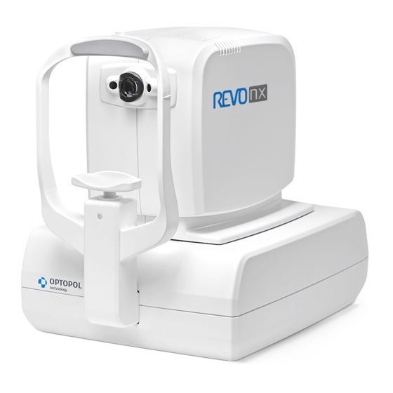
REVO nx
REVO nx 130
SOCT Copernicus REVO
SOCT Copernicus
REVO 60
REVO 80
REVO FC
Make sure you read this manual before using
the instrument. Keep this manual in a safe
place so that you can use it in the future.
User Manual
Software Version 10.0
User Manual Rev. A
0197
SOCT User Manual Version 10.0 rev. A
Manufacturer:
OPTOPOL Technology Sp. z o. o.
42-400 Zawiercie
www.optopol.com
info@optopol.com.pl
ul. Żabia 42
POLAND
Advertisement
Table of Contents

Related Manuals for Optopol REVO FC


Summarization of Contents
1 DESCRIPTION OF THE DEVICE
1.1 Intended use
Explains the intended use of the SOCT system for diagnostic aid in ocular diseases.
1.2 Intended User
Details the qualifications and skills required for the user, including Ophthalmologists and Optometrists.
1.6 Contraindication
Lists situations where the SOCT should not be used, such as photodermatosis history.
1.7 Protective Measures for IT Systems
Outlines cybersecurity measures for IT systems, including antivirus and firewalls.
4 UNPACKING AND INSTALLATION
4.1 Unpacking
Describes how to unpack the device shipped from the factory, including handling instructions.
4.2 Connecting cables
Details the USB 3.0 cable connection to the PC and power supply.
5 SOCT SOFTWARE
5.1 Running SOCT application
Explains how to start and load the SOCT application after system initialization.
6 PATIENT WINDOW
6.2 Registering new patients
Details the process of adding a new patient and required fields like name and DOB.
7 EXAMINATION ACQUISITION TAB
7.1 Selection of scan pattern mode
Explains how to select scan modes like Retina, Disc, Anterior, Central for different examinations.
7.2 Selection of scanning program
Describes how to select scan programs like 3D, Radial, B-scan, Cross, Raster.
7.3 Selection of protocol
Explains how to use predefined scanning programs for specific diseases and anatomy.
7.4 Device head movement controls
Details how to operate the movement control panel using mouse or touch screen.
8 CONDUCTING EXAMINATION
8.1 Preparation for examination
Covers patient preparation, pupil size, refraction, and device positioning before examination.
8.2 Acquisition modes description
Describes the Full Auto, Semi Auto, and Manual modes of examination acquisition.
8.3 Scanning programs description
Details various scanning programs like Retina, Central, Disc, Angiography, Biometry, Topography.
9 RESULT REVIEW
9.1 Type of View Mode
Describes different analysis views like Single, Both Eyes, Comparison, Progression.
10 POSTERIOR ANALYSIS
10.1 Retina Thickness Analysis
Details the analysis of retinal thickness, including different views and graphs.
10.2 Optic Nerve Head Analysis
Describes the analysis of NFL and ONH parameters for the optic nerve head.
10.3 Advanced View – Retina and Optic Nerve Head analysis
Explains the combined report with visual field data for glaucoma summary.
11 ANTERIOR SEGMENT ANALYSIS
11.1 Anterior Radial
Describes the corneal thickness analysis performed on anterior segment images.
11.1.5 Edit anterior surface
Explains how to manually edit surface boundary lines for better fit.
11.1.6 AOD measurement
Describes the tool for evaluating the angle opening of the anterior chamber.
11.1.7 Angle measurement tool
Details how to measure an angle using the tool, with options for visibility.
11.1.8 Caliper tool
Explains the tool for measuring lengths of various structures within the anterior chamber.
12 FULL SCREEN WINDOW
12.1 Fundus reconstruction, eye preview or pSLO
Explains how to toggle between fundus reconstruction, eye preview, or pSLO views.
12.3 Measurement tools and annotations
Explains how to use measurement tools and add annotations on fundus or tomogram images.
12.4 Brightness and contrast adjustment
Details how to adjust brightness and contrast using sliders for optimal image quality.
12.7 Edition of recognized layers
Explains how to manually correct layer recognition for accurate analysis.
12.8 Manual disc contour edition
Describes how to manually edit the disc contour for accurate analysis.
13 PRINT
13.1 Posterior segment examination reports/outputs
Covers reports for Retina 3D, Disc 3D, Optic Nerve Head, Topography, and Fundus examinations.
14 OUTPUT
15 SELECTING FUNDUS PHOTO
Describes how to import and display fundus images instead of reconstruction.
16 EXAMINATIONS CORRELATION
Details OCT-OCT and Fundus correlation for accurate comparison.
16.1 OCT-OCT Registration
Explains automatic and manual correlation of OCT scans using blood vessel shapes.
17 ANGIOGRAPHY OCT
17.1 RETINA OCT-A
Covers Retina OCT-A, Angio OCT Analysis, and Quantification Tools for vascular imaging.
17.2.1 FAZ tool
Explains the Foveal Avascular Zone measurement tool for angiographic analysis.
17.2.2 VFA tool
Describes the Vascular Flow Area measurement tool for detecting vessel areas.
17.2.3 NFA tool
Explains the Non-Flow Area measurement tool for quantifying non-perfused areas.
17.2.4 Quantification Maps [Density and Skeleton]
Provides quantification of vasculature in sectors and heat maps.
17.2.5 Angio OCT Analysis Table
Presents analysis tables for single, comparison, and both eyes views.
17.4 DISC OCT-A
17.4.1 [Single] view
Details the standard and detailed views for Disc OCT-A analysis.
17.4.1.2 Detailed
Allows viewing large objects and quantifying results for Disc Angio.
17.4.3 Angio Disc Comparison view
Compares two examinations of one eye on the same side from different dates.
17.4.4 [Progression] view
Compares four examinations over time for Disc Angio.
18 BIOMETRY OCT
18.1 Biometry Acquisition Mode
Explains the acquisition modes for biometry measurements.
18.2 Result review
Describes how to review biometry results and verify scanned structures.
18.1.1 Full Auto mode
Describes the Full Auto mode for biometry, including patient guidance.
18.1.2 Semi Auto mode
Describes the Semi Auto mode for biometry, requiring manual signal optimization.
18.1.3 Acceptance screen
Explains the acceptance screen for biometry results, requiring verification.
18.1.4 White to White
Details the White to White (WTW) measurement of limbus to limbus distance.
19 IOL CALCULATION TAB
19.1 Performing IOL calculation
Guides through calculating IOL power using patient data and selected formulas.
19.2 Marking implemented lenses
Explains how to mark selected IOLs for each eye.
19.3 IOL Editor
Describes the IOL Editor for importing, exporting, and editing IOL data.
20 TOPOGRAPHY OCT
20.1 Topography - Safety Notes
Provides safety precautions for using the topography module.
20.2 Topography Acquisition Mode
Describes the acquisition modes for topography scanning.
20.2.1 Full Auto mode
Describes the Full Auto mode for topography acquisition.
20.2.2 Semi Auto mode
Describes the Semi Auto mode for topography acquisition.
20.2.3 Acceptance screen
Explains the acceptance screen for topography results, checking measurement parameters.
20.3 Result review
Covers reviewing topography results and evaluation representations.
20.3.1.1 Enlarged detailed map view
Details enlarged map views for topography analysis.
20.3.3 [Comparison] view
Compares two topography examinations of one eye on the same side.
20.3.4 [Progression] view
Shows topography analysis results comparing six examinations over time.
20.4 Analysis
Details Central Keratometry, Keratometry (Meridian), Keratometry (SemiMeridian), Keratoconus screening.
20.4.4 Keratoconus screening
Explains the process of classifying keratoconus occurrence using KPI.
20.5 Map Types
Describes Axial, Tangential, Refractive Power maps and their properties.
21 CALIBRATION
21.1 Calibration procedure preparation
Explains how to prepare for calibration by installing the calibration tool.
21.2 Topography calibration
Details initial and standard topography calibration procedures.
21.2.1 Intitial Topography calibration procedure
Outlines the steps for the initial topography calibration process.
21.2.2 Standard calibration
Explains the standard calibration process when initial calibration is not required.
21.2.3 Calibration process
Details the steps involved in the calibration process, including tool installation.
21.3 Axial length (biometry) calibration
Covers biometry calibration to ensure high precision of measurements.
21.3.2 Biometry calibration with the IOL calculation tab activated
Explains biometry calibration when the IOL calculation tab is active.
21.3.3 Calibration process
Details the steps involved in the calibration process, showing progress and results.
21.3.4 Entering biometry calibration parameters
Explains how to enter calibration parameters provided with the calibration tool.
21.3.5 Common calibration
Describes how to perform common calibration for Biometry, Topography, and WTW.
22 SETUP WINDOW
22.1 General
Allows clinic details, language selection, and software skin layout settings.
22.2 Database
Covers database handling, location selection, and networking parameters.
22.3 Storage
Allows locating and adding storage folders for examination data.
22.4 Users accounts
Manages SOCT software users, including adding, removing, and editing accounts.
22.4.1 Creating user accounts
Details the process of creating new user accounts with different privilege levels.
22.4.2 LDAP Settings
Explains LDAP configuration for central account management.
22.5 Preferences
Allows customization of device and software settings.
22.5.1 CMDL Interface
Describes using Command Line Interface for data exchange with external systems.
22.5.2 Devices setup
Configures device settings and command line interface.
22.5.2.1 Protocols tab
Allows creating, editing, and deleting examination protocols.
22.5.2.2 Creating New Protocol
Explains how to create new protocols by adding scan programs.
22.5.2.3 Editing a Protocol
Details how to edit existing protocols, add/delete scan programs, or change order.
22.5.3 Parameters tab
Allows changing parameters like default eye, working position, and load program.
22.5.4 Voice messages
Customizes voice guide support, enabling or disabling patient guidance.
22.5.5 Results settings
Configures results display settings for review tabs.
22.5.6 Anonymization
Configures anonymization for personal data during output operations.
22.5.7 Visual Field
Configures PTS database connection and data transfer settings.
22.5.8 Input settings window
Sets input restrictions for patient data like ID, suffix, and prefix.
22.7 Output settings
Describes how to create, modify, and remove output sets for examination data.
22.7.4 DICOM C-storage output set
Configures DICOM C-storage output settings for data storage.
22.8 Backup
Covers database and examination backup options, including manual and automatic.
22.9 Recovery
Explains data recovery from internal and external storage.
22.10 DICOM
Details DICOM interface modules (SCU) and system settings.
22.10.1 System settings
Configures DICOM system settings like AE Title, Station Name, and Location.
22.10.2 MWL settings
Configures Modality Work List settings for patient data integration.
23 MAINTENANCE AND CLEANING PROCEDURE
23.1 Routine Cleaning
Details cleaning of device casing, applied parts (forehead/chinrest), and lens.
23.3 Hard Disk Defragmentation
Explains the necessity and process of hard disk defragmentation for performance.
24 NETWORK CONFIGURATION
24.1.1 SOCT network
Explains SOCT software network setup and viewing station configuration.
24.1.2 SOCT software - viewing station configuration
Details configuration steps for SOCT software on viewing stations.
25 REMOTE CONNECTION
25.1 PTS application for Structure & Function report
Configures PTS application for remote communication with the database.
31 PRODUCT COMPLIANCE
31.3 EMC INFORMATION
Provides guidance on electromagnetic emissions and immunity.
Need help?
Do you have a question about the REVO FC and is the answer not in the manual?
Questions and answers