Table of Contents
Advertisement
Quick Links
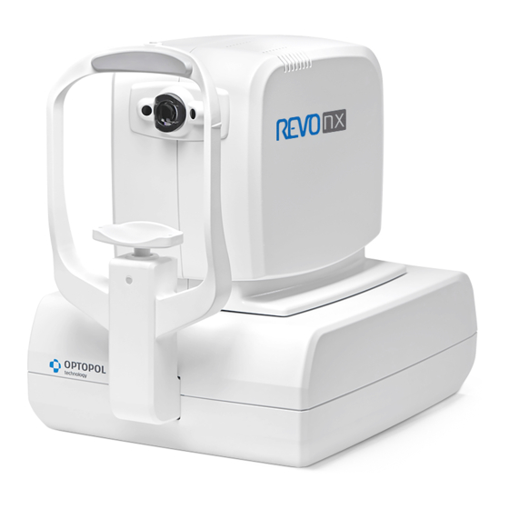
REVO nx
REVO nx 130
SOCT Copernicus REVO
SOCT Copernicus
REVO 60
REVO 80
REVO FC
Make sure you read this manual before using
the instrument. Keep this manual in a safe
place so that you can use it in the future.
User Manual
Software Version 10.0
User Manual Rev. A
0197
SOCT User Manual Version 10.0 rev. A
Manufacturer:
OPTOPOL Technology Sp. z o. o.
42-400 Zawiercie
www.optopol.com
info@optopol.com.pl
ul. Żabia 42
POLAND
Advertisement
Table of Contents

Related Manuals for Optopol REVO 60


Summarization of Contents
Description of the Device
Intended Use
Explains the device's purpose in diagnosing and managing ocular diseases.
Intended User
Defines the professionals qualified to operate the SOCT system.
Places of Use
Specifies the recommended locations for operating the SOCT device.
Proper Instrument Use
Provides guidelines for correct and safe operation of the SOCT instrument.
Contraindication
Lists conditions or patient histories that contraindicate SOCT use.
Protective Measures for IT Systems
Details cybersecurity measures for IT systems and network safety.
Cybersecurity Functions
Summarizes cybersecurity controls for the SOCT system with Windows 10.
Technical Data
Technical Data REVO NX 130 / REVO NX
Provides detailed technical specifications for REVO NX models.
SOCT Copernicus REVO/ SOCT Copernicus / REVO 60/REVO 80
Lists technical specifications for SOCT Copernicus and REVO 60/80 models.
Technical Data REVO FC
Details technical specifications for the REVO FC model, including fundus camera.
Device Classification
Classifies the device according to laser and protection standards.
Minimum Computer System Requirements
Specifies the hardware and software requirements for capture and review stations.
Safety
Safety Information
Provides essential safety information and hazard symbols used in the manual.
Product Label
Illustrates and describes the marks and indications found on the product label.
Safety Standards
Details safety standards and compliance requirements for the SOCT system.
Warnings
Highlights potential hazards that could result in property damage, injury, or death.
Cautions
Indicates hazards that, if not avoided, may result in property damage or injury.
General Notes
Provides important general notes and disclaimers regarding system operation and data.
Notes on Use
Offers essential notes for proper use before and after operating the SOCT device.
Unpacking and Installation
Unpacking
Instructions on how to unpack the SOCT device and its components.
Connecting Cables
Details on how to connect the SOCT device to a PC using USB 3.0 cable.
Device Connection
Explains the electrical connection scheme for the SOCT system.
Factory Default Calibration and Configuration
Information on factory settings and the need for user installation or configuration.
SOCT Software
Running SOCT Application
Step-by-step guide on how to launch the SOCT application after system initialization.
SOCT Application Structure
Overview of the user-friendly application structure and its main modules.
Patient Window
Patient List View
Describes how to view, sort, and filter the patient list for easy management.
Registering New Patients
Guide on how to register new patients, including required and optional fields.
Editing Personal Data
Instructions on how to edit existing patient data through the patient list.
Unregistering Patients
Details on how to delete patient records and their associated data from the system.
Examination List
Describes the information displayed for each examination in the list.
Deleting Exam/s
Procedure for deleting specific examinations or exams from a visit.
Connecting Scans Connected to the Wrong Patient
Explains how to move examinations or visits to the correct patient association.
Exporting Examinations
Guide on exporting examinations in raw .opt format for import into SOCT.
Export with Anonymization
Details on exporting examinations with anonymization settings applied.
Importing Examinations
Instructions on importing .oct and .opt formatted examinations into the system.
Filter
Explains how to use the filter window to find examinations by various criteria.
Output
Describes how to output results for single exams, visits, or all patient results.
Work List
Explains the functionality of the Work List tab for external software integration.
Examination Acquisition Tab
Selection of Scan Pattern Mode
Guides on selecting scan modes like Retina, Disc, Anterior, and Central.
Selection of Scanning Program
Details on selecting scanning programs such as 3D, Radial, B-scan, Cross, and Raster.
OCT Biometry Programs
Describes OCT Biometry programs for measuring axial length and other parameters.
OCT Topography Programs
Explains OCT Topography programs for analyzing corneal curvatures.
OCT Angiography Programs
Information on OCT Angiography programs for 3D scan visualization.
Fundus Camera Programs
Details on Fundus Camera programs for capturing color fundus photos.
OCT + Fundus Photography
Describes combined OCT and Fundus Photography scan programs.
Selection of Protocol
Guides on selecting predefined scanning programs for specific diseases or anatomies.
Device Head Movement Controls
Explains how to operate the device head using mouse or touch screen controls.
Eye Preview
Describes the eye preview window showing views from two cameras for patient pupil detection.
pSLO Live Fundus Preview
Details on the pSLO live image showing enface view of fundus for alignment.
IR Preview
Instructions for optimizing the image on the IR preview for optimal fundus position.
Operation on the Fundus Preview
Guidance on manipulating scanning area, angle, fixation target, and scan width.
Fixation Target Change
Information on selecting different sizes of internal and external eye fixation targets.
Customizing Scan Parameters
Explains how to adjust scan parameters and save custom settings as defaults.
Conducting Examination
Preparation for Examination
Steps to prepare the patient and check pupil size before conducting an examination.
Acquisition Modes Description
Describes the available acquisition modes: Full Auto, Semi Auto, and Manual.
Scanning Programs Description
Details on scanning programs like Retina, Central, Disc Area, and Angiography.
OCT Angiography Examination
Procedure for performing OCT Angiography examinations and understanding acceptance screens.
OCT Angiography Mosaic Mode
Explains predefined and manual modes for creating mosaic angiograms.
Anterior Measurement
Guides for conducting anterior segment examinations including cornea and angle scans.
Wide Anterior Programs
Details on wide anterior scan programs for REVO FC and REVO & NX models.
Biometry Program
Information about the optional OCT Biometry program for measurements.
Topography Program
Information about the optional OCT Topography program for corneal analysis.
Fundus Image REVO FC (REVO FC only)
Describes the OCT/Fundus and Fundus modes specific to the REVO FC device.
Full Range Examination Mode
Explains the Full Range examination mode for increased scanning depth.
External Fixation
Describes the method of using an external target light for patient fixation.
Chorioretinal/Vitreoretinal Mode
Explains C-gate mode settings for enhancing information above or around the RPE.
Examination Tips
Provides tips for handling patients and troubleshooting common issues during examinations.
Critera for Exam Acceptance
Criteria for ensuring OCT and Fundus reconstruction images are suitable for diagnosis.
Result Review
Type of View Mode
Describes different analysis views like Single, Both Eyes, Comparison, and Progression.
Lock Function
Enables synchronized manipulation of tomograms across different tabs.
Types of Analysis
Outlines the available analysis types, including Single Retina analysis.
AI DeNoise
Explains the AI DeNoise algorithm for filtering noise and improving image quality.
3D Tab
Details the availability and usage of AI DeNoise function in the 3D tab.
Posterior Analysis
Retina Thickness Analysis
Analysis of retinal thickness, including maps, graphs, and normative data.
Optic Nerve Head Analysis
Analysis of NFL thickness and optic nerve head shape parameters.
Advanced View – Retina and Optic Nerve Head Analysis
Provides glaucoma summary reports for 3D Retina and Disc scans.
Structure & Function – Combined OCT and VF Report
Combines visual field data with OCT analysis for anatomical relationship visualization.
Structure & Function – VF Locations Layer
Allows overlaying VF results and locations onto OCT maps for analysis.
Anterior Segment Analysis
Anterior Radial
Corneal thickness analysis based on recognized OCT structures in anterior scans.
Edit Anterior Surface
Enables manual correction of corneal surface boundary lines for better fit.
AOD Measurement
Tool to evaluate the angle opening, measuring distances and angles.
Angle Measurement Tool
Enables measurement of angles in degrees, with visibility options.
Caliper Tool
Used to measure lengths of structures within the anterior chamber.
Tomogram Review Analysis
Analysis of tomogram images and defined thickness of specified parts.
Full Screen Window
Fundus Reconstruction, Eye Preview or pSLO
Drop-down list to toggle between fundus reconstruction, eye preview, pSLO, or fundus photo.
Imaging Tools
Includes tools for changing color, tomogram proportions, and image settings.
Measurement Tools and Annotations
Allows using measurement tools for area, distance, and angle on images.
Brightness and Contrast Adjustment
Adjusts brightness and contrast using sliders for optimal image visibility.
Full Screen Mode Exit
Exits full screen mode and returns to exam analysis.
Tomogram Window Manipulation
Provides options for displaying and manipulating tomogram previews.
Edition of Recognized Layers
Enables manual correction of automatically recognized layers in tomograms.
Manual Disc Contour Edition
Allows manual editing of the disc shape and position for analysis.
Redraw the Disc Contour
Procedure for redrawing the disc contour manually using markers.
Print
Posterior Segment Examination Reports/Outputs
Describes how to print reports and outputs for posterior segment examinations.
Disc 3D
Details the examination report format for Disc 3D scans.
Optic Nerve Head Analysis Reports
Covers the reports generated for optic nerve head analysis.
Topography Examination Reports
Details the examination reports generated by the topography module.
Fundus Examination Reports
Describes the examination reports generated for fundus imaging.
Angiography Examination Reports
Disc OCT-A Examination Reports
Provides reports for Disc OCT-A examinations.
Biometry Examination Reports
Details the examination reports generated by the biometry module.
Multi B-scan Report
Explains the procedure for printing multi B-scan reports.
Single Tomogram Report
Instructions on how to print a single tomogram on a full page.
Select Desired Printer
Guides on selecting the desired printer before printing reports.
Output
Selecting Fundus Photo
Guides on importing and selecting fundus photos for examination.
Fundus Image Registration Correction
Procedure for correcting the position of imported fundus images.
Linking a Fundus Photo to an Examination
Explains how to link a single fundus photo to multiple OCT exams.
Examinations Correlation
OCT-OCT Registration
Details automatic and manual correlation of OCT examinations based on vessel shape.
Fundus Correlation
Explains automatic and manual correlation of fundus photographs with OCT.
Extracting Tomograms from a 3D Exam
Describes how to display follow-up tomograms for comparison and tracking.
FUNDUS CAMERA – RESULT REVIEW
Functionality for reviewing results from Fundus Camera photography acquisition.
Angiography OCT
Retina OCT-A
Procedure for acquiring Angio OCT data and using Standard and Detailed views.
Angio OCT Analysis Table
Presents analysis tables for FAZ, VFA, and NFA measurements.
Quantification Tools: FAZ, VFA, NFA
Tools for quantifying vascular parameters like FAZ, VFA, and NFA.
Motion Correction
Algorithm to eliminate eye movement artifacts in Angio OCT and 3D exams.
Disc OCT-A
Details on performing and analyzing Disc OCT-A scans in various views.
Mosaic
Feature for presenting wider field of view with that same level of details.
Biometry OCT
Biometry Acquisition Mode
Steps for acquiring biometry data, including initial calibration.
Full Auto Mode
Describes the fully automated biometry acquisition process.
Semi Auto Mode
Explains the semi-automatic mode for optimizing signal in challenging cases.
Acceptance Screen
Post-capture screen for verifying scanned ocular structures in biometry.
White to White
Measurement of distance from limbus to limbus and pupil diameter.
Result Review
Recommendations for reviewing biometry results for validity and manual correction.
IOL Calculation Tab
Performing IOL Calculation
Guides on selecting biometry exams and entering keratometry and refraction data.
Marking Implemented Lenses
Procedure for marking specific IOLs for calculation and editing.
IOL Editor
Window for importing, exporting, and editing IOL data and constants.
Importing IOL Data
Instructions on importing IOL data from various file formats.
Exporting IOL Data
Procedure for exporting the full database or single lens data.
Adding Lenses Manually
Guide on manually adding new IOL lenses to the system.
Deleting Lenses Manually
Instructions on how to delete single lenses or all lenses by a manufacturer.
Viewing the List of Lenses
How to unfold and view available lenses sorted by manufacturer.
Editing IOL Data
Allows personalization of IOL data, including manufacturer, model, and revisions.
Adding Additional Power Ranges and Increments
Procedure for creating custom power ranges and increments for IOL calculations.
Topography OCT
Topography - Safety Notes
Provides safety notes and recommendations for using the topography module.
Topography Acquisition Mode
Guides on selecting scan programs and operating the topography acquire window.
Full Auto Mode
Describes the fully automated topography acquisition process.
Semi Auto Mode
Explains the semi-automatic mode for optimizing signal in topography scans.
Acceptance Screen
Checks measurement parameters for acceptability after topography acquisition.
Total Quality Factor
Defines the summary factor for trusting measurement reliability.
Result Review
Provides an overview display of evaluation representations for the anterior eye segment.
Analysis
Details Central Keratometry, Keratometry (Meridian), Keratometry (SemiMeridian), and Keratoconus screening.
Map Types
Explains different map types like Axial, Tangential, Refractive Power, and Elevation maps.
Color Scale - Standards
Guides on changing scale types, steps, and units for map visualization.
Calibration
Calibration Procedure Preparation
Instructions on installing and preparing the calibration tool.
Topography Calibration
Procedure for initial and standard topography calibration.
Calibration Process
Details the steps involved in the calibration process.
Axial Length (Biometry) Calibration
Ensures high precision of measurements by calibrating the Biometry module.
Entering Biometry Calibration Parameters
Instructions to enter calibration parameters provided with the calibration tool.
Common Calibration
Allows simultaneous calibration of Biometry, Topography, and WTW modules.
Setup Window
General
Allows customization of clinic details, language, and software skin layout.
Database
Tools for handling database, setting networking parameters, and selecting locations.
Storage
Allows locating the database in various folders and adding storage space.
Users Accounts
Manages SOCT software users, including adding, removing, and editing accounts.
LDAP Settings
Configuration for central account management using Lightweight Directory Access Protocol.
Preferences
Tab for customizing device and software settings.
CMDL Interface
Enables independent operation and data exchange with external systems.
Devices Setup
Configuration tabs for setting up connected devices and protocols.
Parameters Tab
Allows changing device and software parameters like eye selection and Angio settings.
Voice Messages.
Customization options for voice guide support during patient alignment and acquisition.
Results Settings.
Settings for controlling how results are displayed in review tabs.
Anonymization.
Configuration of personal information anonymization for data output.
Visual Field
Configuration for data transfer between PTS software and SOCT.
Input Settings Window
Settings for patient ID, suffix, prefix, and disease list management.
Edit Disease List Window
Allows creating and managing a list of diseases for patient records.
Output Settings
Describes how to create, modify, and remove output data sets.
Graphic File Standard
Information on viewing outputted data in standard graphic file formats.
Maintenance and Cleaning Procedure
Routine Cleaning
Guidelines for cleaning the device casing, applied parts, and lens.
Software Maintenance Activities
Essential activities for keeping the SOCT software in good condition.
Hard Disk Defragmentation
Procedure to defragment the OCT PC hard disk for optimal performance.
Network Configuration
SOCT Network
Details on locating examination data on network locations and sharing the database.
SOCT Software - Viewing Station Configuration
Instructions for configuring SOCT software on viewing stations for database access.
Remote Connection
PTS Application for Structure & Function Report
Configures PTS application for remote communication with the database.
Environmental Conditions
Environmental Conditions of Use
Specifies temperature, humidity, and pressure requirements for operation.
Storage Conditions
Lists environmental conditions for storing the SOCT device.
Transport Conditions
Details environmental conditions and shock/bump limits for transporting the device.
Patient Environment
Describes the required patient environment for installation and comfort.
Measurement Units
Specifies that measurements are in SI format, primarily in micrometers.
Warranty and Service
Service Life
Information regarding the service life of the SOCT product.
About Repairs
Procedure to follow if a problem cannot be solved and contact information for repairs.
Utilization
After Usage Period
Instructions for contacting the distributor for utilization instructions after usage period.
Troubleshooting
Q1: Shadow at Horizontal Tomogram Edge
Addresses shadows and diagonal images in horizontal tomograms due to beam centering.
Q2: Diagonal Horizontal Retina Cross Section
Troubleshooting for diagonal horizontal retina cross section images.
Q3: Shadow at Vertical Tomogram Edge
Addresses shadows and diagonal images in vertical tomograms due to beam centering.
Q4: Diagonal Vertical Tomogram Image
Troubleshooting for diagonal vertical tomogram images.
Q5: Fuzzy and Upside Down Tomogram Images
Resolves issues with fuzzy and upside down tomogram images due to C-gate position.
Q6: Software Communication Errors
Troubleshooting communication errors after starting the software.
Q7: No Signal Detected Message
Resolves 'No signal detected' messages by running Skantest and calibration.
Q8: Bad Quality Tomograms
Addresses issues with bad quality tomograms, suggesting lens cleaning and calibration.
Q9: Connection Error with Device
Troubleshooting connection errors between SOCT application and device, checking USB.
Q10: OCT Live Windows Become Black
Addresses black OCT live windows by checking device status and restarting the application.
Q11: No Connection to Remote Database
Troubleshooting remote database connection issues by verifying network settings.
Q12: Analyze Window Displayed in Quarter Screen
Resolves analyze window display issues by adjusting screen and Windows resolution.
Product Compliance
Radio Interference
Details FCC compliance for REVO nx digital device regarding radio frequency energy.
Canadian Regulations
States compliance with Class A limits for radio noise emissions in Canada.
EMC Information
Guidance on electromagnetic emissions and immunity for SOCT usage environments.
Need help?
Do you have a question about the REVO 60 and is the answer not in the manual?
Questions and answers