Advertisement
Table of Contents
- 1 Table of Contents
- 2 Overview
- 3 Microscope Lens List
- 4 System Start up
- 5 Graphical User Interface Description
- 6 Locate Specimen, Focus, Find Cells / Region of Interest
- 7 Confocal Imaging Set up
- 8 Saving an Image File
- 9 Removing Sample from Microscope Stage
- 10 Z-Stack Set up
- 11 Airyscan Imaging Set up
- 12 Tile / Position Set up (Simple
- 13 Tile / Position Advanced Setup
- 14 Time Series Set up
- 15 Oil Immersion Lens Clean Procedure
- 16 System Power down
- Download this manual
Advertisement
Table of Contents

Related Manuals for Zeiss LSM 800


Summary of Contents for Zeiss LSM 800
- Page 1 Imaging and Flow Cytometry Core CPOS LSM 800 SOP LSM 800 Confocal Microscope Standard Operation Protocol ver. 2.1 (Updated: 11/13/2020)
-
Page 2: Table Of Contents
Imaging and Flow Cytometry Core CPOS LSM 800 SOP Contents Overview ................................A-3 Microscope Lens list ............................... B-3 System start up ..............................C-4 Graphical User Interface Description ........................D-6 Locate specimen, focus, find cells / region of interest ..................E-7 Confocal imaging set up ............................ -
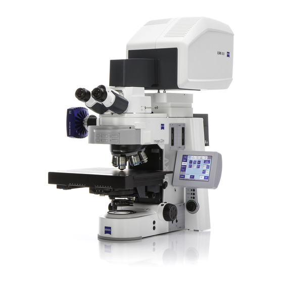
Page 3: Overview
LSM 800 SOP A. Overview This guide will cover operation of the Carl Zeiss GmbH LSM 800 scanner paired with an inverted microscope setup and ZEN Blue edition ver. 2.3 driving software. For specific operations of the ZEN Blue software please read http://med.hku.hk/corefac/downloads/ZEN_BLUE2.3SOP_20200415.pdf... -
Page 4: System Start Up
○ Turn ON computer Double click on the program icon on desktop. click on Start | All Programs | Carl Zeiss Microscopy | ZEN | ZEN (blue edition) entry (blue icon). Click on “ZEN system” button. - Page 5 Imaging and Flow Cytometry Core CPOS LSM 800 SOP iii. Select desired objective. Put on lens heater by first unravelling Velcro tape and then re-attach around the objective lens. See image for reference. Mock-up on-stage incubator and adjust motorized stage position to confirm objective lens is centred with sample frame.
-
Page 6: Graphical User Interface Description
Imaging and Flow Cytometry Core CPOS LSM 800 SOP D. Graphical User Interface Description The terms will be used throughout this document to locate different buttons. Reference to this part from time to time when you read the rest of this SOP. -
Page 7: Locate Specimen, Focus, Find Cells / Region Of Interest
Imaging and Flow Cytometry Core CPOS LSM 800 SOP E. Locate specimen, focus, find cells / region of interest You should be on "Locate" tab if the software has just finished calibration. If not navigate to "Locate" tab. Select objective lens with software / on microscope control panel. -
Page 8: Confocal Imaging Set Up
Imaging and Flow Cytometry Core CPOS LSM 800 SOP F. Confocal imaging set up Prerequisites: Region of interest is centred in field of view and have clicked 'Acquisition' tab. Select one of the pre-set made by Imaging and Flow Cytometry Core. - Page 9 Imaging and Flow Cytometry Core CPOS LSM 800 SOP Saturation ..fix this later 15. Activate all channels you need by ticking checkboxes. 16. Highlight a track you wish to adjust. 17. You may use the '1 AU' hotkey to adjust pinhole size.
- Page 10 Imaging and Flow Cytometry Core CPOS LSM 800 SOP F-10 25. 'Digital Offset' for mathematically subtract apparent intensity of image typical setting = 0. All pixel values will appear as: (original value) - (Digital Offset value) = image pixel intensity.
-
Page 11: Saving An Image File
Applicable if no such folder exists and you are the first one in your lab using LSM 800. Navigate into the folder you have just created. Choose file format as '.czi' acronym for 'Carl Zeiss Image' Type in name (preferably no spacing). Click 'Save'... -
Page 12: Removing Sample From Microscope Stage
Imaging and Flow Cytometry Core CPOS LSM 800 SOP H-12 H. Removing sample from microscope stage. On control panel press 'load position' button. Objective lens should be moved to lowest position allowing clearance for you to operate with the stage area. -
Page 13: Z-Stack Set Up
Imaging and Flow Cytometry Core CPOS LSM 800 SOP I-13 I. Z-stack set up Pre-requisite: you have setup all necessary channels according to previous chapters including laser power, master gain, pin hole size. **Note: image intensity might increase beyond saturation if the z-position you chose to setup laser power / master gain is not the brightest structure in the z-volume. - Page 14 Imaging and Flow Cytometry Core CPOS LSM 800 SOP I-14 12. Most application “keep interval” is useful. 13. If you select 'optimal' in 'Z-stack' window. LSM will perform optical sections where each section will have 50% overlap with the previous section (2x Nyquist sampling).
-
Page 15: Airyscan Imaging Set Up
Imaging and Flow Cytometry Core CPOS LSM 800 SOP J-15 J. Airyscan imaging set up Follow air to oil immersion lens change procedure in training. Switch to 63x objective. Select pre-set with prefix '0IFCore_Airyscan...' Click 'Smart setup' select fluorescent dyes and then click '+' button. - Page 16 Imaging and Flow Cytometry Core CPOS LSM 800 SOP J-16 19. Adjust laser power so that max image intensity reach ~ 5000 - 8000 intensity units (around 50% of detector dynamic range) under 16-bit mode or ~30 intensity units under 8-bit mode.
-
Page 17: Tile / Position Set Up (Simple
Imaging and Flow Cytometry Core CPOS LSM 800 SOP K-17 K. Tile / Position set up (Simple) (With multi-position function included in this chapter) click checkbox to activate 'Tile' module. Tile Simple set up Scroll to “tile Set up” window. - Page 18 Imaging and Flow Cytometry Core CPOS LSM 800 SOP K-18 12. Reference to Chapter [E] 'locate specimen, focus, find cells/region of interest' 13. Go to 'Locate' tab. 14. Turn on fluorescence channel for navigation. 15. Use xy-motorized control to navigate your specimen.
-
Page 19: Tile / Position Advanced Setup
Imaging and Flow Cytometry Core CPOS LSM 800 SOP L-19 L. Tile / Position Advanced Setup Advanced set up (Preview scan) Common steps = Establish a preview scan -> determine region of interest (R.O.I.) either in area or in points -> verify positions -> Start Experiment. - Page 20 Imaging and Flow Cytometry Core CPOS LSM 800 SOP L-20 Go to 'Tile Setup' and draw an R.O.I. that can contain the whole specimen. Rectangular / circular / free-hand draw. Preview scan tab. For maximum speed, disable 'Use Existing Experiment Settings'.
- Page 21 Imaging and Flow Cytometry Core CPOS LSM 800 SOP L-21 20. If no Support points were selected, then Tile will be acquired according to current z- height. Support points are useful when the specimen plane is uneven / tilted. One should always ensure their specimen is level before attempting to rescue with “support points”...
- Page 22 Imaging and Flow Cytometry Core CPOS LSM 800 SOP L-22 to scan. User x-y control knob to move to area you would like to mark and then press 33. Repeat step L28 to step L32 until you have selected all sites you wish to image.
- Page 23 Imaging and Flow Cytometry Core CPOS LSM 800 SOP L-23 40. Use definite focus (DF) or Autofocus (AF) on the remaining points. Select 'Autofocus AF' in 'Select Verification Helper Method'. 41. When ‘DF’ or ‘AF’ previously ‘Set Z and move to next will be changed to ‘Run DF / AF and...
-
Page 24: Time Series Set Up
Imaging and Flow Cytometry Core CPOS LSM 800 SOP M-24 M. Time series set up Pre-requisite: you have setup a series of actions z-stack, multichannel, tile, or any combination. 44. Scroll to 'Experiment Information' to check estimated time required to perform such actions. -
Page 25: Oil Immersion Lens Clean Procedure
• [N.B.] Use only lens cleaning tissue instead of Kimwipe or any coarser material to prevent scratches on optical surfaces. • [N.B.] Quote Carl Zeiss Cleaning guide "The natural tendency to minimize waste is misdirected, considering the relative cost of lens tissue compared to the potential of damaging an expensive objective."... -
Page 26: System Power Down
Imaging and Flow Cytometry Core CPOS LSM 800 SOP O-26 O. System Power down Prerequisite: You have cleaned immersion objective lenses according to previous chapter [N]. [Optional] if you have turned on live cell incubator follow these steps. Turn off CO2 supply by turning gas tank valve clockwise. - Page 27 Imaging and Flow Cytometry Core CPOS LSM 800 SOP O-27 17. Turn laser emission key from 'I' back to '0' 18. Turn off number 4 and 3 power supply units. 19. Check that LED beside number [7] is no longer blinking indicating computer has finished shut down.
Need help?
Do you have a question about the LSM 800 and is the answer not in the manual?
Questions and answers