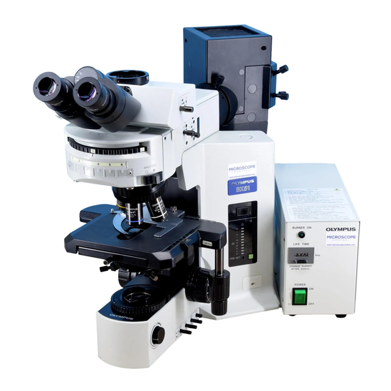
Olympus BX51 Instructions Manual
Polarizing microscope
Hide thumbs
Also See for BX51:
- Instructions manual (40 pages) ,
- Operating manual (17 pages) ,
- Operating manual (11 pages)
Table of Contents
Advertisement
INSTRUCTIONS
BX51/52-P
POLARIZING MICROSCOPE
This instruction manual is for the Olympus Polarizing Microscope Model BX51/52-P. To obtain optimum
performance and to familiarize yourself fully with the use of your microscope, we recommend that you
read this manual together with the instruction manual for the BX51/BX52 microscope thoroughly before
use. Retain this instruction manual in an easily accessible place near the work desk for future reference.
A X 6 3 0 7
Advertisement
Table of Contents

Subscribe to Our Youtube Channel
Related Manuals for Olympus BX51


Summary of Contents for Olympus BX51
- Page 1 BX51/52-P POLARIZING MICROSCOPE This instruction manual is for the Olympus Polarizing Microscope Model BX51/52-P. To obtain optimum performance and to familiarize yourself fully with the use of your microscope, we recommend that you read this manual together with the instruction manual for the BX51/BX52 microscope thoroughly before use.
-
Page 2: Table Of Contents
BX51/52-P CONTENTS IMPORTANT — Be sure to read this chapter for safe use of the equipment. — 1 NOMENCLATURE 2 ASSEMBLY 2-1 Assembly Diagram ................................................. 3 2-2 Detailed Assembly Procedure ........................................ 4-9 3 CONTROLS 10-11 4 USING THE CONTROLS 12-15 4-1 Stage ...................................................... -
Page 3: Important - Be Sure To Read This Chapter For Safe Use Of The Equipment
1. A microscope is a precision instrument. Handle it with care and avoid subjecting it to sudden or severe impact. 2. The BX51/52 series microscope can be used with up to two additional intermediate attachments (e.g., U-DO3 dual- viewing attachment, U-CA or U-ECA magnification changer, etc.). When using an additional intermediate attachment, please make sure with your Olympus representative or the latest brochure. -
Page 4: Nomenclature
BX51/52-P NOMENCLATURE Eyepiece Observation tube BX51/52 Microscope frame * Polarizing intermediate attachment * Objective * Centerable revolving (Strain-free objective series) eyepiece * Polarizing rotatable stage * Strain-free Lamp housing condenser *Components for polarized light observation... -
Page 5: Assembly
The diagram below shows how to assemble the various components. The numbers indicate the order of assembly. }For details on the BX51/52 microscope frame, consult the BX51/52 manual. # When assembling the components, make sure that all parts are free of dust and dirt, and avoid scratching any parts or touching glass surfaces. -
Page 6: Detailed Assembly Procedure
· Mount the mechanical stage so that the positioning pins on the under- side fit into the positioning holes … on the stage top surface. Using the Allen screwdriver provided with the BX51/52 microscope frame, tighten the clamping screw. …... - Page 7 Mounting the Condenser (U-POC-2) (Fig. 3) ² 1. Turn the coarse adjustment knob @ to raise the stage to its upper limit. 2. Turn the condenser height adjustment knob ² to lower the condenser holder to its lower limit. 3. Loosen the condenser clamping screw ³. 4.
- Page 8 BX51/52-P Mounting the Objectives (Fig. 4) # Insert the 10X or 20X objective into the primary hole (position where black rubber plugs are inserted into the centering holes on the nose- piece). Mount the other objectives in such a manner, that the magnification in-...
- Page 9 Mounting the Intermediate Attachments (Fig. 6) 1. Using the Allen screwdriver, loosen the observation tube clamping screw @ on the microscope frame. 2. Insert the circular dovetail mount at the bottom of the intermediate attachments into the opening on the microscope frame and clamp by tightening the clamping screw @.
- Page 10 BX51/52-P Mounting the Observation Tube (Fig. 7) 1. Using the Allen screwdriver, fully loosen the observation tube clamping screw @ on the intermediate attachment. 2. Insert the circular dovetail mount at the bottom of the observation tube into the opening on the intermediate attachment, placing the observation tube to point the binocular eyepieces towards the front.
- Page 11 Mounting Rotatable Analyzer (U-AN360P-2) (Fig. 9) 1. Place a desired ND filter (30 mm diam.) in the empty hole @ as required. 2. Insert the rotatable analyzer (U-AN360P-2) ² as far as the click-stop position. Then screw in the stopper knob ³. (Fig. 10) When using the fixed analyzer U-ANT instead of the rotatable analyzer U-AN360P-2, place the fixed analyzer in the test plate adapter U-TAD.
-
Page 12: Controls
BX51/52-P CONTROLS The illustration shows the U-CPA set. Other components are intro- duced on the following pages. Bertrand lens focusing knob Bertrand lens knob Analyzer Slider clamping knob Slot for compensators Mechanical stage Vernier scale Stage rotation Aperture iris diaphragm lever... - Page 13 Intermediate Adapter for AN360P (U-OPA) Rotatable Analyzer (U-AN360P-2) Empty hole (ND filter insertion possible) Analyzer slot Clamping knob Stopper knob Alignment polarizer Rotation dial Test Plate Adapter (U-TAD) Sensitive Tint Plate (U-TP530) Quarter Wave Plate (U-TP137) Test plate clamping knob Hole for the fixed analyzer (U-ANT) Slot for compensator plate...
-
Page 14: Using The Controls
BX51/52-P USING THE CONTROLS 4-1 Stage Specimen Placement When Using the Stage Clips (Fig. 11) Position the specimen in the center, and clamp the specimen with the stage clips. When Using the Mechanical Stage (U-FMP) (Fig. 12) Open the spring-loaded finger @ and place the specimen on the stage. - Page 15 Stage Rotation (Fig. 13) When the stage rotation clamping knob @ is loosened, the stage can be rotated horizontally through 360°. Fig. 13 Using 45° Click Stop Lever (Fig. 14) When the 45° click stop lever @, located at the right side of the stage, is moved toward the observer, and the stage is moved from this position to the first click-stop position, the specimen is moved 45°...
- Page 16 BX51/52-P Stage Height Adjustment (Figs. 15 & 16) }By lowering the position of the substage, the microscope will accom- modate specimens with a maximum height of 40 mm. This is useful when observing metallurgical specimens and other thick objects. 1. Lower the stage to its lower limit, then remove the stage from the microscope.
-
Page 17: Intermediate Polarizing Attachment
4-2 Intermediate Polarizing Attachment Using the Bertrand Lens (U-CPA only) (Fig. 17) By manipulating the Bertrand lens knob @ on the front, the Bertrand lens is inserted into or removed from the light path. At the pushed-in ({) position, the lens is engaged. At the pulled-out (¦) position, the lens is removed from the light path. -
Page 18: Polarized Light Observation
BX51/52-P POLARIZED LIGHT OBSERVATION 5-1 Adjustments Before Observation In polarized light microscopy ultimate performance is not obtainable unless optical adjustments are made correctly. Always performed the following adjustments before observation. Remove the quarter wave plate and sensitive tint plate from the light path. - Page 19 AN360P (U-OPA) | until complete extinction is obtained. At this point, tighten the intermediate attachment clamping screw ƒ. (Fig. 20) 6. Remove the provided alignment polarizer. 7. Attach the condenser. 8. Center the condenser. (Refer to page 18 in the BX51/52 instruction manual.) ƒ Fig. 20 9.
- Page 20 1. Pull out the Bertrand lens knob @ to the OUT (¦) position. (Fig. 23) 2. Center the condenser. (Refer to page 18 in the BX51/52 instruction manual.) 3. Follow steps 9 and 10 in “When Using the Intermediate Attachment for AN360".
- Page 21 3. Adjust the position of the intermediate tube (Refer to page 7.) 4. Center the condenser. (Refer to page 18 in the BX51/52 instruction manual.) 5. Push in the Bertrand lens knob @ to the IN ({) position for conoscopic observation.
- Page 22 BX51/52-P 6. To brighten and facilitate observation of the conoscopic image during optical axis adjustment, rotate the analyzer slightly away from the position of total extinction. (Fig. 26) Conoscopic image Fig. 26 7. Insert the two provided centering wrenches ² into the centering holes for the primary hole with the 20X or 10X objective on the nosepiece.
- Page 23 10. Centering the rotatable stage (Figs. 28 & 29) (1) Place the specimen. ³ (2) Focus on the specimen and look for an easily recognizable detail ³ in the field. Move this detail in the center of the eyepiece cross lines. (3) When the stage is rotated, the detail moves in circle |.
- Page 24 BX51/52-P Adjusting for Extinction (Fig. 30) # Remove specimen, test plate, compensator, etc. from the light path. 1. Swing out the top lens of the condenser, and engage the 10X objective. 2. Insert the rotatable analyzer into the light path and set vibration direction scale at the 0°...
- Page 25 Adjusting the Eyepiece Cross Lines (Fig. 31) }To align the eyepiece cross lines and the vibration direction, an orientation plate (B2-PJ) is required. Orientation plate (B2-PJ) Fig. 31 # Remove the test plate adapter and the compensator from the light path.
- Page 26 BX51/52-P 3. Coincide the orientation plate’s center portion with the intersection of the eyepiece cross lines. Engage the analyzer into the light path (in case of U-AN360P-2, set the analyzer to position 0°) to obtain crossed filters position (extinction). 4. While observing, rotate the stage to locate the position where the orientation plate is darkest. At this point, clamp the stage.
-
Page 27: Orthoscopic Observation
5-2 Orthoscopic Observation }In principle, polarized light enters the light path, parallel to the optical axis, to enable observation of the optical characteristics of the specimen. Therefore, swing out the top lens of the condenser. Use 4X to 100X objectives. 1. -
Page 28: Conoscopic Observation
BX51/52-P 5-3 Conoscopic Observation }Use 20X to 100X objectives. 1. Engage the analyzer and adjust for extinction position. 2. Swing the condenser top lens into the light path. 3. When using the polarizing intermediate attachment U-CPA, push in the Bertrand lens knob to engage the Bertrand lens into the light path. -
Page 29: Specifications
SPECIFICATIONS Specifications Item Polarizing intermediate Intermediate attachment attachment (U-CPA) for AN360P (U-OPA) 1. Polarizing intermediate Field No. attachment Bertrand lens Focusable (U-CPA and U-OPA) Bertrand aperture stop Fixed diameter Bertrand lens knob Pushed in position for switch position: { IN between orthoscopic Pulled out and conoscopic... - Page 30 BX51/52-P Specifications Item Polarizing intermediate Intermediate attachment attachment (U-CPA) for AN360P (U-OPA) 4. Stage (U-SRP) Type: Polarizing rotatable stage with 3-point centering mechanism 360° horizontal rotation, clamps at any desired position 360° scale (minimum division: 1°; minimum reading 6' by means of vernier scale) 45°...
-
Page 31: Optical Characteristics
Magnification right). NOTE Numerical aperture (N. A.) Mechanical Refer to the latest catalogue or consult your local Olympus tube length Cover glass thickness representative for the updated information on the eyepieces and objectives that can be combined with this microscope. -
Page 32: Troubleshooting Guide
Under certain conditions, performance of this unit may be adversely affected by factors other than defects. If problems occur, please review the following list and take remedial action as needed. If you cannot solve the problem after checking the entire list, please contact your local Olympus representative for assistance. Trouble... - Page 33 MEMO...
- Page 34 2-43-2,Hatagaya, Shibuya-ku, Tokyo, Japan Postfach 10 49 08, 20034, Hamburg, Germany 2 Corporate Center Drive, Melville, NY 11747-3157, U.S.A. 491B River Valley Road, #12-01/04 Valley Point Office Tower, Singapore 248373 2-8 Honduras Street, London EC1Y OTX, United Kingdom. 104 Ferntree Gully Road, Oakleigh, Victoria, 3166, Australia This publication is printed on recycled paper.
Need help?
Do you have a question about the BX51 and is the answer not in the manual?
Questions and answers