
Related Manuals for Motic PantheraTEC Series


Summary of Contents for Motic PantheraTEC Series
- Page 1 PantheraTEC Metallurgy Instrument Operation Manual If the equipment is used in a manner not specified by the manufacturer, the protection provided by the Note equipment may be impaired. WWW.MOTIC.COM MOTIC HONG KONG LIMITED...
- Page 2 English: Please familiarize yourself with the Instruction Manual provided in English language. Other Language versions are available as download on Motic web services under the Address: https://moticeurope.com/en/resources#section-instructions...
-
Page 3: Table Of Contents
TABLE OF CONTENTS Introduction Welcome 1.2 PantheraTEC Features 1.3 Genral Notes On Instrument Safety 1.3.1 General safety notes and Instruction 1.3.2 Instrument safety, FCC and EMC conformity 1.3.3 Transporting, unpacking, storage of the Instrument 1.3.4 Instrument Disposal 1.3.5 Use of the Instrument 1.3.6 Intended use of the Microscope 1.3.7... - Page 4 4.6 Eyepiece tube 4.7 Intermediate tube 4.8 Eyepieces 4.9 Filters 4.10 Power cord Digital Parts (Setup and Operation) 5.1 How to use QR-Link (PantheraTEC-BF/BD-TD, PantheraTEC-POL/EpiPOL Digital) 5.2 ImagingOnDevice 5.2.1 PantheraTEC Digital Series Backside Connection Panel 5.3 TeachingOnDevice (PantheraTEC-BF/BD-TD, PantheraTEC-POL/EpiPOL Digital) 5.3.1 PantheraTEC ImagingOnDevice realtime sharing ImagingOnDevice with QR-Link clients...
- Page 5 6.10 Use of Aperture Diaphragm (For PantheraTEC-BF/BD/PantheraTEC-EpiPOL) 6.11 Use of Polarizer and Analyzer (For PantheraTEC-BF/BD/PantheraTEC-EpiPOL) 6.12 Polarization and Conoscopy Adjustment (For PantheraTEC-POL/EpiPOL) 6.12.1 Centering the objective 6.12.2 Performing Polarization Examinations with Transmitted Light 6.12.3 Performing Polarization Examinations with Reflected Light (For PantheraTEC-EpiPOL) 6.12.4 Focusing and centering the Bertrand lens 6.12.5 Compensators...
-
Page 6: Introduction
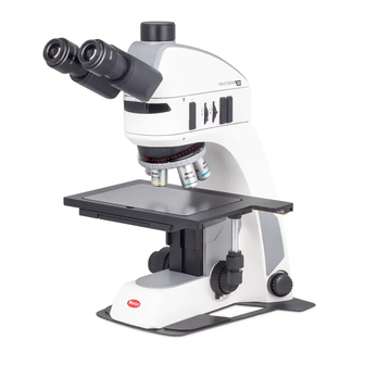
1. INTRODUCTION 1.1 Welcome Welcome to the PantheraTEC User Documentation. PantheraTEC is the new family of upright microscope from Panthera. Its design and intuitive controls result in a simple, robust, easy-to-use yet powerful microscope that helps you inspect a wide range of samples. -
Page 7: Pantheratec Features
PantheraTEC Features Four types of PantheraTEC microscopes are available. The different microscopes have different features and are suitable for different applications: Feature PantheraTEC–POL PantheraTEC–EpiPOL Model POL Digital EpiPOL EpiPOL Digital Reflected Light Transmitted Light Condenser Stage 360º Rotatable 360º Rotatable Centering of objectives Main feature... -
Page 8: Genral Notes On Instrument Safety
Please familiarize yourself with this Operation Manual before starting to use the Instrument. In case additional information or support is needed, please contact Motic after sales Service. To ensure safe operation and optimal function of the Instrument, strictly observe the precautions and warnings given in this Operation manual. -
Page 9: Instrument Safety, Fcc And Emc Conformity
Risk of burn – Do not touch the lamp during or immediately after period of operation. Don’t pick the microscope up from the bottom during equipment operation. Proper handling of the microscope will ensure years of trouble free service. If repair become necessary, please contact your Motic agency or our Technical Service directly. -
Page 10: Transporting, Unpacking, Storage Of The Instrument
Please always observe the following safety notes when using the microscope: Motic cannot assume any liability for other applications then the intended use, including the included modules and components. This includes to service or repair work that is not carried out by authorized Motic service personnel. - Page 11 LED source. Wait for the lamp to cool down before replacing it and do not touch the new bulb. The instrument may only be opened by qualified Motic service staff. The operation of the instrument in explosion-risk environments is not allowed.
-
Page 12: Intended Use Of The Microscope
If possible defects Motic must be notified immediately and steps are taken to minimize damage. ● If notified of such a defect Motic will evaluate the defect and if within warrant, rectify it at his discretion, either by repairing the instrument or by delivering a replacement. -
Page 13: Nomenclature
2. NOMENCLATURE Overview This chapter describes the main components and controls of PantheraTEC microscopes, as well as the types of illumination supported. Types of Illumination PantheraTEC microscopes support both transmitted light and reflected light illumination. Transmitted Light In transmitted light illumination, the light source is below the sample. The light passes through the sample before being focused into the eyepieces. -
Page 14: Main Models
Main Models 2.3.1 PantheraTEC-BF... -
Page 15: Pantheratec-Bd
2.3.2 PantheraTEC-BD... -
Page 16: Pantheratec-Pol
2.3.3 PantheraTEC-POL... -
Page 17: Pantheratec-Epipol
2.3.4 PantheraTEC-EpiPOL Objective Lenses: PantheraTEC-BF Magnification N.A. W.D (mm) Plan Achromat LD 5X 0.13 20.3 Plan Achromat LD 10X 0.25 17.5 Plan Achromat LD 20X 0.40 Plan S-APO 50X 0.80 Plan S-APO 100X (optional) 0.90... - Page 18 PantheraTEC-BD Magnification N.A. W.D (mm) Plan Achromat BD 5X 0.13 17.3 Plan Achromat BD 10X 0.25 16.3 Plan Achromat BD 20X 0.40 Plan S-APO BD 50X 0.80 Plan S-APO BD 100X (optional) 0.90 PantheraTEC-POL Magnification N.A. W.D (mm) Strain-free Plan UC 4X 30.5 Strain-free Plan UC 10X 0.25...
- Page 19 Working distance WD: The distance from the cover glass top surface to the nearest point of the objective. Numerical aperture NA: The NA is a performance indicator. The higher NA, the higher the resolving power. It describes the ability of the lens to collect light under steep entry angles.
-
Page 20: Setting Up The Instrument
3. SETTING UP THE INSTRUMENT Avoid placing the instrument in locations exposed to direct sunlight, dust, vibration, high temperature, high humidity and where it is difficult to unplug the power supply cord. 3.1 Operating environment ● Indoor use ● Altitude: Max 2000 meters ●... - Page 21 Interchangeable 3W LED (Optional) and 30W HAL (Standard); Full Koehler illumination Please familiarize yourself with the instructions given in this Operation Manual. In case of unresolved questions, please contact Motic after sales Service or consult Motic Webservices for further instructions.
-
Page 22: Assembling The Microscope
In order to prevent electric shock, always turn the power switch on the power supply off before connecting the power cord. 4.2 Metal Support ● To make sure the PantheraTEC series more stable, every TEC series of microscopes comes with the metal support. ● Screw the metal support from the underneath with the provided clamp screws. -
Page 23: Stage
4.3 Stage ● Lower the stage so that the sample can fit below the objectives. The condenser automatically moves together with the stage. Ensure that the stage is low enough so that none of the objectives collide with the sample when rotating the objective nosepiece. ●... -
Page 24: Way To Install The 6X4 Stage (For Pantheratec-Bf/Bd)
4.3.1 Way to install the 6X4 stage (For PantheraTEC-BF/BD) ● To protect the 6X4 stage with long travel range, the 6X4 stage is taken from the stand and packed separately in the package. ● Take the 6X4 stage from the package and use the provided Allen key and screws to lock the stage from the underneath. -
Page 25: Objectives
4.4 Objectives ● Lower the stage completely. Screw the objectives into the revolving nosepiece so that clockwise rotation of the nosepiece brings the next higher magnification objective into position. Note: The PantheraTEC-POL/EpiPOL microscope allows you to center three objective positions in relation to the reference objective position. - Page 26 Strain-free condenser used for PantheraTEC-POL/EpiPOL (Fig. 4-1) (Fig. 4-2) LED Condenser used for PantheraTEC-BF/BD (Fig. 4-3)
-
Page 27: Eyepiece Tube
4.6 Eyepiece tube ● Loosen the eyepiece clamp screw (Fig. 5-2). Insert the round dovetail mount on the eyepiece tube into the round dovetail mounts on the intermediate tube (Fig. 5-1). Tighten the eyepiece tube clamp screw to secure the eyepiece tube in place. (Fig. 5-2) (Fig. -
Page 28: Eyepieces
4.8 Eyepieces ● Use the same magnification eyepieces for both the eyes. ● To secure the eyepiece in the eyepiece sleeve, tighten the clamp screws. ● Twist the eyepiece (anti-clockwise or clockwise) with 20~30 degree (Fig. 7.1) and pull the eyepieces gently out when removing the eyepiece. -
Page 29: Power Cord
● Filter selection: Filter Function Blue Filter (Colour Balancing Filter) For routine microscopy and photomicrography Green Interference (546nm) For retardation measurement and contrast adjustment ● A diffuser is built into the base of the microscope. 4.10 Power cord ● Connect the socket of the of the power cord to the AC inlet on the rear of the base of the microscope. -
Page 30: Digital Parts (Setup And Operation)
In case you do not have the Panthera APP yet, please scan the QR-Code on the backside Type Label to download Panthera APP in Android or IOS system, and get connected to the correct APP store via Motic servicing Servers. In case of any issue, please visit the Website http://www.motic.com/Panthera/app.html Connect the same network with Panthera. -
Page 31: Imagingondevice
Panthera Series is providing the user with an unseen combination of build in Digital Capabilities. “Plug in your HDMI Screen, mouse and keyboard and start enjoy the simplicity of working with Motic Panthera Series.” ● Direct Connect to Screen ● Projector ●... -
Page 32: Teachingondevice (Pantheratec-Bf/Bd-Td, Pantheratec-Pol/Epipol Digital)
● USB Keyboard, simply connect the Keyboard of your choice ● 1Gbit Lan connection port for high speed remote consultaion on Motic DssStore or simply network sharing ● Plug your personal USB stick to transport your imagery ●... -
Page 33: Pantheratec Imagingondevice Realtime Sharing Imagingondevice
PantheraTEC ImagingOnDevice stores relevant Data into the EXIF-Header of each Image being made. User can recall the same Illumination settings using the ImageRecall feature in order to allow a easy reconstruction of the before Experiment. Motic EXIF Header ■ Objective Lens Type with Specs ■... -
Page 34: Usage Of Microscope Components
6. USAGE OF MICROSCOPE COMPONENTS 6.1 Coarse and fine focusing (Fig. 9-1) ● Focusing is carried out with the coarse and fine focus knobs at the left and right of the microscope stand. ● The direction of vertical movement of the stage corresponds to the turning direction of the focus knobs. -
Page 35: Coarse Focus Quick Stop
6.3 Coarse focus quick stop (Fig. 10) ● The coarse focus quick stop makes the stage can fixed at any position at which the specimen is in focus i.e. by using the handle to lock the coarse focus knob. ● With the specimen in focus, turn the handle to fix the knob. -
Page 36: Interpupillary Distance Adjustment
● This adjustment will enable the user to observe the specimen with both eyes PantheraTEC Series Microscopes are equipped with a swivelling Trinocular to provide the flexibility to adjust the view height for individual viewers convenience (Fig. 12) (only for PantheraTEC-BF/BD series). -
Page 37: Diopter Adjustment
6.6 Diopter adjustment Every human eye is different, to adjust the Instrument to best performance adjustment can be Necessary ● Set the diopter on both eyepieces to the “0” position. ● Change to 10X Magnification and the image of the specimen into focus using one eye only. ●... -
Page 38: Illumination Properties Adjustment
6.7 Illumination Properties Adjustment You can adjust the following illumination properties: ● Reflected illumination and transmitted illumination switch ● Brightness of transmitted illumination ● Brightness of reflected illumination ● Color of reflected illumination ● Oblique illumination for reflected illumination INFO Reflected illumination is not available for PantheraTEC-POL 6.7.1 How to switch the reflected illumination and transmitted illumination Switch to the reflected illumination, push and turn up the intensity knob, then the reflected illumination... -
Page 39: Using Oblique Reflected Illumination (For Pantheratec-Bf/Bd)
6.7.3 Using Oblique Reflected Illumination (For PantheraTEC-BF/BD) You can adjust the reflected illumination (if available) so that the light is projected obliquely onto the sample. This enhances the appearance of height differences on its surface. Procedure: To change the direction of the oblique illumination, insert the BF oblique slider into the vertical slot on the intermediate tube and turn the wheel to change the illumination angles. -
Page 40: Pantheratec Without Smartcam (Free And Store Mode)
Procedure: To adjust the luminous field diaphragm, turn the corresponding ring. Adjust the diaphragm such that it just disappears from the field of view when looking through the eyepieces. 6.7.5 PantheraTEC without smartCAM (Free and Store mode): To adjust the Brightness, turn the Light Intensity Control Knob and observe the brightness change until the desired brightness has been reached. -
Page 41: Restore Factory Default For Intensity (For Pantheratec Smartcam Version)
Free and Store is the preset standard Illumination mode. (Fig.17-1) (Fig.17-2) 6.7.7 Restore factory default for intensity (For PantheraTEC smartCAM version) ● Turn the Instrument off by switching the main switch on the backside of the Instrument. Keep the intensity knob pressed and switch instrument on. Keep holding the intensity control knob for 10s. -
Page 42: Smart Epibd Segmental Illumination Adjustment (For Pantheratec-Bd)
6.8 Smart EpiBD segmental illumination adjustment (For PantheraTEC-BD) PantheraTEC-BD provides the segmentable Smart Ringlight mode selection for quick inspections at the Darkfield, defects detecting and quality control in industrial manufacturing process which can be performed quicker, easier and more efficient with the further improved contrast by this new Darkfield observation method. -
Page 43: Use Of Field Diaphragm (For Pantheratec-Bf/Bd/Pantheratec-Epipol)
One-sixth-circle: one more press, it comes to the one-sixth-circle mode, the button 2 will twinkle on time, button 3 keeps lighting 2-opposite one-sixth-circle: one more press, it comes to the 2-opposite one-sixth-circle mode, the button 2 will twinkle 2 times, button 3 keeps lighting 3-opposite one-sixth-circle: one more press, it comes to the 3-opposite one-sixth-circle mode, the button 2 will twinkle 3 times, button 3 keeps lighting Half (another half version): one more press, it comes to half mode, the button 2 will twinkle 3... -
Page 44: Use Of Aperture Diaphragm (For Pantheratec-Bf/Bd/Pantheratec-Epipol)
(Fig. 20-1) (Fig. 20-2) 6.10 Use of Aperture Diaphragm (For PantheraTEC-BF/BD/PantheraTEC-EpiPOL) ● The condenser aperture diaphragm is provided for adjusting the numerical aperture (N.A.) of the illuminating system of the microscope, it decides the resolution of the image, contrast, depth of focus and brightness. -
Page 45: Use Of Polarizer And Analyzer (For Pantheratec-Bf/Bd/Pantheratec-Epipol)
6.11 Use of Polarizer and Analyzer (For PantheraTEC-BF/BD/PantheraTEC-EpiPOL) ● Insert the polarizer (marked with “P”) into the front slot of EPI. ● Insert the Analyzer (marked with “A”) into the side slot of EPI. ● Analyzer is rotatable and the color of specimen with polarization will be changed when rotating. (Fig. -
Page 46: Performing Polarization Examinations With Transmitted Light
Rotating stage *** Please Note : Usually the user is adviced to turn the nosepiece turret using the knurled nosepiece ring. Due to some user would use the body of the Objective to turn the nosepiece turret, there could be a gradually de-centering effect depending in the number of incidents. - Page 47 Procedure: Turn the polarizer ring under the sample to the 0° position. (Fig. 23-1) (Fig. 23-2) (Fig. 23-3) (Fig. 23-4) If desired, observe the sample in the polarized light, for example to determine the fracture direction of a material. Insert the fixed analyzer into the horizontal slot of the intermediate tube. Alternatively, insert the rotatable analyzer, set to the 0°...
-
Page 48: Performing Polarization Examinations With Reflected Light
Dark cross image is formed on the exit pupil of the objective Notes ● Rotate the Bertrand lens turret to “B” position and bring the Bertrand lens in the optical path to enable the exit pupil of the objective to be seen through the eyepiece. Rotate the polarizer so that a dark cross image is formed on the exit pupil as shown in the figure above. -
Page 49: Focusing And Centering The Bertrand Lens
6.12.4 Focusing and centering the Bertrand lens ● Rotate the Bertrand lens turret to “B” position and bring the Bertrand lens in the optical path. ● Bring 40x Objective into optical path. ● Use the Bertrand lens focus ring under the Bertrand lens turret to focus on the image of the condenser aperture diaphragm that is stopped down to 70 –... -
Page 50: Measuring Retardation From 1Λ To 4 Λ
c. Quartz Wedge – This device has a range of 4 orders and is commonly employed for qualitative retardation measurements of petrographic specimens or other birefringent materials whose retardation value falls within the wedge limit. 6.12.5.1 Measuring Retardation from 1λ to 4 λ ●... -
Page 51: 45° Click-Stop
6.12. 6 45° Click-stop ● Use 2.5mm allen key to lose the screw in the stage 45°orientation knob. ● Rotate the 45°orientation knob clock wise and tighten the 45°orientation knob screw with 2.5mm allen key. ● At this time, the rotary stage has a click-stop. Set the rotary stage at “0” position and each rotation will click-stop at every 45°. -
Page 52: Photomicrographic Procedure
7. PHOTOMICROGRAPHIC PROCEDURE ● To ensure vibration free operation, set the microscope on a sturdy vibration free table or a bench with a vibration proof device. ● Pull the optical path selection lever of the trinocular eyepiece tube all of the way out to the limit, theratio of light entering the observation tube and standard phototube will be 20:80. -
Page 53: Care And Maintenance
Store the objectives, eyepieces and filters in a container or desiccators with drying agent. ● Proper handling of the microscope will ensure years of trouble free service. ● If repair becomes necessary, please contact your Motic agency or our Technical Service direct. -
Page 54: Bulb Replacement
Note: ● If equipment is used in a manner not specified by the manufacturer, the warranty may be void. ● To avoid getting wet, do not use the microscope near water. 8.5 Bulb replacement The lamp and the lamp house become very hot during and after a period of operation. Risk of burn –... -
Page 55: Electrical Specifications
(Fig. 26-3) (Fig. 26-4) 8.5.2 Electrical Specifications: PantheraTEC–BF Input: AC 100~240V 50~60Hz 0.55A Reflected Lamp: 3W LED Transmitted Lamp: 3W LED PantheraTEC–BD Input: AC 100~240V 50~60Hz 0.55A Reflected Lamp: 3W LED Transmitted Lamp: 3W LED PantheraTEC–POL Input: AC 100~240V 50~60Hz 1.2A Transmitted Lamp: 3W LED or 6V 30W Hal PantheraTEC–EpiPOL Input: AC 100~240V 50~60Hz 1.2A... -
Page 56: Troubleshooting Table
9. TROUBLESHOOTING TABLE As you use your microscope, you may occasionally experience a problem. The troubleshooting table below contains the majority of frequently encountered problems and the possible causes. Optical Problem Possible Cause Lamp not installed properly Lamp not centred Diffuser is in intermediate position Condenser not mounted correctly Condenser is not centred... -
Page 57: Electrical
Stage installed on inclined plane Unequal focus Specimen holder not fixed securely on stage Specimen not secured in position Lamp voltage is set too low Image tinged yellow Blue filter is not being us Slide is upside down Focusing is not possible with high magnification objectives Cover glass is too thick Slide is upside down... -
Page 58: Selection Of The Power Supply Cord
Motic Instruments are certified and tested for safety and environmental conformity. Only power supply cords which are conform with below listed certification marks and countries are applicable. CAUTION: Do not use non-approved power cord for Motic products, Motic can no longer warrant the electrical safety of the equipment. - Page 59 Table 2 HAR Flexible Cord Approval Organizations and Cordage Harmonization Marking Methods Alternative Marking Utilizing Printed or Embossed Black-Red-Yellow Thred Harmonization Marking (May (Length of color section in Approval Organization be located on jacket or insulation of internal wiring) Black Yellow Comite Electrotechnique Belge CEBEC...
-
Page 60: Microscope Terminology
MICROSCOPE TERMINOLOGY Abbe Condenser Magnification A two-lens sub-stage condenser located below The number of times by which the size of the the stage of a microscope and functions to collect image exceeds the original object. Lateral light and direct it onto the object being examined. magnification is usually meant. - Page 61 Diopter adjustment Resolving Power The adjustment of the eyepiece of an instrument A measure of an optical system's ability to to provide accommodation for the eyesight produce an image which separates two points or differences of individual observers. parallel lines on the object. Depth of Focus Resolution The axial depth of the space on both sides of the...
- Page 62 Tel: 86-0592-562 7866 Fax: 86-0592-562 7855 © 2002-2019 Motic China Group Co., Ltd. All rights reserved. Motic is a registered trademark and service mark of Motic China Group Co., Ltd. Microsoft Windows logo is a registered trademark of Microsoft Corporation.
Need help?
Do you have a question about the PantheraTEC Series and is the answer not in the manual?
Questions and answers