Advertisement
Quick Links
REV
R-3
Applied Standard
Product Name
Model/Type
Manufacture Name
Manufacture's
Address
Trade Mark
Comment
R&D Project Leader Jin Wook, Shon
Prepared By
Reviewed By
Q.M Manager Hyung Min, Heo
Q.M Leader Lee Chang-Soo
Approved By
Date of issue
Q-I-300-02(R0)
Product Description
Optical Coherence Tomography
HOCT-1F, HOCT-1
[Summary]
MDD 93/42/EEC
Optical Coherence Tomography
Optical Coherence Tomography
HOCT-1F, HOCT-1
Huvitz Co.,Ltd.
38, Burim-ro 170beon-gil, Dongan-gu, Anyang-si
Gyeonggi-Do, 14055, Republic of Korea
Homepage: www.huvitz.com
Huvitz
2020. 01.30 .
DOCUMENT
HQI-J-OT1-001
ES_DATE
2017-11-01
RE_DATE
2020-01-30
PAGE
1 / 52
2020.01.30
2020.01.30
2020.01.30
Huvitz Co.,Ltd
Advertisement

Related Manuals for Huvitz HOCT-1F


Summary of Contents for Huvitz HOCT-1F
- Page 1 DOCUMENT HQI-J-OT1-001 Product Description ES_DATE 2017-11-01 RE_DATE 2020-01-30 Optical Coherence Tomography HOCT-1F, HOCT-1 PAGE 1 / 52 [Summary] Applied Standard MDD 93/42/EEC Optical Coherence Tomography Product Name Optical Coherence Tomography Model/Type HOCT-1F, HOCT-1 Manufacture Name Huvitz Co.,Ltd. 38, Burim-ro 170beon-gil, Dongan-gu, Anyang-si Manufacture’s...
- Page 2 DOCUMENT HQI-J-OT1-001 Product Description ES_DATE 2017-11-01 RE_DATE 2020-01-30 Optical Coherence Tomography HOCT-1F, HOCT-1 PAGE 2 / 52 ■ REVISION HISTORY DATE REVISED REASON REVISED CONTENT 2017-11-01 First issued Corrected typo error of intended use in 2018-08-20 Periodic Review clause 5.2...
- Page 3 Intended Use The HOCT-1F, HOCT-1 is intended for use as a diagnostic device to aid in the check and management of ocular diseases such as macular holes, cystoid macular edema, diabetic retinopathy, aged related macular degeneration, and so on, which are occurred at a macular, an optic disk, an inner structure of retina, and a cornea.
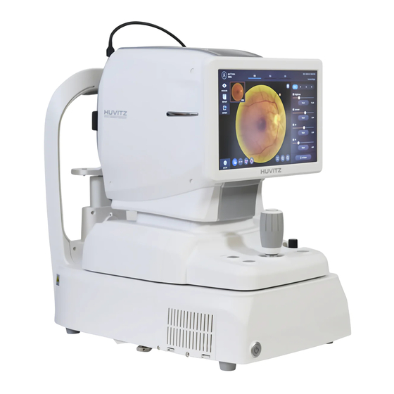
- Page 4 DOCUMENT HQI-J-OT1-001 Product Description ES_DATE 2017-11-01 RE_DATE 2020-01-30 Optical Coherence Tomography HOCT-1F, HOCT-1 PAGE 4 / 52 3. Photo View (1) External Picture -. Front -. Rear Q-I-300-02(R1):2018/06/04 Huvitz Co.,Ltd...
- Page 5 DOCUMENT HQI-J-OT1-001 Product Description ES_DATE 2017-11-01 RE_DATE 2020-01-30 Optical Coherence Tomography HOCT-1F, HOCT-1 PAGE 5 / 52 -. Left -. Right -. Bottom Q-I-300-02(R1):2018/06/04 Huvitz Co.,Ltd...
- Page 6 DOCUMENT HQI-J-OT1-001 Product Description ES_DATE 2017-11-01 RE_DATE 2020-01-30 Optical Coherence Tomography HOCT-1F, HOCT-1 PAGE 6 / 52 (2) External Description 1) Front View Part Name Description Display Monitor for displaying captured image and user interface icon. Chin rest Button for moving chin rest up and down.
- Page 7 DOCUMENT HQI-J-OT1-001 Product Description ES_DATE 2017-11-01 RE_DATE 2020-01-30 Optical Coherence Tomography HOCT-1F, HOCT-1 PAGE 7 / 52 RGB port Port for external display device. DP port Port for communicating external DP device. LAN port Port for external network (2 ports)
- Page 8 DOCUMENT HQI-J-OT1-001 Product Description ES_DATE 2017-11-01 RE_DATE 2020-01-30 Optical Coherence Tomography HOCT-1F, HOCT-1 PAGE 8 / 52 3) Bottom View Part Name Description Base Packing lock Lock for fixing body and base during transportation. (2 points) Q-I-300-02(R1):2018/06/04 Huvitz Co.,Ltd...
- Page 9 RE_DATE 2020-01-30 Optical Coherence Tomography HOCT-1F, HOCT-1 PAGE 9 / 52 4. GUI Interface ① OCT/Fundus mode (HOCT-1F only) Observation screen Name Description Patient Show patient name and ID number. information Can be moved to select/resist patient mode by clicking.
- Page 10 DOCUMENT HQI-J-OT1-001 Product Description ES_DATE 2017-11-01 RE_DATE 2020-01-30 Optical Coherence Tomography HOCT-1F, HOCT-1 PAGE 10 / 52 Reset scan Reset scan position to center. position Focus Move in accordance with focus of patient’s eye. Ref.M Move reference mirror position for OCT scan.
- Page 11 DOCUMENT HQI-J-OT1-001 Product Description ES_DATE 2017-11-01 RE_DATE 2020-01-30 Optical Coherence Tomography HOCT-1F, HOCT-1 PAGE 11 / 52 OCT mode Observation screen (Refer. OCT/Fundus mode – Observation screen’ for uncommented item) Name Description Signal Level Mode can be changed Confirmation screen (Refer OCT/Fundus mode –...
- Page 12 2017-11-01 RE_DATE 2020-01-30 Optical Coherence Tomography HOCT-1F, HOCT-1 PAGE 12 / 52 Fundus mode (HOCT-1F only) Observation screen (Refer OCT/Fundus mode – Observation screen’ for uncommented item) Name Description Select capture mode. Capture mode - Single: Capture one fundus image.
- Page 13 DOCUMENT HQI-J-OT1-001 Product Description ES_DATE 2017-11-01 RE_DATE 2020-01-30 Optical Coherence Tomography HOCT-1F, HOCT-1 PAGE 13 / 52 △ Angio Mode (Optional) Observation screen ❶ (Refer. OCT/Fundus mode – Observation screen’ for uncommented item) Name Function Signal Level Mode can be changed Confirmation screen ❶...
- Page 14 Shows the patient list saved in the HUVITZ WebViewer database currently. New patient gets added into the patient list when new patient information is transmitted at the HUVITZ’s ophthalmic device or when patient is added on using ‘Add Patient’ in the WebViewer. When mouse is placed on top of the patient list, URL copy icon that enables move to the applicable patient immediately, gets activated.
- Page 15 ES_DATE 2017-11-01 RE_DATE 2019-07-02 Optical Coherence Tomography HOCT-1F, HOCT-1 PAGE 15 / 52 examination. The button is shown when choosing patient. Information on the software version is displayed. When user icon is pressed on, ID logged-in currently is displayed, and it is possible to log-out when log-out button is pressed on.
- Page 16 DOCUMENT HQI-J-OT1-001 Product Description ES_DATE 2017-11-01 RE_DATE 2019-07-02 Optical Coherence Tomography HOCT-1F, HOCT-1 PAGE 16 / 52 5. Accessories ①Chinrest paper ②Touch pen ③Power cable ④Fuse ⑤Dust cover ⑥External LED ⑧User manual ⑨Packing lock cap ⑩Model Eye ⑦Blower ⑫ Anterior ⑪...
- Page 17 DOCUMENT HQI-J-OT1-001 Product Description ES_DATE 2017-11-01 RE_DATE 2019-07-02 Optical Coherence Tomography HOCT-1F, HOCT-1 PAGE 17 / 52 User manual Instruction for use volume a cap that unlocks and ф17.0 x 4.6mm Packing lock cap 1 unit covers the hall Self-diagnosis using Model...
- Page 18 Angiography Range X:3 ~ 12mm, Y:3~9mm (Optional) △ △ Angiography Analysis FAZ, Vessel density Tools (Optional) Fundus Camera (HOCT-1F/HOCT-1FA) Type Non-mydriatic fundus camera Resolution 60 line pair/mm (centre), 40 line pair/mm (middle), 25 line pair/mm (periphery) Angle of view 45˚...
- Page 19 DOCUMENT HQI-J-OT1-001 Product Description ES_DATE 2017-11-01 RE_DATE 2019-07-02 Optical Coherence Tomography HOCT-1F, HOCT-1 PAGE 19 / 52 6.2 △ Essential Performance Model Essential Associated Failure Mode Risk Control Performance unacceptable risk HOCT-1 The scanning of the Measurement error The tomographic...
- Page 20 DOCUMENT HQI-J-OT1-001 Product Description ES_DATE 2017-11-01 RE_DATE 2019-07-02 Optical Coherence Tomography HOCT-1F, HOCT-1 PAGE 20 / 52 7.1 Functional block diagram 7.2 Part’s Function General Name Function Drawing Part name Part Code / and critical Revision No. feature part(*) INLET(*)
- Page 21 DOCUMENT HQI-J-OT1-001 Product Description ES_DATE 2017-11-01 RE_DATE 2019-07-02 Optical Coherence Tomography HOCT-1F, HOCT-1 PAGE 21 / 52 Retina Camera camera for preview fundus PCB ASSY(CMOS M2EPCB0007- image AR0130) NIR LED illumination source for cornea IR LED(740NM) 71010040005-A and retina camera...
- Page 22 DOCUMENT HQI-J-OT1-001 Product Description ES_DATE 2017-11-01 RE_DATE 2019-07-02 Optical Coherence Tomography HOCT-1F, HOCT-1 PAGE 22 / 52 Area Number Working Required Required Designed Designed Test point and type Voltage creepage clearance creepage clearance of Means (mm) (mm) (mm) (mm) Protection:...
- Page 23 DOCUMENT HQI-J-OT1-001 Product Description ES_DATE 2017-11-01 RE_DATE 2019-07-02 Optical Coherence Tomography HOCT-1F, HOCT-1 PAGE 23 / 52 Between secondary 2 MOOP IIIb circuit and un- earthed enclosure 1) The record was that obtained as the result of tests of certified SMPS (Report Ref. No. E356265-A11- 2) linear interpolation used △...
- Page 24 2017-11-01 RE_DATE 2019-07-02 Optical Coherence Tomography HOCT-1F, HOCT-1 PAGE 24 / 52 To form retinal vessel maps, the system main body scans several times at the same position, and gets the same number of tomography images. And then the system calculates the variance among those tomography, finally projects those variances into 2D image.
- Page 25 DOCUMENT HQI-J-OT1-001 Product Description ES_DATE 2017-11-01 RE_DATE 2019-07-02 Optical Coherence Tomography HOCT-1F, HOCT-1 PAGE 25 / 52 8.2 Operator’s work flow Flow Description and Critical Features(*) 1. Turn on the device 2. Prepare the patient 1. Turn On the device 0.
- Page 26 DOCUMENT HQI-J-OT1-001 Product Description ES_DATE 2017-11-01 RE_DATE 2019-07-02 Optical Coherence Tomography HOCT-1F, HOCT-1 PAGE 26 / 52 8.3 Optical System 8.3.1 Optical Path ⑫ ③ ⑤ ⑬ ④ ⑪ ⑨ ⑭ ⑩ ⑥ ② ⑧ ⑦ ○ ① ⑮ ○...
- Page 27 DOCUMENT HQI-J-OT1-001 Product Description ES_DATE 2017-11-01 RE_DATE 2019-07-02 Optical Coherence Tomography HOCT-1F, HOCT-1 PAGE 27 / 52 ○ ○ ⑥ ○ ○ ⑦ ○ ① ② ○ ○ ○ ○ ○ Q-I-300-02(R1):2018/06/04 Huvitz Co.,Ltd...
- Page 28 DOCUMENT HQI-J-OT1-001 Product Description ES_DATE 2017-11-01 RE_DATE 2020-01-30 Optical Coherence Tomography HOCT-1F, HOCT-1 PAGE 28 / 52 8.3.2 List of optical components Name and Part Code / critical feature Function Drawing Part name Revision No. part(*) Patient’s Eye The Patient’s eye...
- Page 29 DOCUMENT HQI-J-OT1-001 Product Description ES_DATE 2017-11-01 RE_DATE 2020-01-30 Optical Coherence Tomography HOCT-1F, HOCT-1 PAGE 29 / 52 Color LED or IR LED to the ASSY Illumination The Light Size Transformer for W2_OT-ILLUM-MASK2- W2MASSY074- Mask Small Pupils ASSY To reflect the IR LED and to...
- Page 30 DOCUMENT HQI-J-OT1-001 Product Description ES_DATE 2017-11-01 RE_DATE 2020-01-30 Optical Coherence Tomography HOCT-1F, HOCT-1 PAGE 30 / 52 (○ à ○ à ○ à ○ à ○ à ○ à ○ à ○ △ 9. Calibration Method 9.1 OCT Spectrometer waveform...
- Page 31 ES_DATE 2017-11-01 RE_DATE 2020-01-30 Optical Coherence Tomography HOCT-1F, HOCT-1 PAGE 31 / 52 .- This test at this page is done at a scan speed 68k. .- To prevent a saturation, set the offsets 4.5, 1.1. .- Set a control pattern to a ‘point’.
- Page 32 DOCUMENT HQI-J-OT1-001 Product Description ES_DATE 2017-11-01 RE_DATE 2020-01-30 Optical Coherence Tomography HOCT-1F, HOCT-1 PAGE 32 / 52 9.3 OCT Scan Pattern Patterns Settings A scanning should be done as the following settings, and a tomography image should be shown clearly.
- Page 33 HQI-J-OT1-001 Product Description ES_DATE 2017-11-01 RE_DATE 2020-01-30 Optical Coherence Tomography HOCT-1F, HOCT-1 PAGE 33 / 52 9.4 OCT Scan Pattern for Cornea Patterns Settings A scanning should be done as the following settings, and a tomography image should be shown clearly.
- Page 34 DOCUMENT HQI-J-OT1-001 Product Description ES_DATE 2017-11-01 RE_DATE 2020-01-30 Optical Coherence Tomography HOCT-1F, HOCT-1 PAGE 34 / 52 .- Pattern Display 9.6 Optic disc AngioScan Pattern for Angiography(Optional) Patterns Settings Angiography Settings: Range: 3~9, A scan points : Scan .- Pattern...
- Page 35 HQI-J-OT1-001 Product Description ES_DATE 2017-11-01 RE_DATE 2020-01-30 Optical Coherence Tomography HOCT-1F, HOCT-1 PAGE 35 / 52 9.7 OCT Scan Range Patterns Settings .- Place a jig with +33D M/E and -33D M/E on a ‘Chinrest’ .- Align & focus with + 33D M/E.
- Page 36 .- Auto tracking function should be checked 2 positions including at a front and at a rear. 4mm Model Eye Tested Example 9.9 Resolution (HOCT-1F only) Patterns Settings .- Place a jig to check a fundus resolution. .- Align and focus with a resolution M/E Procedures .- Illuminate a back side of the M/E with an external fixation LED without a LED...
- Page 37 37 / 52 Resolution Model Eye Tested Example 9.10 Diopter Range (HOCT-1F only) Patterns Settings .- Place a jig with +33D M/E and -33D M/E on a ‘Chinrest’ .- Click (+) button at an additional lens icon to insert a plus lens.
- Page 38 38 / 52 High Diopter Model Eye Tested Example 9.11 Small Pupil (HOCT-1F only) Patterns Settings .- Place a jig with pupil size 3.2mm on a ‘Chinrest’ .- Align & focus with a M/E that has a pupil size 3.3mm.
- Page 39 Small pupil Model Eye Tested < at normal mode > Example < at small pupil mode > 9.12 F.O.V (HOCT-1F only) Patterns Settings .- step 1: Fasten a FOV jig into an objective lens. .- step 2: Adjust a fundus flash 0.
- Page 40 DOCUMENT HQI-J-OT1-001 Product Description ES_DATE 2017-11-01 RE_DATE 2020-01-30 Optical Coherence Tomography HOCT-1F, HOCT-1 PAGE 40 / 52 FOV Model Eye Tested Example 9.12 FoV of IR Image Patterns Settings .- step 1: Fasten a FOV jig into an objective lens.
- Page 41 HQI-J-OT1-001 Product Description ES_DATE 2017-11-01 RE_DATE 2020-01-30 Optical Coherence Tomography HOCT-1F, HOCT-1 PAGE 41 / 52 9.13 IR Image Patterns Settings .- step 1: Align and focus with a human’s eye. .- step 2: Look at an IR live image and check whether an optic disk and vessels are visible Procedures or not.
- Page 42 If working dots isn’t fine after auto tracking, the calibration for auto tracking is needed. . Calibration is not allowed by a user because of the technical issues. Contact to the Huvitz’s authorized distributor or Huvitz Co.,Ltd. If the period of calibration is resticted by the local law, follow up the local law.
- Page 43 43 / 52 camera images to PC If the problem is persisted, turn off Board, when boot up. the device and contact Huvitz or your authorized distributor. Failed to initialize the pupil .- Keep operating temperature. mask motor, which is used .- Turn off and turn on again...
- Page 44 If the problem is persisted, turn off motor not initialized” myopia/hyperopia, when the device and contact Huvitz or your boot up. authorized distributor. Failed to initialize the color .- Turn off and turn on, check it again.
- Page 45 Do not use the solvents such as strongly volatile substance, thinner, benzene, etc Do not use a sponge or cloth soaked in water because the water might leak into the equipment. Clean headrest rubber and chinrest with an alcohol before sending device to authorized agent or Huvitz for maintenance.
- Page 46 Product Description ES_DATE 2017-11-01 RE_DATE 2020-01-30 Optical Coherence Tomography HOCT-1F, HOCT-1 PAGE 46 / 52 Replacing fuse (1) Ensure the power switch of device off (O). (2) Remove power cable from inlet. (3) Pull out fuse holder in the inlet with a tweezers.
- Page 47 Observation & photographing of the fundus : HOCT-1F ONLY Type of photography Color, Red-free& IR Color & Red-free, IR 45°...
- Page 48 DOCUMENT HQI-J-OT1-001 Product Description ES_DATE 2017-11-01 RE_DATE 2020-01-30 Optical Coherence Tomography HOCT-1F, HOCT-1 PAGE 48 / 52 Characteristic Optical Coherence Tomography Optical Coherence Tomography Observation & photographing of the fundus image/fundus tomogram Internal fixation target: Dot matrix type organic The display position can be changed and adjusted.
- Page 49 The HOCT-1F, HOCT-1 is The CIRRUS™ HD-OCT with intended for use as a diagnostic It is intended for use as a diagnostic Retinal Nerve Fiber Layer (RNFL),...
-
Page 50: Main Board
DOCUMENT HQI-J-OT1-001 Product Description ES_DATE 2017-11-01 RE_DATE 2020-01-30 Optical Coherence Tomography HOCT-1F, HOCT-1 PAGE 50 / 52 Characteristic Optical Coherence Tomography Optical Coherence Tomography Optical Coherence Tomography 3 μm) Optical transverse ~15 microns 15 microns 20 microns resolution OCT axial imaging... - Page 51 DOCUMENT HQI-J-OT1-001 Product Description ES_DATE 2017-11-01 RE_DATE 2020-01-30 Optical Coherence Tomography HOCT-1F, HOCT-1 PAGE 51 / 52 X,Z: Citizen micro, RB- 2 times per 1 patient * 25 patients 35CGM-NCA09- W2MASSY014- per 1 day* 260 days per 1year * 7...
-
Page 52: Impulse Noise
DOCUMENT HQI-J-OT1-001 Product Description ES_DATE 2017-11-01 RE_DATE 2020-01-30 Optical Coherence Tomography HOCT-1F, HOCT-1 PAGE 52 / 52 (-10±2)℃ 4H ~(55±2)℃ 4H, No malfuction and performance Discharge Power 10CYCLE On 1min / Off 9min Input deterioation On/Off & slow Voltage 90/110/132/180/220/264V...
Need help?
Do you have a question about the HOCT-1F and is the answer not in the manual?
Questions and answers