Advertisement
Quick Links
Cancer Institute
Microscopy Core Facility
Z
A
I
A1
M1
EISS
XIO
MAGER
AND
A U
G
SERS
UIDE
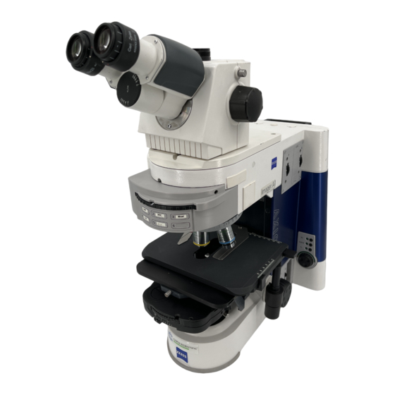
This is intended to provide a basic coverage of use of the Zeiss Axio Imager M1 for multi-
channelled fluorescent acquisition. For more complex usages please contact
Fig.1 – Front View of Microscope
Viewing
Slide
Microscope
Ocular Viewer
control
touch-screen
Mercury Lamp
(Reflected light)
Main Stage
Control Box
Microscope Power
st
1
Focusing
Supply Box
Wheel
V1.0
Advertisement

Related Manuals for Zeiss Axio Imager M1


Summarization of Contents
Locate Focal Plane of Sample
Select Magnification
Choose the correct magnification level for sample viewing and focusing.
Image Processing and Annotation
Viewing Images or Z-Stacks
How to view collected images and Z-stacks within the software interface.
Deconvolution of Z-Stack
Using deconvolution to clean up images and remove aberrations.
Orthoview Processing
Averaging and overlaying Z-stack images into a single 2D view.
Image Annotations
Adding scale bars, text, and geometric shapes for publication.
Need help?
Do you have a question about the Axio Imager M1 and is the answer not in the manual?
Questions and answers