
Related Manuals for Hoefer HE33


Summary of Contents for Hoefer HE33
- Page 1 Hoefer HE 33 mini submarine electrophoresis unit HE33-IM/Rev. H0/05-04...
-
Page 3: Table Of Contents
Page finder Mini submarine electrophoresis unit function . . Important information ....Unpacking ......Specifications . - Page 4 •...
-
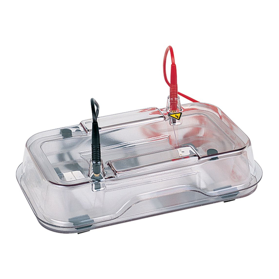
Page 5: Mini Submarine Electrophoresis Unit Function
Mini submarine electrophoresis unit function The Hoefer ™ HE 33 horizontal agarose unit is intended for rapid electrophoresis of small quantities of nucleic acids in agarose gels. A gel is cast in the gel caster, which holds one or two combs. (Eight different combs are avail- Fig 1. -
Page 6: Important Information
• If this equipment is used in a manner not specified by the manufacturer, the protection provided by the equipment may be impaired. • Only accessories and parts approved or supplied by Hoefer, Inc. may be used for operating, maintaining, and servicing this product. •... - Page 7 équipent cet appareil peuvent être rendues inéfficaces. • Seulement les accessoires et piéces detachées approuvés ou fournis par Hoefer, Inc. sont recommandés pour l’utilisation, l’entretien et réparation de cet appareil. •...
-
Page 8: Unpacking
Inspect all components • used as delivered from for damage that may have occurred while the Hoefer, Inc. except for unit was in transit. If any part appears damaged, alterations described in the User Manual, and contact the carrier immediately. -
Page 9: Operating Instructions
Operating instructions Agarose gels are fi rst prepared using the gel cast- ing kit. Samples are then loaded into wells and electrophoretically separated. The fl uorescent dye ethidium bromide can be added to the gel or electrophoresis buffer or both to track separa- tion progress. - Page 10 Prepare solutions Caution: Ethidium bromide is a Prepare 250 ml of running buffer. (Refer to p. 12 for known mutagen. Always wear recipes of commonly used electrophoretic running gloves when handling. buffers.) Prepare the sample loading buffer. (Refer to p. 13 for a recipe and a table of volume requirements for each comb size.) ...
- Page 11 Cast the gel Install the running tray. Firmly grasp the casting tray with one hand. With the other hand, place one end of the running tray against the foam pad at the bottom edge, press the tray against the pad, and then lower it to rest on the bottom of the casting tray, seating the other end of the tray against the opposite foam pad.
- Page 12 Remove the running tray and gel by grasping the handles of the tray and pressing against one of the foam pads. Once the tray clears the opposite pad, lift it out. Transfer the running tray and gel to the chilled Fig.
- Page 13 The electrophoresis run Refer to the notes, buffers, and volumes section for additional information and guidelines. Chill the base before use, especially when higher voltage settings will be used or when the separation will require more than 30 minutes. Note: To monitor separation progress, either add 0.5 µg/ml (final conc.) of ethidium bromide to the running buffer now, or add 50 µg/ml (final conc.) ethidium...
- Page 14 Quick, high-voltage runs Certain applications, such as screening samples or checking sample purity, can be accomplished quickly under high voltage conditions. Chill the base (-20 °C) and limit the run to 5 minutes or less at 500 V. Slower, lower voltage runs Note: To calculate the voltage A voltage gradient of 12 V/cm (150 V) separates 0.1 to 23 kb fragments of a Hind III digest of λ...
-
Page 15: Care And Maintenance
Repeat with second pad on the wall opposite the fi rst pad. Replacing the electrode It is recommended that electrodes be replaced only by Hoefer technicians. Call your local representative for advice. •... -
Page 16: Troubleshooting
Troubleshooting problem solution Deformed sample well Allow the gel to set for a minimum of 30 minutes and make sure it is at room temperature before removing the comb. When removing the comb, hold it at a slight angle and lift very slowly to prevent the gel from breaking. -
Page 17: Notes, Buffers, And Volumes
Notes, buffers, and volumes Agarose gel electrophoresis notes Agarose gel electrophoresis can be used to separate DNA fragments as small as 0.1 kb or less. Polyacrylamide gels are usually used for fragments smaller than 1 kb. DNA mobility Suggested agarose concentration for sepa- rating fragments of various sizes is given in Table 2 below. - Page 18 RNA samples usually require longer runs or buffers that are easily depleted, and so require circulation. The Hoefer HE 100 horizontal unit is recommended for this application rather than the HE 33. Running buffers for DNA in agarose gels...
- Page 19 —or— 1X, to yield 89 mM Tris base, 89 mM boric acid, and 2 mM EDTA. 2. 10X Tris-acetate-EDTA (TAE) stock buffer † (0.4 M Tris, 0.2 M acetic acid, 10 mM EDTA, pH ~8.4, 1000 ml) Tris base (FW 121.1) 0.40 M 48.4 g Acetic acid (99.5%)
- Page 20 Note 2: Xylene cyanol (0.25%), which migrates more slowly than bromophenol blue, can be added as an additional marker if desired. The agarose concentration determines the position of the dye bands relative to a polynucleotide. † Tracking dyes may be omitted to eliminate obscuring and dragging effects caused by comigration with smaller nucleic acids.
-
Page 21: Bibliography
H2O) for 15 to 60 minutes and then view or photograph the sample on a UV transilluminator. To photograph the gel, either place the running tray on the transilluminator surface or slide the gel onto the surface for maximum expo- sure. -
Page 22: Ordering Information
Mini Submarine Electrophoresis Unit, basic. Includes gel running tray, HE33B gel casting tray and bubble level. (Order comb and comb back separately.) Mini Submarine Electrophoresis Unit, kit. Same as above plus HE33-8-1.5 one 8-well, 1.5 mm thick comb, comb back, and screws. Replacement parts Buffer chamber assembly... - Page 23 HE31A-P-1.0 HE31A-8-1.0 HE31A-12-1.0 HE31A-16-1.0 Preparative HE31A-P-1.5 HE31A-8-1.5 HE31A-12-1.5 HE31A-16-1.5 Comb back with 2 screws HE31-BK Replacement screws for comb backs (pk/12) HE31-S Companion products Hoefer EPS 2A200 power supply PS2A200 MacroVue UV-20 Transilluminator 115 V~ UV20-115V 230 V~ UV20-230V •...
- Page 24 Printed in the USA Hoefer, Inc. 953 Indiana Street San Francisco, CA 94107 USA www.hoeferinc.com...
Need help?
Do you have a question about the HE33 and is the answer not in the manual?
Questions and answers