Sign In
Upload
Download
Table of Contents
Contents
Add to my manuals
Delete from my manuals
Share
URL of this page:
HTML Link:
Bookmark this page
Add
Manual will be automatically added to "My Manuals"
Print this page
×
Bookmark added
×
Added to my manuals
Manuals
Brands
AmScope Manuals
Microscope
490 Series
Operator's manual
AmScope 490 Series Operator's Manual
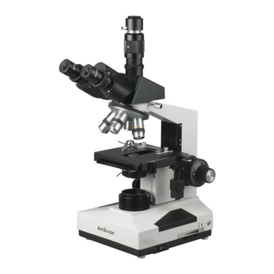
Compound microscope
Hide thumbs
1
2
Table Of Contents
3
4
5
6
7
8
9
10
11
12
13
14
15
16
17
18
19
20
21
22
23
24
25
26
27
28
29
30
31
32
33
34
35
36
37
38
39
40
41
42
43
44
45
46
47
48
49
50
51
52
53
54
55
56
57
58
59
60
61
62
63
64
page
of
64
Go
/
64
Contents
Table of Contents
Troubleshooting
Bookmarks
Table of Contents
Table of Contents
Before Use
Introduction
Precautions
1 Microscope Parts
2 Operation
Unpacking
Assembly
Adjusting the View
Specimen Set up
Focusing
Using the Trinocular Port
Attaching a Camera
Setting the Stage's Stop-Limit
Adjusting Focus Tension
Setting the Condenser Lens Adjustment Knob
Adjusting the Iris
Changing a Filter
Changing the Halogen Light Bulbs
Changing the Fuse
Using Oil Immersion
Using Darkfield Condenser Lenses (Optional Accessories)
Using a Dry Darkfield Condenser Lens (Optional Accessory)
Using Oil Darkfield Condenser Lens (Optional Accessory)
Using a Phase Contrast Kit (Optional Accessory)
Using a Simple Phase Contrast Kit (Optional Accessory)
Using a Turret Phase Contrast Kit (Optional Accessory)
Microscope Maintenance
Darkfield Condenser Kit & Phase Contrast Kit Maintenance
3 General Specifications
General Model Comparison
Objectives
Eyepieces
Darkfield Condensers
Phase Contrast Kits
4 Parameters
Electrical System
Microscope Parameters
5 Recommended Accessories
Eyepieces
Darkfield Condensers
Objective Lenses
Phase Contrast Kits
Cameras and Accessories
Halogen Light Bulbs and Fuses
Cases and Bags
Stage Warmer
Slides
Cleaners
Books
6 Troubleshooting
Optical Issues
Mechanical Issues
Electrical Issues
7 General Microscopy Guide
Interpupillary Distance
Mechanical Stage
Phase Contrast
Advertisement
Quick Links
1
Microscope Parts
2
Unpacking
3
Assembly
Download this manual
OPERATOR'S
MANUAL
(Please Read This Manual Before Using the
Microscope
)
490 Series
Compound Microscope
Table of
Contents
Previous
Page
Next
Page
1
2
3
4
5
Advertisement
Table of Contents
Need help?
Do you have a question about the 490 Series and is the answer not in the manual?
Ask a question
Questions and answers
Related Manuals for AmScope 490 Series
Microscope AmScope 150 Series User Manual
(17 pages)
Microscope AmScope B100 Operation Instruction Manual
B series biological microscope (16 pages)
Microscope AmScope MU USB3.0 User Manual
Mu series microscope camera (147 pages)
Microscope AmScope 120 Series User Manual
(20 pages)
Microscope AmScope AM-1 Series User Manual
(19 pages)
Microscope AmScope SM-4 Series Operator's Manual
Stereo microscope (37 pages)
Microscope AmScope LED-144 Series User Manual
(7 pages)
Microscope AmScope DM756 Series Manual
(25 pages)
Microscope AmScope DM745-HDM11 Manual
Digital-integrated microscope (23 pages)
Microscope AmScope UHM210-11 Manual
(19 pages)
Microscope AmScope PM200 Series User Manual
(16 pages)
Microscope AmScope AFDM1080 Manual
Auto-focus digital microscope (19 pages)
Microscope AmScope DM745-HDM9-3MP Product Manual
Digital microscope for industrial inspection with 0.7x-4.5x zoom and 9" screen (15 pages)
Microscope AmScope DM140 User Manual
Portable digital microscope (12 pages)
Microscope AmScope B110C Product Manual
40x-1000x dual led, ergonomic, lab binocular compound microscope with 3d two-layer mechanical stage (10 pages)
Microscope AmScope DM750-2MP Manual
Digital-integrated microscope (16 pages)
This manual is also suitable for:
T490
B490-30w
B490
T490-30w
T490-led
B490-led
Table of Contents
Print
Rename the bookmark
Delete bookmark?
Delete from my manuals?
Login
Sign In
OR
Sign in with Facebook
Sign in with Google
Upload manual
Upload from disk
Upload from URL


Need help?
Do you have a question about the 490 Series and is the answer not in the manual?
Questions and answers