
Related Manuals for Maquet FLOW-I


Summary of Contents for Maquet FLOW-I
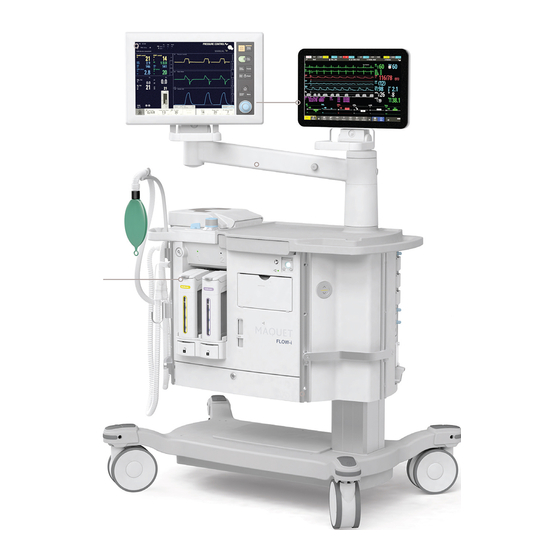
- Page 1 POCKET GUIDE POCKET GUIDE FLOW-i version 2.1 FLOW-i MODES OF VENTILATION WAVEFORMS AND LOOPS...
- Page 3 EMPTY Table of contents Introduction Background and basic concepts Waveforms and loops Capnogram, end-tidal CO monitoring Other factors References and suggested reading...
- Page 4 Scope This document is intended to function as a guide when working with the FLOW-i anesthesia system. The main focus lies on understanding and analyzing waveforms and loops presented on the control panel. Other pocket guides describe other aspects of the FLOW-i system.
- Page 5 INTRODUCTION Waveforms and loops Waveforms and loops are used to visually display the curr ent lung status of the patient. Their appearances are based on real-time values and are continuously updated. Specifically, this pocket guide will provide the user with the following: A background on the real-time values used primarily to illustrate the breathing cycle;...
- Page 6 Operators of the FLOW-i system control ventilation by having the system deliver gas using pressure. This pressure can either be set directly to create a flow into the lungs (pressure control), or indirectly so by defining a target volume to be administered with every breath (volume control).
- Page 7 BACKGROUND AND BASIC CONCEPTS Estimated values and set parameters This chapter describes three important estimated values that are used to generate waveforms and loops on the contr ol panel: Pressure Flow Volume Pressure Volume Flow Flow over time is the administered volume.
- Page 8 BACKGROUND AND BASIC CONCEPTS Linear and exponential change Linear change A linear type increase, or decrease, is characterized by a straight line in a graphical diagram (A). The change is constant irr espective of the current value. This is the case with the volume waveform in volume contr ol. The increase in volume is constant when comparing any section of the inspiratory phase.
- Page 9 This movement of gas is r eferred to as 'flow'. The pressure waveform on the control panel describes the external pressure used to deliver gas to the patient. It is measur ed inside the FLOW-i.
- Page 10 BACKGROUND AND BASIC CONCEPTS Pressure control - pressure waveform In pressure control, pressure is constant during the entire inspiration. Volume control - pressure waveform In volume control, pressure increases during inspiration, thus always maintaining a pressure gradient between delivery pressure and lung pressure.
- Page 11 BACKGROUND AND BASIC CONCEPTS Flow Flow, or movement of gas, is the r esult of a pressure gradient. The value of flow is proportional to the pressure difference. As the difference, i.e. pressure gradient, approaches zero, flow decreases. The flow waveform on the control panel describes the amount of gas per unit of time that moves into, or out of, the patient.
- Page 12 BACKGROUND AND BASIC CONCEPTS Pressure control - flow waveform In pressure control, the numeric value for flow is highest at the beginning of inspiration and at the beginning of expiration. This is when the pressure gradient is largest. Inspiration and expiration can both be described by an exponential function.
- Page 13 BACKGROUND AND BASIC CONCEPTS Volume The delivered volume is either specified by setting the tidal volume/minute volume in volume control mode, or is a subsequent effect of setting the pressure level above PEEP in pressure control mode. In pressure control, lung and thorax characteristics such as resistance and compliance effect the final delivered volume.
- Page 14 BACKGROUND AND BASIC CONCEPTS Pressure control - volume waveform Delivered volume increases exponentially in pressure control, slowing down as the pressure gradient becomes smaller. There is no inspiratory pause, expiration follows immediately after inspiration. Volume control - volume waveform Delivered volume increases linearly in volume control. The inspiratory pause is evident from the plateau between inspiration and expiration.
- Page 15 WAVEFORMS AND LOOPS Waveforms Waveforms allow for a quick assessment of how ventilation parameters interact and how patient dependant factors contribute to their appearance. Changes in resistance and compliance are immediately reflected in the waveform (and loop) appearance.
- Page 16 WAVEFORMS AND LOOPS Pressure control, waveform example...
- Page 17 WAVEFORMS AND LOOPS Pressure-Time waveform. Points and regions of interest Inspiration Start of Inspiration Expiration Early inspiratory pressure End inspiratory pressure Early expiratory pressure End expiratory pressure Flow-Time waveform. Points and regions of interest Inspiration Peak inspiratory flow Expiration Decelerating flow Peak expiratory flow The slope represents the decreasing flow from the patient during expiration...
- Page 18 WAVEFORMS AND LOOPS Volume control, waveform example...
- Page 19 WAVEFORMS AND LOOPS Pressure-Time waveform. Points and regions of interest Start of Inspiration Inspiration Inspiratory rise time Pause time Peak inspiratory pressure Expiration Early inspiratory pause pressure End inspiratory pause pressure Early expiratory pressure End expiratory pressure Flow-Time waveform. Points and regions of interest Peak inspiratory flow Inspiration Zero flow phase...
- Page 20 WAVEFORMS AND LOOPS Pressure control - summary time (s) Inspiratory volume time (s) Expiratory volume time (s) Expiration Inspiration phase phase Pressure remains constant during the inspiratory phase. Expiration starts when the valves open, removing the delivery pressure. Flow decreases exponentially during inspiration and expiration. The rate is dependant on the pr essure difference between the system and lungs.
- Page 21 WAVEFORMS AND LOOPS Volume control - summary resistance compliance time (s) Inspiratory volume time (s) Expiratory volume time (s) Inspiration Expiration phase phase Pressure increases linearly during the inspiratory phase to maintain a constant flow as lung pressure builds up. During the inspiratory pause, the pressure distributes evenly across the upper and lower airways, causing the measured pressure in the breathing circuit to decrease and stabilize at a level below the peak pr essure, i.e.
- Page 22 WAVEFORMS AND LOOPS Loops Loops provide another means of illustrating the relationships between pressure, flow and volume. Loops are updated with every breath. With the possibility of storing a reference loop, the development of compliance and r esistance can be monitored. Additionally, when selecting the 'overlay loops' option, the last two displayed loops are kept on the control panel.
- Page 23 WAVEFORMS AND LOOPS Volume Control Volume - Pressure Slope Expiration starts Inspiration starts A completed loop represents one breathing cycle, divided into inspiration (blue area) and expiration (pink area). The volume vs pressure loop is a combination of the volume and pressure waveforms.
- Page 24 WAVEFORMS AND LOOPS Volume Control Flow - Volume Expiration starts Inspiration starts A completed loop represents one breathing cycle, divided into inspiration (blue area) and expiration (pink area). The flow vs volume loop is a combination of the flow and volume waveforms.
- Page 25 WAVEFORMS AND LOOPS Volume control Pressure control When comparing volume control and pressure control, differences in loop appearance are most prominent during inspiration. Expiration follows the same exponential pattern in both types of modes, and the loops look similar. Volume-pressure loop Flow-volume loop In volume control, pressure and volume In volume control, the constant flow during...
- Page 26 CAPNOGRAM, END-TIDAL CO MONITORING The capnogram The FLOW-i continuously monitors the level of CO in the breathing circuit. Enabling the CO waveform allows for quick assessment of the status of the patient. time (s) time (s) time (s) time (s)
- Page 27 CAPNOGRAM, END-TIDAL CO MONITORING The capnogram will not appear to be in synchrony with the other displayed waveforms with respect to start of inspiration and start of expiration. This is a consequence of two factors: - At the end of inspiration, the upper respiratory tract is filled with the administered gas containing no CO (ideally).
- Page 28 CAPNOGRAM, END-TIDAL CO MONITORING The capnogram makes it possible to monitor the ventilation of the lungs and acquire information on circulation, pulmonary blood flow and metabolism. The following table lists a few examples of capnogram anomalies. The gray area symbolizes the range 3.5% - 4.5% CO (~24-35 mmHg) concentration.
- Page 29 CAPNOGRAM, END-TIDAL CO MONITORING Condition Effect on capnogram Esophageal intubation, disconnection, obstruction of the endotracheal tube, extubation etc. time (s) No apparent sampling of , levels drop to zero. Leakage Lower total measured CO levels. Abnormal shape of waveform depending on the severity of leakage.
- Page 30 OTHER FACTORS Other factors The appearance of waveforms and loops ar e not only affected by system settings. Other, less predictable, factors also contribute to their appearance. Relevant to interpretation and analysis of waveforms and loops ar e: Resistance Compliance Leakage The following overview shows which waveforms ar e affected by these factors depending on the chosen ventilation mode.
- Page 31 OTHER FACTORS Resistance Resistance, as measured in mechanical ventilation, is a measur e of the friction gas encounters when flowing through the breathing circuit (ET tube and patient tubings) and patient airways. In other words, it is a measure of the difficulty gas has when moving through the tubings and patient airways.
- Page 32 OTHER FACTORS Example 1 Volume control, pressure - time Observations: Ppeak has increased. Ppause remains unchanged. The difference in pressure between Ppeak and Ppause has increased. Conclusion: These symptoms are indicative of an increase in inspiratory resistance. Possible causes include partial occlusion in the endotracheal tube, or increased intrathoraic pressure due to CO insufflation.
- Page 33 OTHER FACTORS Example 2 Volume control, flow - time Inspiratory peak flow is unchanged. Expiratory peak flow has decresed. The decrease in expiratory flow appears linear. End expiratory flow does not reach zero before the next inspiration starts = Auto PEEP (red circle). Conclusion: During volume control, inspiratory flow remains unaffected by changes in resistance and compliance.
- Page 34 OTHER FACTORS Example 3 Volume control, volume - time Inspiratory tidal volume unchanged. Stable plateau. The decrease in volume is slow. Expiratory volume curve does not reach baseline and is truncated. Conclusion: A slower decrease in volume is indicative of flow limitation. A truncated expiratory volume curve is in itself indicative of leakage or air-trapping.
- Page 35 OTHER FACTORS Example 4 Pressure control, flow - time Decreased inspiratory- and expiratory peak flow. Inspiratory and expiratory decrease appears linear. Slower decrease in inspiratory and expiratory flow. Inspiration stops before baseline is reached. Conclusion: Increased inspiratory and expiratory resistance resulting in smaller volumes being delivered to the patient (volume = ar ea under curve in a flow-time waveform).
- Page 36 OTHER FACTORS Example 5 Pressure control, volume - time Inspiratory tidal volume has decreased. The increase in volume is slow and appears linear. The volume does not reach a plateau before expiration starts. The decrease in volume is slow. Conclusion: A slow increase in volume during inspiration indicates incr eased inspiratory resistance.
- Page 37 OTHER FACTORS Compliance Compliance is an estimate of how an inflatable object, e.g. a lung, increases in size as a result of increased inner pressure. A common unit is ml/cmH A compliance of 50 ml/cmH O means that for every increase in pressure by one cmH O, the volume increases by 50 ml.
- Page 38 OTHER FACTORS Example 6 Volume control, pressure - time Peak inspiratory pressure has increased, but the difference between P and P remains unchanged. peak plateau Conclusion: The increase in peak pressure combined with a similar increase in pause pressure indicate decreased compliance. The overall increase in pressure ensures that the defined target volume is delivered despite the decreased elasticity of the lung.
- Page 39 OTHER FACTORS Example 7 Volume control, flow - time Inspiratory peak flow is unchanged. Expiratory peak flow has increased. The decrease in in expiratory flow appears linear rather than exponential. The decrease in flow is faster and the baseline is r eached quicker. Conclusion: During volume control, inspiratory flow remains unaffected by changes in resistance and compliance.
- Page 40 OTHER FACTORS Example 8 Volume control, volume - time Inspiration is unaffected. Volume decrease during expiration is rapid and the baseline is quickly reached. Conclusion: A rapid decrease during expiration is indicative of decr eased lung compliance. The decreased elasticity of the lung creates a higher expiratory flow, resulting in a rapid volume decrease.
- Page 41 OTHER FACTORS Example 9 Pressure control, flow - time Peak flow remains unaffected, or slightly decreased. Rapid linear decrease of flow during inspiration and expiration; baseline is quickly reached. Decrease in delivered volume. Conclusion: The rapid decrease in flow is an indication of decr eased compliance. As a consequence, the delivered volume decreases.
- Page 42 OTHER FACTORS Example 10 Pressure control, volume - time Delivered volume decreases. Rapid decrease during expiration; baseline is quickly r eached. Conclusion: In pressure control, a decrease in delivered volume is indicative of decreased compliance or increased resistance. If the waveform reaches a plateau before expiration, decreased compliance becomes the more likely candidate.
- Page 43 Leakage Leakage can be caused by numerous issues with the breathing circuit or airways. The FLOW-i has several systems monitoring for signs of leakage, but it is nevertheless of value to be awar e of the change in waveform/loop appearance associated with leakage.
- Page 44 OTHER FACTORS Example 11 Normal appearance during inspiration. Expiratory volume as depicted by the area under curve is smaller than the inspiratory volume. Conclusion: The difference in inspiratory and expiratory volume is suggestive of a leak in the system. In volume contr ol, the inspiratory pressure, flow and volume waveforms are unaffected by leakage.
- Page 45 OTHER FACTORS Example 12 Volume control, volume - time Normal appearance during inspiration. Volume decrease during expiration stops before the baseline is reached. The waveform is truncated before the next inspiration. Conclusion: The premature stop of volume decrease during expiration is a consequence of delivered gas leaking out of the system.
- Page 46 OTHER FACTORS Example 13 Pressure control, volume - time The last section of inspiratory flow does not r each baseline, or reaches baseline but produces a 'bump' immediately prior to expiration. The inspiratory volume is larger than the expiratory volume, as depicted by the area under waveform.
- Page 47 OTHER FACTORS Example 14 Pressure control, volume - time Delivered volume increases. Volume decrease during expiration stops before the baseline is reached. The waveform is truncated before the next inspiration. Conclusion: The increase in delivered volume, combined with an expired volume that does not reach baseline suggests a leakage.
- Page 48 OTHER FACTORS Loops Selecting loops to be displayed on the contr ol panel introduces another means of monitoring resistance and compliance. The shape of the loops are ultimately defined by the ventilator settings. However, the change in appearance of loops ar e dependant on changes in resistance and compliance (and leakage).
- Page 49 OTHER FACTORS Volume control, volume - pressure Observations: Inspiratory section (red) is more bow shaped. Angle of slope (blue dotted line) shifted towar d the x-axis. Conclusion: The shift in slope angle, combined with the change in appearance of the inspiratory section of the loop, is indicative of a decr ease in dynamic characteristics, i.e.
- Page 50 OTHER FACTORS Volume control, flow - volume Observations: Inspiratory section does not change. Peak expiratory flow has decreased and has a spike (arrow). Linear decrease in expiratory flow section during expiration (amber). The loop is not closed (magnification), flow does not r each baseline.
- Page 51 OTHER FACTORS Pressure control, volume - pressure Δ Δ Observations: Angle of slope (blue dotted line) shifted towar d the x-axis. Volume at P has decreased (red delta). peak Conclusion: The shift in slope angle indicates a decr ease in dynamic characteristics.
- Page 52 OTHER FACTORS Pressure control, flow - volume Observations: Peak inspiratory flow has decreased. Decrease in inspiratory flow is slower (amber) . Inspiratory flow does not reach baseline, i.e. it is interrupted and the system switches to expiration (arrow). Peak expiratory flow has decresed. Linear decrease in expiratory flow during expiration (purple) .
- Page 53 REFERENCES AND SUGGESTED READING References and suggested reading Tobin M.J.: Principles and Practice of Intensive Care Monitoring. McGraw-Hill of the McGraw-Hill Companies Gravenstein J.S., Jaffe M.B., Paulus D.A.: Capnography Clinical Aspects. Cambridge University Press...
- Page 56 We operate under the three brands of ArjoHuntleigh, MAQUET Medical Systems USA GETINGE and MAQUET. ArjoHuntleigh focuses on patient mobility and 45 Barbour Pond Drive wound management solutions. GETINGE provides solutions for infection Wayne, NJ 07470 control within healthcare and contamination prevention within life sciences.
Need help?
Do you have a question about the FLOW-I and is the answer not in the manual?
Questions and answers