Table of Contents
Advertisement
Quick Links
Advertisement
Table of Contents

Related Manuals for CellaVision DM1200


Summary of Contents for CellaVision DM1200
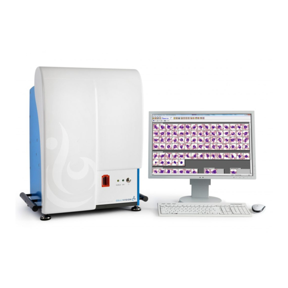
- Page 1 CellaVision ® DM1200 USER’S MANUAL...
- Page 3 All other trademarks used in this document are property of their respective owners. No part of this document or the products it describes may be reproduced or transmitted by any means or in any form without prior consent in writing from CellaVision AB. U.S. patents no. 6268611, 6341180, 7034883, 7327901, 7450762, 8914255 Swedish patents no.
-
Page 4: Table Of Contents
1.2 Warnings and precautions ..................1 1.2.1 Safety alerts in this document ..............1 1.2.2 Symbols on the system ................. 2 1.2.3 General safety information................3 1.3 Intended use of CellaVision ® DM1200 ..............4 1.3.1 Peripheral Blood Application ................ 4 1.3.2 Body Fluid Application................... - Page 5 CellaVision ® DM1200 4.1.3 Red blood cell characterization using the Advanced RBC Application....................39 4.1.4 Estimating platelets..................45 4.1.5 Comments ..................... 49 4.1.6 Pathology review..................50 4.1.7 Order data ..................... 52 4.1.8 Signing a slide ....................52 4.2 Body fluids ........................ 54 4.2.1 Body fluids differential .................
- Page 6 CellaVision ® DM1200 7.1.1 Slide requirements ..................77 7.2 Scan overview image....................77 7.2.1 Overview......................78 7.2.2 Navigating ..................... 79 7.2.3 Tagging regions of interest................80 7.2.4 Copying regions of interest to disk............. 80 7.2.5 Adding comments ..................80 7.2.6 Order data ..................... 81 7.3 Database view ......................
- Page 7 CellaVision ® DM1200 10.3 Remedial maintenance..................113 10.3.1 Change immersion oil pack ..............113 10.3.2 Remove stuck slide from the gripper............. 116 10.4 Database performance..................116 10.4.1 Check database size ................117 11 TROUBLESHOOTING ....................118 11.1 General processing problems................118 11.2 LIS errors ......................120...
- Page 8 CellaVision ® DM1200 F.3 Recommended staining recipes ................159 F.3.1 MGG stain ....................160 F.3.2 Wright stain ....................161 F.3.3 Wright-Giemsa stain ..................161 F.4 Slide preparation for body fluids ................162 F.4.1 Sample......................162 F.4.2 Preparing the sample.................162 F.4.3 Staining ......................162 APPENDIX G GLOSSARY ....................163 INDEX ............................165...
-
Page 9: Introduction
This User's manual will guide you step-by-step through the activity sequence of ® normal use of CellaVision DM1200 (also referred to as the system), aiming to give you a good understanding of the system and its features. References are made to appendices providing additional information. -
Page 10: Symbols On The System
Introduction CellaVision ® DM1200 1.2.2 Symbols on the system These symbols can be found on the system. Make sure that you understand the meaning of each symbol. Symbol Explanation Caution. Indicates the need for the user to consult the instructions... -
Page 11: General Safety Information
• Don’t install or run any software not supplied with the system. It is allowed to install an anti virus program, but it is not recommended to scan the database files. • To maintain electromagnetic compatibility, always use original CellaVision ® listed spare parts and their specified components. -
Page 12: Intended Use Of Cellavision
DM1200 automatically locates and presents images of blood cells on peripheral blood smears. The operator identifies and verifies the suggested classification of each cell according to type. DM1200 is intended to be used by skilled operators, trained in the use of the device and in recognition of blood cells. 1.3.1... -
Page 13: Peripheral Blood Application
Introduction 1.4.1 Peripheral Blood Application The Peripheral Blood application is included in the CellaVision DM Software. General functionality • Presents an image on a screen of every located cell or object; • Organizes and suggests cell classification (preclassification) for white blood cells;... -
Page 14: Advanced Rbc Application
Introduction CellaVision ® DM1200 RBC characterization Besides the RBC morphology characteristics mentioned above, the operator can characterize to Target cells, Schistocytes, Helmet cells, Sickle cells, Spherocytes, Elliptocytes, Ovalocytes, Tear drop cells, Stomatocytes, Acanthocytes, Echinocytes, Howell-Jolly bodies, Pappenheimer bodies, Basophilic stippling, Parasites, and 10 user defined characteristics. -
Page 15: Body Fluid Application
CellaVision ® DM1200 Introduction 1.4.3 Body Fluid Application The Body Fluid application is an optional application. General functionality • Presents an image on a screen of every located cell or object; • Organizes and suggests cell classification (preclassification) for the located nucleated cells;... -
Page 16: Components And Mechanical Operation
– Immersion oil unit – Robot gripper unit – Barcode reader – Control unit – Casing System computer The system computer is a PC running Microsoft Windows and CellaVision DM Software. Slide scanning unit 1. Motorized microscope 5. Barcode reader 10.Status LED 2. - Page 17 CellaVision ® DM1200 Introduction Warning! Never tamper with sensors or other safety devices. These make sure that the system can operate without any risk of personal injury. Motorized microscope The motorized microscope is an upright light microscope with a LED illumination system.
-
Page 18: References To Scientific Literature
Introduction CellaVision ® DM1200 1.6 References to scientific literature Bain, B. J., Bates, I., Laffan, M. A., & Lewis, S. M. (2012). Dacie and Lewis Practical Haematology (11th ed.). London: Churchill Livingstone. Galagan, K. A., Blomberg, D., Cornbleet, P. J., & Glassy, E. F., (Eds.). (2006). -
Page 19: Operating Procedures
CellaVision ® DM1200 Operating procedures 2 Operating procedures 2.1 Starting the system The system computer is configured with a Windows policy restricting access to the operating system for the normal user. When starting the system computer the user will automatically be logged on to Windows and then the software logon window will be displayed. -
Page 20: Magazines And Magazine Id
Operating procedures CellaVision ® DM1200 Information in toolbar System indicators show oil level, hood position etc. For a description of these indicators, see Appendix C Buttons and indicators. System status is shown as a text: Idle, Analyzing, Stopped, Paused or Error. -
Page 21: Loading Slides Into A Magazine
1-12 from the bottom up. Make sure that slides are fully inserted in the magazine before using the magazine. Note: A magazine can be used 100 times. The CellaVision DM Software will display a message when a magazine has been used more than 100 times. 2.6 Processing slides View the processing in the animated tutorial. -
Page 22: Adding Magazine
Operating procedures CellaVision ® DM1200 2.6.1 Adding magazine Slide the magazine into the magazine holder. The magazine must have the barcode turned upwards and the open end of the magazine inwards. When all slides in a magazine have been processed, the magazine is automatically ejected. -
Page 23: Starting The Slide Processing
CellaVision ® DM1200 Operating procedures 2.6.2 Starting the slide processing The slide processing starts automatically when you insert a magazine into the system. You have to manually resume the processing if the system has been stopped, restarted or if an error has occurred. -
Page 24: Magazine Status
Operating procedures CellaVision ® DM1200 2.6.3 Magazine status The following information is available for each magazine: Magazine Magazine ID (barcode) status PB2345678912 Explanation of symbols Processing All the slides in the magazine have been Finished processed with no warnings or errors. -
Page 25: Ejecting Magazine
CellaVision ® DM1200 Operating procedures Slide information dialog Double-click on a slide to open the Slide information dialog. Additional information on the processed slide, for example the cause of an error, is displayed here. 2.7 Ejecting magazine You can eject the magazine that is currently being processed. -
Page 26: Quality Control
Quality control CellaVision ® DM1200 3 Quality control 3.1 Cell location test for peripheral blood The cell location test verifies that the quality of the slide preparation process is good enough to allow the system to locate the cells needed for the analysis. It also verifies the system’s ability to locate cells. - Page 27 2. In the Cell location slides list, click the cell location slide that you want to examine. The CellaVision DM Software will open the first image from that cell location slide. 3. In the overview image, count any nucleated cells that have not been located by the system and enter the number in the WBCs + NRBCs missed text box.
- Page 28 Quality control CellaVision ® DM1200 To evaluate the cell location results 1. Under Total result, check that the Ratio of WBCs + NRBCs found is within your laboratory's established limits. For performance characteristics for the system, when using standardized staining and smear preparation procedures, see A.2 Performance specification.
-
Page 29: Explanation Of Markings
CellaVision ® DM1200 Quality control 3.1.1 Explanation of markings The color of the box indicates how the systems has preclassified the located object. It is possible to compensate for objects incorrectly preclassified as nucleated cells, see 3.1.2 Manual correction. However, the crucial factor in the cell location test is that every cell is located, not that each cell is correctly preclassified. - Page 30 Quality control CellaVision ® DM1200 If a cell is near the edge of the overview image, double-click it to view a magnified image of the area around it. The associated box may be outside the overview image. This object has been located but the box is outside the overview image.
-
Page 31: Manual Correction
CellaVision ® DM1200 Quality control 3.1.2 Manual correction If the system incorrectly preclassified any objects as nucleated cells (that is, marking them with a green box instead of a blue box), you can manually correct the total result. Do not correct for objects marked with a black box. Those are already excluded from the calculation. - Page 32 Quality control CellaVision ® DM1200 To prepare and process a cell location slide 1. Use a freshly stained body fluid sample with a total number of nucleated cells less than 12 000. 2. Process the slide like a regular body fluid slide.
-
Page 33: Explanation Of Markings (Body Fluid)
CellaVision ® DM1200 Quality control 7. If you want to print the results of the cell location test, click the Print results button. 8. Check that the result of the cell location test is within your laboratory's established limits. For performance characteristics for the system, when using standardized staining and smear preparation procedures, see A.2 Performance... -
Page 34: Self Tests
Quality control CellaVision ® DM1200 The box is not always centered over the cell. It may cover part of the cell or may even be completely separate from the cell. As long as there is a box associated with a cell, that cell has been located. -
Page 35: Verifying Processed Slides
CellaVision ® DM1200 Verifying processed slides 4 Verifying processed slides 4.1 Peripheral blood Opening an unsigned slide leads directly to the Verification view, where various tabs can be selected in order to review WBC, RBC and PLT and to sign the slide. - Page 36 Verifying processed slides CellaVision ® DM1200 All cell classes handled by the system are displayed in this figure. WBCs and non-WBCs automatically preclassified by the system are marked with a small dot or an arrow. 1. Arrow indicating a cell class that is automatically forwarded 2.
- Page 37 Reference cells: A library of reference cells for different cell classes is provided with the system. These cells are marked with a CellaVision ® logo. The main gallery always displays WBCs from the slide, while the others show reference cells when the checkbox Reference cells is selected.
- Page 38 Verifying processed slides CellaVision ® DM1200 WBC full screen view: Click WBC full screen view to display all WBCs sorted by class. WBC full screen view Note: Cell classes that have been fully displayed in WBC Full Screen view will also be tick marked.
- Page 39 CellaVision ® DM1200 Verifying processed slides Adjusting magnification: Click Zoom in or Zoom out to change the magnification in all galleries and in the WBC full screen view. Zoom in Zoom out You may also double-click on a WBC to enlarge it, and use the scroll wheel to zoom in or out.
- Page 40 Verifying processed slides CellaVision ® DM1200 Splitting cells: Occasionally the system fails to separate WBCs that are close to each other, and more than one cell in an image will be outlined by the cell marker (green square). These cells should be split so that each can be identified by the operator.
- Page 41 3. Forwarded from another cell class 4. Cell comments exist 5. Reclassified cell ® 6. The image has been manually captured on a CellaVision Image Capture System WBC attributes are shown by default. Click WBC attributes to show/hide them. Note: ®...
- Page 42 4. Split cell or remove split cell. 5. Select cell for e-mail. 6. Save cell as custom reference cell. 7. Save images to disk. Note: All alternatives are not available for images captured on a CellaVision ® Image Capture System. E-mail You may send cell images by e-mail.
-
Page 43: Red Blood Cell Characterization
CellaVision ® DM1200 Verifying processed slides Save cell images to disk You can save cell images from any order to, for example, your local hard drive or a USB flash drive. For more information on how to save cell images, see 6.1.8 Save cell images to disk. - Page 44 Verifying processed slides CellaVision ® DM1200 RBC panel: The RBC panel is used for characterization of the RBC morphology. All morphologies handled by the system are listed. Morphologies precharacterized by the system are marked with a small dot. The columns labeled 0 to 3 grade the morphology characteristics.
- Page 45 CellaVision ® DM1200 Verifying processed slides Customizing the red blood cell overview image Change the magnification of the image by using these buttons: Zoom in Zoom out Entire RBC image - Shows the entire RBC image. Navigate the image by switching between different control modes. The mouse pointer changes accordingly.
- Page 46 Verifying processed slides CellaVision ® DM1200 Characterizing red blood cell morphology There are two ways to report the RBC result: 1. Report all as normal a) Select radio button Report all as 0 - Normal. 2. Use characterization a) Select radio button Use characterization.
-
Page 47: Red Blood Cell Characterization Using The Advanced Rbc Application
CellaVision ® DM1200 Verifying processed slides 4.1.3 Red blood cell characterization using the Advanced RBC Application RBC precharacterization using the Advanced RBC Application is only available for slides processed on a system with the Advanced RBC Application activated. Overview Click Overview to view the RBC overview image. - Page 48 Verifying processed slides CellaVision ® DM1200 the other morphologies in the group are dimmed. Click if you wish to go back to use automatic grading. RBCs automatically precharacterized by the system are marked with a small dot or an arrow.
- Page 49 CellaVision ® DM1200 Verifying processed slides Display names in use: It is possible to edit the display names of RBC morphologies. To do this, contact your service technician. Move the mouse pointer over the morphology in the RBC panel to show the original name.
- Page 50 Verifying processed slides CellaVision ® DM1200 Reclassify Red Blood Cells: Reclassify RBCs by dragging and dropping them from one morphology to another: 1. Place the cursor over the cell image. 2. Click and hold down the left mouse button. 3. Move the cursor to the destination gallery then release the button.
- Page 51 CellaVision ® DM1200 Verifying processed slides Customizing the red blood cell overview image Change the magnification of the image by using these buttons: Zoom in Zoom out Entire RBC image – Shows the entire RBC image. Navigate the image by switching between different control modes. The mouse pointer changes accordingly.
- Page 52 Verifying processed slides CellaVision ® DM1200 Characterizing red blood cell morphology There are two ways to report the RBC result: 1. Report all as normal a) Select radio button Report all as 0 - Normal. 2. Use characterization a) Select radio button Use characterization.
-
Page 53: Estimating Platelets
To show or hide the help lines, click the Help lines button. The help lines are only a visual aid and does not affect the calculation of the platelet concentration. Note: Help lines are not available for images captured on a CellaVision ® Image Capture System. - Page 54 Estimate the average PLT count per grid square and type this value in the field. Note: It is not possible to count PLTs per grid square or to specify an approximate ® PLT count per grid square for images captured on a CellaVision Image Capture System. User’s Manual PM-10829-01 2015-07-10...
- Page 55 CellaVision ® DM1200 Verifying processed slides PLT result: 1. Click Calculate PLT Result in the PLT Count panel. 2. If you wish to report the PLT results calculated from the number of PLTs per HPF, you have two choices: • Select Calculated estimate to report a concentration. The estimate is calculated as [Average PLTs/HPF value] x [PLT estimate factor].
- Page 56 The PLT concentration level can be estimated by setting it to four levels: Significantly decreased, Decreased, Normal, or Increased directly from viewing the image. ® 1. The image has been manually captured on a CellaVision Image Capture System 2. Square Select Concentration level.
-
Page 57: Comments
CellaVision ® DM1200 Verifying processed slides Excluding the platelet analysis Click Exclude PLT Analysis to exclude PLT analysis results from the slide. 4.1.5 Comments For each slide, you can add comments to the WBC, RBC and PLT results. For WBC analyses, you can also add comments to cell classes and individual cells. -
Page 58: Pathology Review
Verifying processed slides CellaVision ® DM1200 Click Comments to add comments to WBC, RBC and PLT. You may write and edit comments in the Comment box. Click Standard Comments to show or hide standard comments. Double-click on a standard comment to add it to the Comment box. You may also select a standard comment and click Append. - Page 59 CellaVision ® DM1200 Verifying processed slides To mark a slide for pathology review 1. Open the slide you want to mark for pathology review. 2. Click the Pathology review button 3. In the Pathology review dialog box, in the lower text box, type a comment describing what you want the pathologist to look for, and then click OK.
-
Page 60: Order Data
Verifying processed slides CellaVision ® DM1200 3. If you want the pathologist to review the slide again, click to clear the Pathology review completed check box, and then, in the lower text box, type a comment describing what you want the pathologist to look for. - Page 61 CellaVision ® DM1200 Verifying processed slides Note: If the slide is part of a multi-slide order, all slides in the order must be signed before the operator is given the option to sign the order. Note: Slide data cannot be changed after signing. Comments may still be added.
-
Page 62: Body Fluids
Verifying processed slides CellaVision ® DM1200 4.2 Body fluids Opening an unsigned slide leads directly to the Verification view, where the tabs for Overview, WBC and Sign Slide are shown. To open a slide, see 6.1.3 Opening an order or a slide. - Page 63 CellaVision ® DM1200 Verifying processed slides All cell classes handled by the system are displayed in the figure below. WBCs and non-WBCs automatically preclassified by the system are marked with a small dot. 1. Display name in use (original name in tooltip) 2.
-
Page 64: Body Fluids Overview Image
Verifying processed slides CellaVision ® DM1200 4.2.2 Body fluids overview image The body fluid overview image displays the entire sample area. See 9.3.6 BF analysis settings for more information. The overview image can be used to find cells of interest and for getting an overall impression of the sample. - Page 65 CellaVision ® DM1200 Verifying processed slides Navigating in the overview image Navigate in the overview image by using the keyboard arrow buttons or the navigation buttons. The image shown in the right part of the screen is an enlargement of the area inside the red rectangle shown in the Mini map.
- Page 66 Verifying processed slides CellaVision ® DM1200 Tagging regions of interest The image shown in the right part of the screen can be saved as a region of interest for later reference. 1. To save the current image, click Tag region.
-
Page 67: Comments
CellaVision ® DM1200 Verifying processed slides 4.2.3 Comments Comments can be added to the Overview, the differential result, each cell class and all individual cells. Note: All comments, except comments on individual cells, are printed in the report. Adding comments To add a comment, click Comment. -
Page 68: Reporting Results
Reporting results CellaVision ® DM1200 5 Reporting results For detailed information on settings, see 9.4 Report settings. Click Report view in the toolbar. Compare slide results and exclude slides from the reported result in the Report view. You can also: •... - Page 69 CellaVision ® DM1200 Reporting results Result panel Here you see the results of each slide in the order. In the Reported Result column you see the summarized results. An unsigned slide has a slide ID written together with slide number in cerise color.
-
Page 70: Report Preview
Reporting results CellaVision ® DM1200 Changing the reported result It is possible to change the RBC results and the PLT concentration for the order, if reported as a level. Changeable results are written in bold text. 1. Click on the result to change. -
Page 71: Signing An Order (Result)
CellaVision ® DM1200 Reporting results 5.3 Signing an order (result) 1. Click Sign order. 2. If not prefilled, type User name and Password. 3. Select whether the order should be sent to the LIS or printed or both. 4. Click OK. -
Page 72: Database
Database CellaVision ® DM1200 6 Database Click Database view in the toolbar. Processed and pending orders are stored in the database. Switch between the two using the tabs Processed orders and Pending orders. 6.1 Processed orders You can search for and open processed orders and slides stored in the system. -
Page 73: Order List
CellaVision ® DM1200 Database 6.1.1 Order list The Order list displays an overview of the orders. Click on the column headers to sort the list. The date and time in the column Analyzed corresponds to the processing date for the last processed slide in an order. - Page 74 Database CellaVision ® DM1200 LIS status Empty field No data sent or received Data received Waiting to send result Result is sent Failed to send result Result is successfully sent Process status Empty field All slides in the order have been processed successfully.
- Page 75 CellaVision ® DM1200 Database Comments Empty field No comments. Comments exist. At least one slide in the order is marked for pathology review. Pathology review has been completed. Multi-slide status Empty field One slide in order. More than one slide in order.
-
Page 76: Slide List
Database CellaVision ® DM1200 6.1.2 Slide list Comments, slide- and process status are indicated in the slide list. 1. Process status 2. Slide status 3. Comments Process status The slide has been processed successfully. The operator has stopped the processing of the slide. No results exist. -
Page 77: Opening An Order Or A Slide
CellaVision ® DM1200 Database 6.1.3 Opening an order or a slide Opening an order automatically opens a slide belonging to it. In the same manner, opening a slide automatically opens the order it belongs to. The currently opened order and slide are always shown in the toolbar. -
Page 78: Searching For An Order Or A Slide
Use the date boxes to specify the time interval. • View BF to see body fluid slides. • View CellaVision Image Capture System to see slides captured with the CellaVision Image Capture System. You can also refine your search by, for example, patient data, order data, and comments. -
Page 79: Save Cell Images To Disk
CellaVision ® DM1200 Database To export orders to an export database 1. In the Database view, on the Processed orders tab, select the orders you want to export. 2. Click Export. 3. In the Export selected orders windows, in the Database list, select the database you want to export the orders to. -
Page 80: Printing Orders
The worklist is available in the Database view and in the Verification view. All slides will be removed from the worklist when you exit the CellaVision DM Software. -
Page 81: Pending Orders
CellaVision ® DM1200 Database To manually add slides to the worklist 1. In the Processed orders tab, do one of the following: • To select a consecutive group of orders, click the first order, press and hold down the Shift key, and then click the last order. -
Page 82: Usage Log
Database CellaVision ® DM1200 STAT mark Empty field Not a STAT order. Order is marked as a STAT order. Order type Empty field Peripheral blood order. Body fluid order. Click Add to add a new pending order. Double-click on a pending order, or click Edit, to view or edit the order data. -
Page 83: Statistics
CellaVision ® DM1200 Database 6.3.1 Statistics The Statistics page contains dates and times of program installation and creation and modification of the Usage log. It also displays the number of successfully processed and failed slides, the number of signed analyses and the total processing time. -
Page 84: Export Log Files
It is important to back up the database often. If a hard disk crash occurs, the whole database will be lost. It will only be possible to recover the data up to the time when the last backup was made. For more information, see CellaVision ®... -
Page 85: Digital Slides
7.1.1 Slide requirements Use slides as specified in A.3.3 Slides and either orange or blue CellaVision slide magazines. You can use cover slips, as long as the total thickness of the slide and the cover slip is within the requirements. If you use cover slips, make sure the scan area is within the cover slip area. -
Page 86: Overview
Digital slides CellaVision ® DM1200 7.2.1 Overview The Mini map shown in the Overview displays the entire sample area. To enlarge a specific area of the sample, click in the Mini map. The magnified area will be displayed in the large image on the screen. On the Mini map, a red rectangle marks the area requested for enlargement. -
Page 87: Navigating
CellaVision ® DM1200 Digital slides 7.2.2 Navigating Move in the Mini map by using the keyboard arrows or the Navigation buttons. The image shown in the right part of the screen is an enlargement of the area inside the red rectangle shown in the Mini map. Click on the Mini map to enlarge another part of the sample. -
Page 88: Tagging Regions Of Interest
Digital slides CellaVision ® DM1200 7.2.3 Tagging regions of interest To save an enlarged area as a region of interest for later reference, click Tag region. Identify your region of interest by adding a comment. 7.2.4 Copying regions of interest to disk You can copy a region of interest to disk. -
Page 89: Order Data
CellaVision ® DM1200 Digital slides 7.2.6 Order data To view information about the order, e.g. scanned area and comments, click Order data in the toolbar. The Order data dialog can also be accessed by right-clicking on an order in the database view. - Page 90 Digital slides CellaVision ® DM1200 To define the scan area for the slide, click Area, then do one of the following: • Enter values in the text boxes to specify the scan area. • Use the mouse to drag a rectangle on the slide. The rectangle must be within the red dotted rectangle on the slide.
-
Page 91: System Information
CellaVision ® DM1200 System information 8 System information In the toolbar, click Help and then System Information for information about your system. The information presented depends on your system and installed software. Some of the following information is present in the System Information dialog: •... -
Page 92: Customizing The System
DM1200 9 Customizing the system Important! Most settings that you can change in the CellaVision DM Software will affect all systems and review stations using the same database. These settings will affect all systems and review stations using the same database: •... -
Page 93: Manage Databases
Manage databases You can create a database on the system computer or connect to a database on a server (running the CellaVision Server Software) or on another system. To view a list of databases and connections to databases • On the Tools menu, click Settings and then click the Database tab. - Page 94 5. In the Remote computer box, type the name of the computer where the remote database is located. If you do not know the name of the remote computer, open the CellaVision DM Software on that computer and then, on the Help menu, click System information.
-
Page 95: Manage Database Size
• The maximum capacity for the database, that is: – 200 GB (approximately 40 000 peripheral blood slides) for a database located on a server running CellaVision Server Software. – 20 GB (approximately 4 000 peripheral blood slides) for a database located on the system computer. - Page 96 Customizing the system CellaVision ® DM1200 Note: The archiving function is not a back up function. It is recommended that you always keep a backup of the database. You will not be able to access orders in the archive if the database is lost.
- Page 97 CellaVision ® DM1200 Customizing the system PB cell images Normal WBCs Abnormal WBCs Non-WBCs Segmented neutrophils Band neutrophils Erythroblast (NRBC) Eosinophils Promyelocytes Giant thrombocyte Basophils Myelocytes Thrombocyte aggregation Lymphocytes Metamyelocytes Megakaryocyte Monocytes Immature eosinophils Smudge cell Immature basophils Artefacts Promonocytes...
-
Page 98: Database Compression
While a database is being compressed, no clients can use or connect to that database. For this reason, you can’t compress the database that you logged on to when you started the CellaVision DM Software. Any users still connected to the database when you start the compression will receive an error message. -
Page 99: User Account Settings
CellaVision ® DM1200 Customizing the system 9.2 User account settings Important! The user account settings apply to all systems that use the same database. This means that your changes may affect other systems. You can create user accounts with different access levels, for everyone who needs access to the system. - Page 100 Customizing the system CellaVision ® DM1200 There are six levels of user authorization: • Observer. Only allowed to view images and results. • User. Can verify WBC, RBC, and PLT results, but can’t sign slides. • Authorized. Can verify WBC, RBC, and PLT results and sign slides and orders.
-
Page 101: Options For Restricted Users
Important! The analysis settings apply to all systems that use the same database. This means that your changes may affect other systems. The RBC analysis area settings will only apply to your CellaVision ® DM1200 and won’t affect other systems. -
Page 102: Default Processing Settings
Customizing the system CellaVision ® DM1200 9.3.1 Default processing settings Important! The default processing settings apply to all systems that use the same database. This means that your changes may affect other systems. You can change how the system processes a slide if the order ID isn’t in the database (in the Pending orders list) or in the LIS, by setting the default analysis values. - Page 103 CellaVision ® DM1200 Customizing the system To enable RBC precharacterization 1. On the Tools menu, click Settings. 2. Depending on your system configuration, do one of the following: • On a system with the Advanced RBC Application, click the Advanced RBC tab.
- Page 104 Customizing the system CellaVision ® DM1200 To change RBC grading and size limits This procedure is valid for systems using the standard RBC functionality. For systems with the Advanced RBC Application, see To change grading, size, and anisocytosis limits for the Advanced RBC Application later in this section.
- Page 105 1. On the Tools menu, click Settings and then click the Advanced RBC tab. 2. Click RBC Limits. 3. To set all limits to CellaVision default values, click Set limits. The default values are based on Gulati, Gene. 2009. Blood Cell Morphology Grading Guide.
-
Page 106: Rbc Analysis Area Settings For The Advanced Rbc Application
Customizing the system CellaVision ® DM1200 9.3.3 RBC analysis area settings for the Advanced RBC Application On a system with the Advanced RBC Application, you can change the aspect ratio of the RBC analysis area so that the RBC overview image fits your monitor. -
Page 107: Plt Settings
CellaVision ® DM1200 Customizing the system 9.3.5 PLT settings Important! The PLT settings apply to all systems that use the same database. This means that your changes may affect other systems. You can change which of these two methods to calculate the PLT concentration you want to use: •... - Page 108 Customizing the system CellaVision ® DM1200 To change the default properties for the PLT view These settings change the way the PLT view looks and functions. You can temporarily change any of these values from the PLT view, while performing the analysis.
-
Page 109: Bf Analysis Settings
CellaVision ® DM1200 Customizing the system 9.3.6 BF analysis settings You need to set the position and the diameter of the BF analysis area to match the position of the sample spot on your BF slides, before you process BF slides for the first time, or if you change your method for preparing BF slides. - Page 110 For example, if you increase the diameter from 6 mm to 8 mm, each BF overview image will take up 78% more space in the database. 8. Click Close and then restart the CellaVision DM Software. User’s Manual PM-10829-01 2015-07-10...
-
Page 111: Worklist Setting
CellaVision ® DM1200 Customizing the system 9. Delete the order with the BF slide from the database. Important! You must delete the order from the database before you process the slide again. If you don’t delete the order, the settings from the previous processing will be used, which means that your changes to the BF analysis area won’t apply. - Page 112 Customizing the system CellaVision ® DM1200 To set a default report template 1. On the Tools menu, click Settings and then click the Report/Sign tab. 2. In the Report template list, click the template you want to use, and then click Set active.
-
Page 113: Order Signing Settings
CellaVision ® DM1200 Customizing the system 9.5 Order signing settings Important! The order signing settings apply to all systems that use the same database. This means that your changes may affect other systems. You can change what is filled in by default in the Sign slide dialog box when it opens. -
Page 114: Reference Cells Settings
Customizing the system CellaVision ® DM1200 To change a standard comment 1. On the Tools menu, click Settings, and then click the Standard comments tab. 2. In the list, click the comment you want to change, and then click Modify. -
Page 115: E-Mail Settings
5. The reference cell you added appears in the Preview frame. 6. If you want to add a comment to the reference cell, click Comment. 7. Click Close and then restart the CellaVision DM Software. The PB Reference cells settings dialog box. -
Page 116: Digital Slides Settings
Note: ® The E-mail setting will be the used for everyone who uses this CellaVision DM1200, even by someone using a different user account or a different database. 9.9 Digital slides settings... -
Page 117: Language Settings
You can use the mouse to drag a rectangle for the area you want to scan. 9.10 Language settings You can change the language for the CellaVision DM Software user interface and the language for the User’s manual. You can choose from languages that were installed during the installation of the system. -
Page 118: Maintenance
Maintenance CellaVision ® DM1200 10 Maintenance Caution! Be careful not to bend or put excessive force on any part of the system interior. 10.1 Weekly maintenance 10.1.1 Clean objectives and LED table 1. Open the hood. 2. Gently wipe the LED table with a soft lint free cloth. - Page 119 CellaVision ® DM1200 Maintenance 3. Use a new lens paper and gently wipe the lens of the dry 10x objective. If necessary, use isopropyl alcohol to clean lens of the dry 10x objective. Discard the lens paper after use. Important! Do not get immersion oil or any other contamination on the dry 10x objective.
-
Page 120: Clean Hood
Maintenance CellaVision ® DM1200 5. Process two slides and then immediately delete the two slides from the database. This will wet the lens of the 100x objective and help to prevent air bubbles between the lens and the immersion oil. -
Page 121: Delete Unsigned Orders
10.1.5 Restart system computer Restart the system computer at least once a week. 10.2 Preventive maintenance No preventive maintenance by the user is necessary. Preventive maintenance is to be performed by CellaVision ® authorized personnel. 10.3 Remedial maintenance 10.3.1 Change immersion oil pack Warning! The oil may cause sensitization by skin contact. - Page 122 Maintenance CellaVision ® DM1200 Caution! Use only the immersion oil specified in A.3.1 Immersion oil. 1. Open the hood. 2. Put a clip on the hose. 3. Push down on the oil hose connection and pull out the hose. User’s Manual...
- Page 123 CellaVision ® DM1200 Maintenance 4. Remove and discard the old oil pack. For information on how to dispose of the old oil pack, see B.3 Disposal information. 5. On the new oil pack, put the clip on the hose. 6. Cut the hose above the line where the arrow points.
-
Page 124: Remove Stuck Slide From The Gripper
4. Open the hood. 5. Remove the slide. 6. Close the hood. 7. Restart the slide scanning unit and CellaVision DM Software. Animated tutorial To open the animated tutorial about how to remove a stuck slide from the gripper, go to Help/Tutorial. -
Page 125: Check Database Size
CellaVision ® DM1200 Maintenance 10.4.1 Check database size 1. On the Help menu, click System information. 2. Check the Database size. 3. Take steps to reduce the size of the database, if the Database size is more than: • 20 000 MB for a local database. -
Page 126: Troubleshooting
Troubleshooting CellaVision ® DM1200 11 Troubleshooting If a problem occurs, follow these steps 1. Observe any unusual circumstances surrounding the problem. 2. Note any error messages displayed. 3. Follow the troubleshooting charts later in this chapter. 4. If the problem remains, contact your vendor for assistance. - Page 127 The database service If you use a local database service, restart slow. needs to be restarted. the system computer. If you use the CellaVision Server Software, restart the server. The database is too Keep the database size below the large.
-
Page 128: Lis Errors
Troubleshooting CellaVision ® DM1200 Problem Cause Action Seemingly normal Too little specimen on Prepare a slide with more specimen. scan slides get slide the slides. status Analysis Wrong area selected. Select the correct area and process the failure, with the slide again. -
Page 129: Cell Location Problems
CellaVision ® DM1200 Troubleshooting 11.3 Cell location problems Cell location problems peripheral blood Problem Cause Action Too many non- Too many smudge Run another slide with less smudge cells. nucleated cells. cells. Verify smear preparation process. Too many artefacts. Increase wash step. - Page 130 Troubleshooting CellaVision ® DM1200 Cell location problems body fluids Problem Cause Action Too many non-WBCs. Too many smudge Run another slide with less smudge cells. cells. Verify sample preparation process. Decrease centrifuge speed. Too many artefacts. Increase wash step. Filter the stain.
-
Page 131: Barcode Problems
The barcode reader is Make sure that the barcode reader cable is not connected. connected to both the barcode reader and the control unit. Restart the CellaVision DM Software. No barcode found. Check positioning and size of the barcode, see A.3.5 Barcodes. -
Page 132: Staining Problems
Troubleshooting CellaVision ® DM1200 11.5 Staining problems Problem Cause Action The RBCs appear Understaining. The fixation and staining time may need bright red, the white to be increased. cells will appear Overwashing. The washing technique may need to be indistinct with pale... -
Page 133: General Startup Problems
CellaVision ® DM1200 Troubleshooting Problem Cause Action Samples have dark The blood smear was Make sure that the blood smear is areas of stain or other not completely dry completely dry before staining it. artefacts. when it was stained. Dirty slides. -
Page 134: Error Message List
Startup error messages Error message Cause Action The program has The CellaVision DM Make sure the CellaVision DM Software already been started Software is already is closed. Then wait 10 seconds and try and you cannot start running or has again. - Page 135 CellaVision DM Software. The hood was open The hood was open. Close the hood. Restart the slide scanning during start-up. unit and the CellaVision DM Software. Jam error occurred At least the motor Restart the slide scanning unit and the during initialization.
- Page 136 10.3.2 Remove start-up. stuck slide from the gripper. Restart the slide scanning unit and the CellaVision DM Software. An unknown error One or more slides are Make sure that the slides are closed.
- Page 137 The oil bag is empty. Change the immersion oil pack, see 10.3.1 Change immersion oil pack. File system or registry The file system or Reinstall the CellaVision DM Software. failure. registry is corrupt. Follow the instructions provided with the CellaVision Software Pack disc.
- Page 138 10.3.2 Remove message reported a stuck slide from the gripper. Restart the positioning failure. slide scanning unit and the CellaVision DM Software. The slide is stuck or Move the gripper to the service position broken.
- Page 139 Check the network connection. If you use a database on the system computer, restart the system computer. If you use the CellaVision Server Software, restart the server. There was a problem The order ID contains Make sure that the order ID follows the when accessing the non-ASCII characters.
- Page 140 User’s Manual PM-10829-01 2015-07-10...
-
Page 141: Appendix A Specifications
A.1 System specification A.1.1 Climate specification The CellaVision ® DM1200 is designed to be safely operated in the following environment: Temperature 18 °C to 31 °C (64 °F to 88 °F) Relative humidity 20% to 80%, non-condensing... -
Page 142: Electric Specification (Monitor Not Included)
Specifications CellaVision ® DM1200 A.1.3 Electric specification (monitor not included) 100 to 240 VAC Voltage input Voltage frequency 50 to 60 Hz Current input, system computer 1.4 to 0.7 A 0.6 to 0.3 A Current input, slide scanning unit Total current input for the system 2.0 to 1.0 A... -
Page 143: Noise Specification
CellaVision ® DM1200 Specifications Warning! Do not use this device in close proximity to sources of strong electromagnetic radiation, as these may interfere with the proper operation. Caution! The electromagnetic environment should be evaluated prior to operation of the device. -
Page 144: Performance Specification
Specifications CellaVision ® DM1200 A.2 Performance specification Performance specification peripheral blood Average WBC cell-location and display of at least 97 % with a standard deviation less than 2 %. Throughput : Approximately 20 slides per hour for complete orders containing RBC, PLT and 100 WBCs. - Page 145 50x overview images (analysis area 6 mm in diameter). To evaluate the Accuracy and Short-term imprecision of the Body Fluid application, a comparison study was conducted at two sites with five operators. The Body Fluid application running on the CellaVision ® DM1200 was...
- Page 146 Specifications CellaVision ® DM1200 Short-term imprecision results for the Body Fluid application Specimen Cell class SD (%) Neutrophils Lymphocytes Eosinophils Macrophages Other cells Serous fluid Neutrophils Lymphocytes Eosinophils Macrophages Other cells Synovial fluid Neutrophils Lymphocytes Eosinophils Macrophages Other cells User’s Manual...
- Page 147 CellaVision ® DM1200 Specifications A reproducibility study was conducted at three different sites on three different instruments, each site processing the samples in duplicates. The study was performed during 20 days at each site and run by in total 10 operators. The results are an extract from the study and based on preclassification data presented in the next table.
-
Page 148: Materials Specification
Specifications CellaVision ® DM1200 A.3 Materials specification A.3.1 Immersion oil Use CellaVision ® Immersion Oil Pack, 150 mL. One oil pack is enough for approximately 3 000 slides. Firefighting procedures Attribute Value Flash point >300ºF / 149ºC Upper explosive limit... -
Page 149: Slides
A.3.4 Magazines Use CellaVision slide magazines. A magazine can be used 100 times. The CellaVision DM Software will display a message when a magazine has been used more than 100 times. Caution! Discard magazines that have been used 100 times. If a magazine is used too many times, it can can cause damage to the system. -
Page 150: Barcodes
Specifications CellaVision ® DM1200 A.3.5 Barcodes All slides must be labeled with an order ID in the form of a barcode. The barcode must contain only the order ID and no other order data. 2343545 Example of slides with barcode. - Page 151 CellaVision ® DM1200 Specifications The barcode must be placed within the measurements specified in this illustration. There must also be at least 1 mm between the edge of the slide and the actual label. Linear barcodes must be aligned so that the bars are parallel to the long edge of the slide, as in this illustration.
- Page 152 Cell size (mil) Data Matrix Code 39 Code 128 Codabar/NW-7 Interleave 2 of 5 These barcode resolutions have been verified by CellaVision AB. Quiet zone Barcode standard Minimum quiet zone, all sides Data Matrix 1 light module 4 light modules Other According to barcode standard specification.
-
Page 153: Appendix B Storage And Handling
10 °C to 40 °C (50 °F to 104 °F), at a maximum relative humidity of 80% with no condensation allowed. B.2 Transporting the system The system shall be packaged, transported and unpacked by CellaVision AB authorized personnel or carrier only. We recommend saving the package for possible future transports. -
Page 154: Appendix C Buttons And Indicators
If you place the mouse-pointer over a button or indicator, you can see a short description of the button in the status bar (in the bottom left-hand corner of the screen). Buttons Button Name Comment CellaVision DM Software System control view Keyboard shortcut F4 Database view Keyboard shortcut F5 Verification view Keyboard shortcut F6 Only available when an order is open. - Page 155 CellaVision ® DM1200 Buttons and indicators Buttons (cont'd.) Button Name Comment Zoom out/Zoom in Scroll mode Color/Brightness Adjusts image color and brightness. Toggle color/brightness Toggles between the default color and brightness settings and your personal settings. Cell marker Shows/Hides square for cell identification.
- Page 156 Buttons and indicators CellaVision ® DM1200 Indicators Indicator Name Comment Automatic start of slide Autostart processing. Indicates an empty oil Oil Level Indicators pack. Indicates need for changing oil pack. Indicates a full oil pack. Indicates all hatches Hatch Indicator closed.
-
Page 157: Appendix D Recommended Workflow
CellaVision ® DM1200 Recommended workflow Appendix D - Recommended workflow Laboratories handle their peripheral blood differential counts in different ways making it hard to suggest one general workflow suitable for all laboratories. See flowcharts of the three workflows below. Note: Before using the shortcuts F6 and F7 an order must be selected. -
Page 158: Workflow For Single Slides And Confirm Cell Counter Results
Recommended workflow CellaVision ® DM1200 D.2 Workflow for single slides and confirm cell counter results User’s Manual PM-10829-01 2015-07-10... -
Page 159: Workflow For Duplicate Slides
CellaVision ® DM1200 Recommended workflow D.3 Workflow for duplicate slides User’s Manual PM-10829-01 2015-07-10... -
Page 160: Appendix E User Access Levels
User access levels CellaVision ® DM1200 Appendix E - User access levels Slide processing and verification Start slide processing ● ● ● ● Verify WBC preclassification ● ● ● ● ● Verify RBC precharacterization ● ● ● ● ● Estimate platelets ●... - Page 161 CellaVision ® DM1200 User access levels Database Find orders by order ID ● ● ● ● ● ● Find orders by patient ID ● ● ● ● ● Find orders by patient name ● ● ● ● Find orders by ordering physician ●...
- Page 162 User access levels CellaVision ® DM1200 Maintenance Export log files ● ● ● ● ● ● ● ● ● Perform a cell location test ● ● ● ● Prime the oil Move gripper to service position ● ● ● ●...
- Page 163 CellaVision ® DM1200 User access levels General settings Change e-mail settings ● ● Change order signing settings ● ● Change user accounts ● Change worklist settings ● ● ● Compress database ● ● Configure auto-delete and archiving ● ● ●...
-
Page 164: Appendix F Slide Preparation Guidelines
Slide preparation guidelines CellaVision ® DM1200 Appendix F - Slide preparation guidelines F.1 Slide preparation for peripheral blood Warning! Always use protective gloves when in contact with blood. F.1.1 Sample Collect blood from a vein or by skin puncture in an EDTA sample tube EDTA or K EDTA, 1.5 ±0.15 mg/mL in liquid or powder form). -
Page 165: Preparing Blood Films
CellaVision ® DM1200 Slide preparation guidelines F.1.2 Preparing blood films 1. Mix the tube (20 complete inversions by hand) before preparation. 2. Use a clean dry microscope glass slide (see A.3.3 Slides for required slide types). Note that glass slides may lose their wetability on exposure to air, resulting in a poor smear. -
Page 166: Example Smears
Example 1 Accepted slides These slides are all prepared according to the specifications. Example 2 Not accepted slides These slides are not prepared according to the specifications and should not be analyzed in the CellaVision ® DM1200. User’s Manual PM-10829-01 2015-07-10... -
Page 167: Recommended Staining Recipes
F.3 Recommended staining recipes ® CellaVision DM1200 is optimized to analyze samples stained with May Grünwald Giemsa (MGG) stain, Wright stain and Wright-Giemsa stain. The staining recipes are only suggestions and may need to be adjusted according to the results of the Cell location test and level of preclassification accuracy that is achieved. -
Page 168: Mgg Stain
Slide preparation guidelines CellaVision ® DM1200 F.3.1 MGG stain Solution Reaction time Change solution May Grünwald stock 5 minutes Twice a week solution a Buffer working solution c Quick rinsing Every day Giemsa working solution e 10 minutes Every day... -
Page 169: Wright Stain
CellaVision ® DM1200 Slide preparation guidelines F.3.2 Wright stain Solution Reaction time Methanol 30 s Wright stain 3 min. Wright stain diluted 1:6 with buffer pH 6 min. 7.15 Rinse in pH adjusted distilled water 2 min. 30 s Reuse the water 5 times only 5 min. -
Page 170: Slide Preparation For Body Fluids
Slide preparation guidelines CellaVision ® DM1200 F.4 Slide preparation for body fluids Warning! Always use protective gloves when in contact with Body Fluids. F.4.1 Sample Quantitative assessment of cell counts and preparation of slides should be performed within 2 hours of collection. -
Page 171: Appendix G Glossary
ID is used throughout the entire analysis to keep track of patient data, images, results, and so on. An order is generally received from the LIS or created in CellaVision DM Software. Order ID. Order identifier, same as slide ID. There can be several slides with the same Order ID but with different slide numbers. - Page 172 Glossary CellaVision ® DM1200 PLT. Platelet, thrombocyte. PLT estimate. Estimation of the PLT concentration. PLT estimate factor. A predetermined factor to calculate the PLT estimate. Poikilocytosis. Presence of abnormally shaped red blood cells. Precharacterization. The characterization that the system suggests for the RBC morphology of a sample.
-
Page 173: Index
CellaVision ® DM1200 Index Index estimate platelets......45 exclude platelet analysis ....49 access levels ........153 exclude red blood cell analysis ..38, archive orders........88 archive status........66 attributes, WBC.......33 auto-delete.........88–89 autostart........15, 148 fire ..........140 full screen, WBC......30 backup, database ......76 barcode......12, 123, 142 galleries ...........29... - Page 174 Index CellaVision ® DM1200 navigate, body fluid ......57 scan digital slide......77 navigate, digital slide ......79 send cells by e-mail....34, 55 settings ........84, 149 shortcut menu........34 shut down ........17 oil, refill.........113 sign order ........63 oil, specification......140 sign slide .........52 order ID........12, 142 sign slide, body fluid.......59...
- Page 175 CellaVision ® DM1200 Index WBC galleries.........29 WBC panel........27, 54 verification view ....27, 54, 146 workflow ........149 worklist .......27, 54, 72, 103 Wright stain........161 Wright-Giemsa stain .....161 WBC attributes........33 User’s Manual PM-10829-01 2015-07-10...
- Page 176 CellaVision AB·Ideon Science Park·SE-223 70 Lund, Sweden Phone +46 (0) 46 286 44 00·Fax +46 (0) 46 286 44 70 info@cellavision.com·www.cellavision.com PM-10829-01 2015-07-10...
Need help?
Do you have a question about the DM1200 and is the answer not in the manual?
Questions and answers
Is there an Interface specification for the CellaVision so we can see how to interface to our LIS?
The CellaVision DM1200 connects with a LIS (Laboratory Information System) using a cable. It supports automatic resending of results when the connection is re-established. The Order ID used in communication should not exceed 24 characters and must not contain leading spaces.
This answer is automatically generated
Can I load a slide on the cellavision DM1200 that has a fixed coverslip?
No, a slide with a fixed coverslip should not be loaded on the CellaVision DM1200.
This answer is automatically generated
Many bar code errors on cellevision. what can I do?
To resolve barcode errors on the CellaVision DM1200:
1. Ensure the barcode reader cable is properly connected to both the barcode reader and the control unit.
2. Restart the CellaVision DM software.
3. If the barcode is printed directly on a slide, make sure it is on a white, smooth, frosted area.
4. Avoid getting immersion oil on the barcode, as it may prevent proper reading.
5. Ensure barcodes are placed within the specified measurements and aligned correctly.
6. Use supported barcode formats: Code39, Code128, Codabar/NW-7, Interleave 2 of 5 (linear barcodes), or DataMatrix and QR (2D barcodes).
If the issue persists, check all connections and restart the system.
This answer is automatically generated
What does it mean that I have a data base error?