Sign In
Upload
Manuals
Brands
Zeiss Manuals
Medical Equipment
ELYRA 7
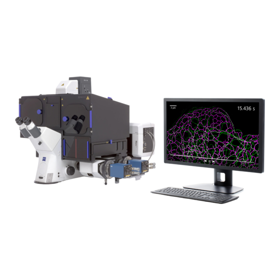
Zeiss ELYRA 7 Super-resolution Microscope Manuals
Manuals and User Guides for Zeiss ELYRA 7 Super-resolution Microscope. We have
1
Zeiss ELYRA 7 Super-resolution Microscope manual available for free PDF download: Operating Manual
Zeiss ELYRA 7 Operating Manual (488 pages)
Brand:
Zeiss
| Category:
Medical Equipment
| Size: 50 MB
Table of Contents
Table of Contents
5
Software and Hardware Options
6
Purpose
11
Software
11
Convention for the Text in this Manual
11
ZEN Configuration Tool
11
Backup
12
Software Operation
12
Hardware Aspects
13
Principles of Superresolution Microscopy (ELYRA)
13
Principle of Structured Illumination Microscopy (SIM)
13
Principle of Photo Activated Localization Microscopy (SMLM)
15
Optical Diagram of ELYRA (Schematic)
17
Performance and Features of ELYRA
19
Microscope Equipment of ELYRA Systems
20
Stage Leveling Using X-Cite or Laser in Fluorescence
20
Computer Hardware and Software
22
Startup and Shutdown of the System
23
Startup of the System
23
Starting ZEN
24
Shutdown Procedure
27
Exiting ZEN Software
27
Switching System Power off
28
Introduction to the Software Application Layout
29
Overview on the Screen Layout
29
Introduction to ZEN
30
Function Elements
33
Application Bar
35
Menu Bar
35
File
37
Maintain
38
Camera
38
Hardware Administrator
38
Test Grid
38
Remote Access Settings
39
Help - about
40
Main Toolbar
41
Status Bar
43
Left Tool Area
44
Tool Groups and Tools
46
Context Menu of the Left Tool Area
47
Center Screen Area
48
Overview of the Center Screen Area
48
Context Menu of the Center Screen Area
50
Right Tool Area
51
Left Tool Area and Hardware Control Tools
53
Locate Tab
54
Microscope Control Tool for Axio Observer
56
Incubation Tool
62
Acquisition Tab
63
Configuration Functions
64
Action Buttons
66
Multidimensional Acquisition Settings
68
Setup Manager - Laser
70
Setup Manager - Imaging Setup
71
Sequential Laser Switching
72
Beam Path Configuration
75
Camera Selection
77
Tube Lens Selection
79
Filter Cube Selection
79
Objective Selection
79
HAL Control
80
Epi Illumination Control
80
Aperture Control
80
Power Density Control
81
Grating Selection
81
Collimator Setting
82
TIRF Angle Setting
82
3D-PALM Slider
85
Acquisition Parameter - Experiment Designer
86
Acquisition Parameter - Acquisition Mode
89
Objective Selection
89
Format and Trigger Selection
90
Acquisition Parameter - Incubation
91
Averaging and Color Depth Selection
91
Acquisition Parameter - Online Processing Options
92
Online Processing SMLM
92
Control Activation Power
93
Acquisition Parameter - Channels
94
Defining Channels and Tracks
96
Ratio Channels
100
Track Control
101
Display Options
103
Acquisition Parameter - Focus
103
Acquisition Parameter - Definite Focus
105
Focus Devices and Strategies for Widefield Imaging Modes
115
Acquisition Parameter - Stage
116
Acquisition Parameter - Shuttle and Find
117
Acquisition Parameter - Regions
124
Multidimensional Acquisition - Z-Stack
128
Performing a Z-Stack Using First/Last Mode
129
Performing a Z-Stack Using Center Mode
131
Performing a Z-Stack Using a Z-Piezo
133
Correction
134
Multidimensional Acquisition - Tile Scan
134
Scan Overview Image for Tiling
137
Multidimensional Acquisition - Time Series
138
Interval Time
139
Marker
141
Start
143
End
145
Multidimensional Acquisition - Positions
146
Position List
146
Sample Carrier
147
Lowering Objective When Moving the Stage
150
Scan Overview Image
150
Multidimensional Acquisition - Information on Experiment
151
Multidimensional Acquisition - Streaming and Auto Save
151
Streaming Tool
152
Auto Save Tool
154
Processing Tab
156
General Structure of the Processing Tab
156
Processing - Airyscan Processing
160
Processing - Maximum Intensity Projection
164
Processing - Color-Coded Projection
165
Processing - Image Calculator
166
Processing - Average
168
Processing - Filter
169
Processing - Linear Unmixing
172
Processing - Ion Concentration
177
Single Wavelength Dyes - Offline Calibration
179
Ratiometric Dyes
180
Processing - Correlation
183
Processing - Modify Series
184
Processing - HDR-Imaging
185
Processing - Stitch
189
Processing - Localization Microscopy Tools
190
Localization Microscopy Tools - Convert to Image
191
Localization Microscopy Tools - Import SMLM Molecules
192
Localization Microscopy Tools - Localization Precision
193
Localization Microscopy Tools - Remove SMLM Outliers
194
Processing - Localization Microscopy
195
Processing - Particle Tracking
203
Processing - Experimental PSF
205
Processing - SIM Tools
209
SIM - Fourier Transform
210
SIM - Modulation Contrast
211
SIM - Stripe SIM
214
SIM - TIRF-Filtered
219
Processing - SIM
221
Processing with Default Setting
222
Processing with Adjust Settings
225
Processing - Channel Alignment
227
Processing - Lightsheet Processing
234
Dual Side Fusion
234
Multiview Processing
237
Online Multiview Processing
254
Processing - Copy
255
Processing - ICS
257
Detrend
257
Remove Structure
258
ICS Correlation
259
Map Filter
260
Nandb Analysis
261
Processing - Adjust
264
Burn in Brightness and Contrast
264
Interpolate Brightness and Contrast
265
Channel Shift
266
Shading Correction
267
Processing - Detrending
268
Maintain Tab
269
Maintain Toul Group - Dual Camera Alignment
270
Maintain Tool Group - Objectives
271
Detection Objective
271
Focus Speed
275
Parfocal Correction
275
Maintain Tool Group - Adjust PALM Slider
277
Options Tool Group - System Options
279
System Options - Camera
280
System Options - Elyra
281
System Options - Load Configurations
282
System Options - Reuse
282
System Options - Hardware
283
System Options - Image Display
283
System Options - Streaming
284
Options Tool Group - Experiment Designer
284
Options Tool Group - Service
284
Center Screen Area / Image Containers - Display and Image Analysis
285
Structure and Functional Concept of the Center Screen Area and the Image Display Container
285
General Structure
285
Container Configuration with the Container Context Menu
289
View
290
Dimensions
290
Display
293
Player
294
Graphics
295
Preview View
298
Split View
299
Gallery View
302
Ortho View
304
Ortho - Select Function
305
Ortho - Distance Function
306
Cut View
307
D View
308
View
312
Shadow Projection
312
Transparency Render Mode
315
Maximum Mode
316
Surface Render Mode
316
Mixed Render Mode
317
Clipping Planes
318
Flying Mode
322
Rendering Settings in Visart
322
Series
324
Interactive Measurements
326
Settings
327
Options
328
View - Basic
329
Histogram View
330
Colocalization View
333
How a Scatter Diagram Is Generated
335
Quantitative Colocalization Parameters Shown in the Data Table
336
Colocalization Coefficients
336
Overlap Coefficient, Overlap Coefficient after Manders
337
Profile View
337
Shuttle and Find View
340
Mean of ROI: Additional View Type for Time Series
343
Kinetic / FRAP View: Additional View Type for Time Series
347
Lambda Coded: Additional View Types for Lambda Mode
353
Unmixing View
354
Automatic Component Extraction
357
FRET View
358
Tools in the FRET View Options Control Block for Acceptor Photobleaching
360
Tools in the FRET View Options Control Block for Sensitized Emission
362
SMLM View
365
Dimensions View Control Tab
366
Display View Control Tab
371
Graphics View Control Tab
372
SMLM-Drift View Control Tab
373
Fiducial Based Drift Correction
375
Model-Based Drift Correction
382
SMLM-Grouping View Control Tab
387
SMLM-Statistics View Control Tab
389
SMLM-Filter View Control Tab
396
SMLM-Render View Control Tab
399
Rendering Data Sets
399
Visualizing Data Sets
406
6.19 Particle Tracking View
410
Particle Tracking Control View Tab
410
Polarization Imaging
412
Information View
413
Right Tool Area, Data Management and Storage
415
General
415
ZEN File Browser
416
Gallery View of the ZEN File Browser
419
Form View of the ZEN File Browser
421
Images and Documents Panel
423
Opening of Files Via the "Open" Command in the File Menu
426
Save
426
Export of Images
427
Index
428
VBA Version
434
Macros
434
Macro Language
434
Macro Control
435
Edit Macro Function
435
Assign Macro to the Macro List
438
Editing and Debugging of Macros
439
Overview of Available Macros
440
Sample Macros
441
Excitationfingerprinting
441
Concatenateimages
444
Object Browser
445
Glossary
446
Programming with the VBA-GUI Interface
447
Introduction
447
Structure of a VBA-GUI Command
448
Recording a Macro
448
Selecting and Managing Image Documents
449
Technical Note
449
Example Macros
450
Access to Images
450
Backup Recording Settings
450
Event Handling
450
Export, Save and Load
450
Vba-Gui Api
450
System Maintenance Tool
452
Calibration Objective
452
Starting the Maintenance Tool
453
General Operation of the Maintenance Tool
455
Software
462
Hardware
464
Courses on "How to Operate the System in an Optimized Way
465
Piezo Focusing Device
466
Specification of Trigger-Interface
468
Level-Adjustable Sample Holder with Insert for Piezo Stage
473
Level-Adjustable Piezo Stage Insert
473
Stage Leveling Using X-Cite in Reflection
478
Stage Leveling Using X-Cite in Reflection and Two Glas Plates
480
Liquid Cooling System of ELYRA
481
Autocorr Objectives - Installation
482
ZEISS Microscopes Networked
483
About the Philosophy of Our ZEISS Microscope System Computers
483
Microscope System Computers and Software
483
Compatibility and Feature List of Components
484
Networking of ZEISS Microscopy Computers
484
Solution 1: Peer to Peer
485
Solution 2A: Storage Shielded
485
Solution 2B: Storage Open
486
Solution 3: Dual Storage
486
ZEISS Microscopes Networked Solution 4: USB & Wifi
487
Advertisement
Advertisement
Related Products
Zeiss PLEX Elite 9000
Zeiss EyeMag Light II
Zeiss ELYRA
Zeiss AxioCam ERc 5s
Zeiss EVO
Zeiss EM 10 A
Zeiss EM 10 B
Zeiss EVO Series
Zeiss 000000-1087-851
Zeiss 000000-1103-022
Zeiss Categories
Microscope
Medical Equipment
Binoculars
Measuring Instruments
Laboratory Equipment
More Zeiss Manuals
Login
Sign In
OR
Sign in with Facebook
Sign in with Google
Upload manual
Upload from disk
Upload from URL