Sign In
Upload
Manuals
Brands
GE Manuals
Measuring Instruments
Precision 500D
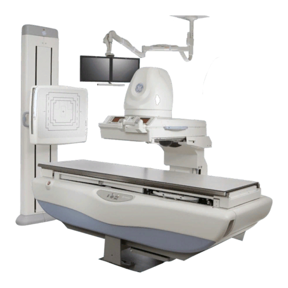
GE Precision 500D Manuals
Manuals and User Guides for GE Precision 500D. We have
1
GE Precision 500D manual available for free PDF download: Operator's Manual
GE Precision 500D Operator's Manual (323 pages)
Brand:
GE
| Category:
Measuring Instruments
| Size: 10.65 MB
Table of Contents
Table of Contents
5
Chapter 1 About this Guide
17
Purpose of this Guide
17
Prerequisite Skills
17
Safety Notices
17
Disposal of Waste
18
Chapter 2 Safety and Regulatory
19
Introduction
19
What Do I Need to Know about
19
X-Ray Protection
19
Certified Electrical Contractor Statement
19
Damage in Transportation
19
Regulatory Requirements
19
Patient Positioning and Control
20
Unsafe Table Region
20
Patient Grasp Handles
20
Safety Harness
20
Reciprocating Bucky
20
Compression Device
20
Myelographic Operation
20
Myelographic Boots (Optional)
20
Cone Operation
20
Travel and Counterbalancing
41
Bucky Grids
41
Oscillating Grid
41
Single High Line/Rate Grid
41
Bucky Grid Alignment
41
Patient Step
42
Foot Rest
43
Shoulder Rest
43
Lead Apron
46
Emergency Stop Button
47
Grounding Kit
48
Purpose
48
Description
48
Instructions
48
Safety Summary
49
Chapter 3 About the P500D
53
Introduction
53
Environmental Requirements
53
Image Storage Capacity
53
Options
54
DICOM 3.0 Interface Option
54
Bar Code Scanner Option
54
International Keyboard Option
54
Power Up/Power down
55
Overview
55
Mouse
57
Radiology Control Interface Module (RCIM)
57
X-Ray Tube Selection
59
Chapter 4 Getting Started
63
Introduction
63
Startup/Shutdown Procedures
63
System Startup
63
Tube Warmup
63
System Shutdown
63
Emergency Stop and System Recovery
63
Emergency Stop Recovery
63
System Recovery
63
Procedure for Tube Warm up
64
Bar Code Scanner Option
64
International Keyboard Option
65
Chapter 5 Integrated User Interface
67
Introduction
67
Power-On
67
Tube Warm-Up Screen
69
Add/Select Patient Screen
71
Start Exam
74
Start Exam Scenario
76
Fluoro Acquisition Screen
81
Angio Acquisition Screen (Option)
83
Video Presentation
84
Fluoro Time
84
View/Film
85
Filming
86
Radiographic Exposure / Review Multitasking
87
Archive to Local CD-RW (OPTION)
88
Emergency Recovery
90
Edit Protocols
91
Protocol Editor
92
Protocols
93
Parameters for Tube 1 (Fluoro) Protocol Step
93
Parameters for Tube 2 (RAD) Protocol Step
94
Protocol Categories
95
Protocols
97
Protocol Steps
104
Retrieve
118
Back up
119
System Configuration Changes
119
User Preferences
120
Default Dose - Fl/Rec
120
Default Pediatric Grid
120
DSA Image Invert
120
Record Image Invert
121
Fluoro Image Invert
121
Auto Send
121
FNR Level
121
Record Edge Enhancement
121
Fluoro Edge Enhancement
121
Printer Destination
121
Auto Send Destination
122
Images Tagged
122
Service User Interface / Effect on IUI
122
Dose Measurement
122
Pulsed Fluoro
122
DSA Package
122
DICOM Ethernet
122
DICOM Print
122
DICOM Worklist
122
Pediatric Option
122
Tone Alert / %HU
123
Number of Tubes
123
Ots
123
Wallstand Location
123
Fluoro Rates
123
Spot Rates
123
Max Record Rate
123
System Log on
123
In-Room Remote Keypad (Option)
126
IR Remote Keypad Commands
127
Chapter 6 Digital Imaging System
129
Menu Displays
129
Disk Space Indicators
130
Edit Series Title
131
Pateint List Screen
133
Auto Send to Reference Image Monitor (with Reference Monitor Installed)
135
Selecting a Patient File - Review Mode
135
Selecting Review Mode
135
Image Acqusition
136
Acquisition Rates
137
Image Subtraction
138
Reregistration of Subtracted Images
139
Image Stacking (and Max/Min Opacification Option)
142
Conrast / Window
146
Brightness / Level
147
Image Invert
147
Default
147
Zoom
148
Edge Enhancement
148
Carry over Processing Features
148
Carry over Digital Shutters
149
Multiple Image Display Formats
149
Delete Image
150
Annotations & Pointers
151
Printing an Image
153
Stenosis Measurment Features and Steps
153
Size
155
Auto Boundary
161
Dicom Worklist (Requires Dicom Option)
163
Chapter 7 Table
167
Introduction
167
Overview
167
Identification and Compliance Plates
168
Travel Specifications
171
Digital Operator Control Panel
172
Types of Radiological Examinations in Significant Zone of Occupancy
173
Operator Controls and Displays
180
Operator Control Locations
180
Imaging Device Operator Controls and Displays - Overview
182
Main Operator Console Screen -Basic Controls and Displays
183
Main Operator Console Screen -Optional Controls and Displays
187
Auxiliary Digital Operator Console
189
Digital Fluoro Presentation Operator Console
191
Digital Multi-Function Operatorcontrol Group
192
Precision 500D Table Operator Controls and Displays
195
System Specifications
198
Precision 500D Table Specifications
198
Precision 500D Table Electrical Requirements
199
Precision 500D Tables, Tubes and Collimators
200
X-Ray Tube Specification
200
Operation
201
Parking (Docking) the Imaging Device Carriage
201
Footswitch
203
Taking Digital Exposures
204
Using the Prep/Record Bar
204
Using the Integrated Fluoro/Expose Switch
204
Equipment Positioning for Table Bucky Procedures
205
Chapter 8 Ots Radiographic Suspension
208
Introduction
208
OTS (Overhead Tube Suspension)
208
Overhead Rail System
210
Telescopic Column and Carriage
211
Tube Support Rotation
212
X-Ray Tube Angulation
213
Over Head Ceiling Suspension User Interface
214
Key Switch on OTS Console
216
Automatic Collimator
216
Locking Lever
218
Display on the Collimator
218
Bottom View of the Collimator
219
Linear LASER Light Localizer
219
Rear View of Collimator
220
Collimator Lamp Changing
220
Changing Lamps on the Collimator (Model 2266999)
220
Changing Lamps on the Collimator (Model 5234954)
222
Rotating the Collimator 90° Around the Vertical Axis
226
Specifications
227
Chapter 9 Sg120 Vertical Bucky Stand
230
Introduction
230
Parts of the Equipment
231
Column Assembly
231
Bucky Assembly
232
Front Panel
232
Vertical Lock Handle
232
Applications of Sg80/120
232
Operation
232
Cassette Loading
233
Cassette Removal
235
AEC Detector Areas
235
Alignment
235
Frontal Panel
236
SG120-Specific Funtionalities
236
Bucky Rotation
236
Bucky Angulation
237
Periodic Maintenance
238
Maintenance Information
238
Labeling
239
Specifications
240
Electrical Requirements
240
Product Dimensions
240
Crate Dimensions
240
SG120 Specifications
241
Wall Stand Bucky
241
Chapter 10 Vertical Bucky Stand
242
Introduction
242
Safe Operation Precautions
244
Operation
244
Vertical Positioning
246
Bucky Rotation
247
Bucky Angulation
248
Hinged Front Panel
248
Manual Cassette Tray
249
AEC Detector Areas
250
Alignment for Automatic Collimation
251
Specifications
252
General Dimensions & Weight
252
Chapter 11 Jedi Generator
256
Introduction
256
Radiation Protection
256
Generator Cabinet Specifications
258
General Input Power Specifications
258
X-Ray Interlock Systems
263
Chapter 12 Accessories
264
Patient Step
264
Foot Rest
265
Optional Table Accessories
266
Shoulder Rest
266
Compression Device
266
Arm Board (Optional)
268
Knee Crutches (Optional)
269
Vertical Cassette Holder (Optional)
270
Maxiray™ 100 Tube Units
272
Application
272
Features
272
Tube Housing
273
Tube Insert
274
Thermal Ratings
274
Radiographic Ratings
275
Fluoroscopic Ratings
275
Weights (Approx
275
Related Publications
275
Warranty
276
User Service and Maintenance
276
Listings
276
Radiographic Tube Ratings
279
Warm-Up Procedure
279
12.5° (218 Mrad) 3Ø, Low Speed, 50 Hz, 2850 Rpm
280
12.5° (218 Mrad) 3Ø, Low Speed, 60 Hz, 3400 Rpm
281
12.5° (218 Mrad) 3Ø, HIGH SPEED, 50/60 Hz, 10,000 Rpm
286
Mammography Ratings
289
12.5° (218 Mrad) 3Ø, 50/60 Hz, 2850/3400 RPM
289
Rapid Film Sequence Ratings
290
0.6, 1.0, 1.25 12.5_ (218 Mrad) 1 Phase, 10,000 Rpm, 50/60 Hz
290
Planned Maintenance
296
Periodic Maintenance
296
Qualified Service
296
User Service and Maintenance
296
Image Quality Testing
297
How to Perform the Quality Assurance Process
299
General
305
Periodic Maintenance Schedule
306
Service
306
User Cleaning and Disinfecting
306
HHS Testing (US Only
307
Recycling
307
Glossary/Terms
308
Operating Modes
308
System Terms
309
Digital Imaging System Terms
310
Symbols
310
Abbrevations
314
Modes of Operation
316
Fluoroscopy Mode
316
Description
316
System Controlled Technique Factors
318
How to Engage/ Disengage
318
Description
318
Example of Specific Clinical Procedures
319
How Mode Should be Used
319
Description
319
System Controlled Technique Factors
319
How to Engage/ Disengage
319
How to Recognize When this Mode Is Selected
319
Advertisement
Advertisement
Related Products
GE Sievers 500 RL
GE 5176/200
GE MAC 5 A4
GE MAC 5 A5
GE 500A436P006
GE keysafe consumer 500
GE 500PAR64/NSP
GE 500PAR64/MFL
GE 500PAR64/WFL
GE 5000 IWS
GE Categories
Refrigerator
Ranges
Microwave Oven
Air Conditioner
Dishwasher
More GE Manuals
Login
Sign In
OR
Sign in with Facebook
Sign in with Google
Upload manual
Upload from disk
Upload from URL