Table of Contents
Advertisement
Quick Links
Advertisement
Chapters
Table of Contents

Related Manuals for Zeiss Axioplan 2


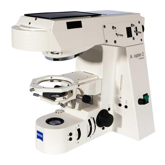
Summary of Contents for Zeiss Axioplan 2
- Page 1 Axioplan 2 imaging and Axiophot 2 Universal Microscopes Operating Manual...
- Page 2 It is not permitted to pass on or copy this document, or to use or forward its contents unless prior consent has been provided. If this regulation is not observed, damages will be claimed. All patenting rights are reserved. For further details, please contact: Carl Zeiss Microscopy D 07740 Jena...
-
Page 3: Table Of Contents
Contents Notes on the safe operation of the instrument ........General . - Page 4 Contents Care, Maintenance ............. . General .
-
Page 5: Notes On The Safe Operation Of The Instrument
Notes on the safe operation of the instrument The Axioplan 2 imaging Universal Microscope has been de Specimens hazardous to health! signed and tested in compliance with EN 61010, part 1 (DIN The Axioplan 2 imaging is not equipped with any... -
Page 6: General
Microscope Components chapter must only be performed by persons expressly authorized by us to do so. The Axioplan 2 imaging is a precise optical instrument which may be impaired in its function or even damaged by inexpert handling. - Page 7 Axioplan 2 imaging. Not all parts described there field of view number must be present on the Axioplan 2 imaging. However, if you system integration are considering adding some of the components to your single lens reflex configuration, the corresponding catalog numbers are listed.
-
Page 8: Purpose
Purpose The Axioplan 2 imaging has been designed as a universal mi Installation conditions croscope and can be used for all areas of light microscopy, pro Dust free environment vided it is configured and equipped appropriately. Depending Maximum relative air humidity 85 %... -
Page 9: Overview And Connections
Axioplan 2 imaging configuration. Setting and adjustment of certain components and modules of the Axioplan 2 imaging sometimes require special tools which are included in the delivery package of the microscope. This particularly concerns the SW 3 ball headed screwdriver and the SW 1.5 screwdriver for Allen head screws. -
Page 10: Putting Into Operation
The Axioplan 2 imaging is handed over to you in a state which will enable the users to attach all the items listed in the Microscope Components chapter themselves. -
Page 11: Fig. 2 Axioplan 2 Imaging (Instrument Back)
Putting into Operation Setting up the microscope The Axioplan 2 imaging microscope features an integrated wide range power unit and can be connected to line voltages ranging from 100...240 V AC, 50 ... 60 Hz. The wide range power unit sets itself automatically to the appropriate line voltage. -
Page 12: Stand
The following stand types exist for the Axioplan 2 imaging: The following description of stands starts with the manual Manual type model. On this basis, only the differences and additional This stand can only be operated manually. -
Page 13: Manual Stand
For optical status checking, the switch lights up in green in position I (for defects see chapter Care, Maintenance). When the Axioplan 2 imaging is switched on, not only the HAL 100 microscope lamp is supplied, but also the Axiophot 2 Photo module, if mounted, and the motorized internal components. - Page 14 Stand 7, 8 Filter wheels for transmitted light Luminous field diaphragm Two rotatable filter wheels (filter magazine) with 4 positions Wheel for the continuous setting of the aperture of the each are equipped with different filters; two different models luminous field diaphragm (transmitted light). are available.
-
Page 15: Fig. 4 Stage Mounting
Pushed in = open diaphragm Pulled out = closed diaphragm. Manual focusing drive The universal microscope Axioplan 2 imaging is focused via coaxial drives on both sides of the stand. Coarse drive (large knob on the inside): 1 revolution = approx. 2 mm, 1 increment = approx. 2 mm. -
Page 16: Fig. 5 Height Adjustment Of The Stage Carrier
(Fig. 6) The condenser carrier is screwed to the stage carrier. All the condensers available for the Axioplan 2 imaging are attached to the condenser carrier. The height of the carrier can be adjusted on both sides and permits the centering of the inserted condensers. -
Page 17: Fig. 7 Objective Nosepiece
Stand Objective nosepiece The objective nosepiece is used for mounting the objectives and changing them quickly. Depending on the application, the customer can choose from nine different nosepieces. Note: Nosepiece positions which are not being used must be covered with dust caps. The nosepieces for polarization is equipped with centering screws (SW 1.5) for the centration of the objectives. -
Page 18: Fig. 9 Back Of Manual Stand, Also See Fig
Stand Back of manual stand Key to Fig. 9 (back of manual stand) Connecting tube for microscope lamps incident light with locking screw for dovetails to fix the lamps Ventilation slots (always keep clear; minimum distance: 15 cm) Sockets for instrument plug with integrated compart ment for instrument fuses (change of fuses →... -
Page 19: Stand E/Stand Mot
Coarse/fine drive of focusing nance). Rapid stage lowering CHANGE (on both sides of the stand) When the Axioplan 2 imaging is switched on, not only the Rapid stage lowering VIEW (on both sides of the stand) Socket for connection of motorized condensers... - Page 20 Using the keys (4) and (5) attached to the right and left of the Note: When the Axioplan 2 imaging is switched on, stand you can operate two of the following motorized instrument initialization is performed to allow recognition of components: the connected modules.
-
Page 21: Fig. 11 Back Of Motorized Stand
CAN bus. Normally, the signals from a PC which must be equipped with an appropriate driver board (CAN bus inter face) are transferred to the Axioplan 2 imaging and read off from there. The second connector also allows cascading. This enables several series switched instruments to be activated by a PC. - Page 22 Coded microscope parts The intermediate tube (coded or motorized) installed is con nected to the microscope via this connector using the cable Various modules of the Axioplan 2 imaging and some supplied. additional microscope components can be coded. Codings is beneficial when the microscope software is used.
-
Page 23: Light Manager
The KÖHLER illumination setting procedure should already have been performed prior to the inital use of your Axioplan 2 imaging (→ page 74). You should also make an occasional check to ensure that this setting has not changed. The factory programmed setting of the light manager... -
Page 24: Microscope Control Software Axiovision Control
Microscope control software AxioVision Control What does AxioVision Control offer? AxioVision Control permits the remote control of the following Carl Zeiss microscopes from your PC: Axioplan 2 imaging / Axiophot 2 Axiovert 100 M Axioskop 2 Remote control with AxioVision Control provides the... -
Page 25: Electrical Connections
Microscope control software AxioVision Control Electrical connections Microscope The coupling of the Axioplan 2 with the PC or the notebook is performed via the RS 232 C serial interface (Fig. 13/1) using a connecting cable. Axiophot 2 photo module The electrical connections are on the back of the Axiophot 2... -
Page 26: System Overview
Achromatic aplanatic condenser 1.4 H Ph DIC, (445325 9901 000) Brightfield insert (445453 0000 000) (445467 0000 000) UV condenser 0.6 Pol for Axioplan 2 imaging Pol (445460 0000 000)) Achromatic condenser 0.8 H Achromatic condenser 0.8 H mot. (445443 0000 000) (445444 0000 000) Achromatic condenser 0.8 H D Ph mot. - Page 27 System Overview Stage carriers, Stages, Condenser carriers Specimen holder with spring clip R (453533 0000 000) Adapter for mineralogy specimen mount (453541 0000 000) Rotary, centerable mechanical stage 75x50 / 240_ R (453502 9904 000) Specimen holder A (000000 1046 521) Specimen holder A (453539 0000 000) Mechanical stage 75x50 R with...
- Page 28 Upper stand part of Axioplan 2 imaging (444053 0000 000) (000000 1050 687) Eyepiece Pl 16x/16 Br. foc. Upper stand part of Axioplan 2 imaging mot. (444054 0000 000) (000000 1050 688) Eyepiece E Pl 10x/25 Br. foc. (444234 0000 000) Binocular tube with Eyepiece E Pl 10x/23 Br.
-
Page 29: Fig. 24 Optovar Intermediate Tube
System Overview Intermediate tubes, Nosepieces, Compensators Optovar intermediate tube Intermediate zoom tube 1.0x ... 2,5x cod. Intermediate tube Pol 1.0x/1.25x/1.6x/2.0x/2.5x cod. (452180 0000 000) (with quartz depolarizer) (452175 0000 000) (452184 0000 000) Fluorescence protection screen (452163 0000 000) 7x nosepiece H W 0.8 (452130 0000 000) 6x nosepiece H DIC W 0.8 Compensator lambda 6x20... - Page 30 System Overview Intermediate tubes, Illumination, Analyzers Intermediate tube for image projection Intermediate assistant's tube 2x (452181 0000 000) (452179 0000 000) HAL 100 lamp housing with collector, lamp mount and heat reflecting filter path-deflecting mirror for (447219 0000 000) 2 illuminators (mot) (447131 9901 000) HBO 50 lamp housing with lamp mount...
- Page 31 System Overview Equipment for Documentation Film cassette 35 mm Mot (456070 0000 000) Camera attachment TV adapter for Axiophot 2 M 4"x5" (452230 0000 000) (MC 80 DX/MC 200 CHIP) TV adapter 60 C 2/3" 1.0x (456060 0000 000) (456105) 0000 000 4"x5"...
- Page 32 (452142 0000 000) Intermediate tube Pol (with quartz depolarizer) (452184 0000 000) Upper stand part of Axioplan 2 imaging (452101 0000 000) 6x nosepiece Pol W 0.8 cod. (452139 0000 000) up to 6 objectives Pol W 0.8 (1x with intermediate ring 444910 0000 000) or 5x W 0.8 + 1 objective HD DIC M 27 and 1 DIC slider as per pricelist...
-
Page 33: Microscope Components
The Axioplan 2 imaging incorporates the System Integration Microscope illuminators (SI) principle. This SI design allows the Axioplan 2 imaging to be configured or converted to meet various requirements. The following part of the manual describes all of the compo... -
Page 34: Microscope Illuminators
Lamp housing 447219 0000 000 halogen illuminator light (with collector and heat reflecting filter, All stands power unit integrated in Axioplan 2 ima ging) 380079 9540 000 12 V/100 W halogen lamp N HBO 103 Epi fluorescence N HBO 103 lamp housing... - Page 35 Microscope Components General notes Thermally sensitive fluorescence filters! Risk of damage to instrument! Fluorescence filters are sensitive to the thermal Before you drape the dust cover over the micro radiation of the microscope lamp. scope, switch off the microscope and the external Therefore, never remove the heat reflecting filter power supply of the microscope illuminator.
-
Page 36: Fig. 15 Hal Microscope Illuminator
Pol applications. The filter is installed in illumination tube using a retaining ring. Electrical supply The microscope Axioplan 2 imaging features a wide-range power unit and can be connected to line voltages ranging from 100 ... 240 V AC, 50 ... 60 Hz. The wide-range power Fig. -
Page 37: Fig. 17 Lamp Housing And Lamp Mount
(2), (3) and (4). Coarse adjustment Remove microscope illuminator. Switch on Axioplan 2 imaging. Direct light ray towards a projection surface (wall, paper) at least 3 m away. Adjust screw (2) until the lamp filament is imaged sharply on the projection surface. -
Page 38: Tubes
Microscope Components Tubes Overview Specification Splitting Application/Combination Cat. no. Binocular phototube 100 : 0 3 switch positions 452143 0000 000 with sliding prism 30_/25 50 : 50 1 eyepiece shutter as light 0 : 100 shutter with 50:50 ratio Binocular phototube 100 : 0 3 switch positions and 452145 0000 000... -
Page 39: Tubes
Microscope Components General All binocular tubes have a viewing angle of 30_. The inter pupillary distance of can be set between 55 and 75 mm by pressing the two halves of the tube together or pulling them apart. Note: If intermediate tubes are used, the tube lens (Fig. -
Page 40: Eyepieces
Microscope Components Eyepieces Overview Specification Application/Combination Cat. no. Eyepieces Eyepiece E Pl 10x/25 Br. foc. Standard eyepiece of research category for large field of view 25 mm 444234 0000 000 Eyepiece E Pl 10x/23 Br.foc. High performance aspheric eyepiece for Epiplan objectives or for use of 444235 0000 000 intermediate tube Pol in field of view Eyepiece Pl 16x/16 Br.foc. - Page 41 Use of reticles Eyepieces featuring a red dot allow the use of reticles. Make sure that the reticle always faces the field stop. Reticles should be inserted by the Zeiss servicing staff in dust free conditions. 11 12 B 40 042 e 09/99...
-
Page 42: Intermediate Tubes
Crossline or iris stop can be inserted in field of 452184 0000 000 view, focusing of Bertrand optics for axial image observation (conoscopy) Removal of individual object details form field of view using iris diaphragm, for Axioplan 2 imaging Pol / Axiophot 2 Pol B 40 042 e 09/99... -
Page 43: Fig. 22 Mounting Of Intermediate Tube
Microscope Components General Intermediate tubes are always attached to the upper part of the stand. To do this, the binocular tube must first be removed and its tube lens removed. Note: Use the cover of the tube lens container to remove the tube lens (4). -
Page 44: Fig. 25 Zoom Intermediate Tube
Microscope Components Optovar intermediate tube (Fig. 24) The Optovar intermediate tube additionally allows a conve nient magnification change in the steps 1.0x, 1.25x, 1.6x, 2.0x and 2.5x. You can read the magnification factors at the projecting disk (1) on the right hand side. To set a different magnification factor, turn the disk to the appropriate click stop position. -
Page 45: Reflector Modules
Microscope Components Reflector modules Overview Specification Application/combination Cat. no. Reflector module H Brightfield, incident light 000000 1046 274 Reflector module D Darkfield, incident light 000000 1046 276 Reflector module DIC Differential Interference contrast, 000000 1046 277 incident light Reflector module DIC Red I Differential interference contrast, 000000 1046 278 incident light,... -
Page 46: Reflector Modules
Microscope Components Attachment of reflector modules Usually, the reflector turret is correctly equipped in the factory. The reflector turret features eight reflector positions and can be equipped as required. To retrofit or change the equipment, proceed as follows (see Fig. 27): Swing the required opening of the reflector turret (1) into the beam path (see window 2). - Page 47 Ideally, the filters and the beam splitter should be assembled by Carl Zeiss service staff. 11 12 B 40 042 e 09/99...
-
Page 48: Objectives
Microscope Components Objectives General Information All the objectives of the Axioplan 2 imaging incorporate the principle of ICS optics, i.e. they project the image to infinity. Only the tube lens produces the intermediate image which can be viewed via the eyepieces... - Page 49 Microscope Components Labelling of objectives Explanation Color Meaning The color of the labelling black Standard marks the contrasting k th t ti Pol/DIC technique intended for this technique intended for this green Ph 1, 2, 3 objective Color coding of the magnifi black 1.25x cation...
-
Page 50: Stages
Microscope Components Stages Overview Specification Comments Cat. no. Stages Mechanical stage 75x50 R with ergo drive stage focusing on right; ab 000000 1063 835 rasion proof coating; height adjustment and adjustment of smoothness of drive controls Mechanical stage 75x50 L with ergo drive stage focusing on left;... - Page 51 Microscope Components Specification Comments Cat. no. Specimen holders Specimen holder with spring clip R 453533 0000 000 Special specimen holder with special fixing device, 453538 0000 000 for mounting 2 specimens Specimen holder for 000000 1046 521 mechanical stages 000000 1063 835/836 Specimen holder A 000000 1070 588 Specimen holder A...
-
Page 52: Fig. 30 Mechanical Stage
Microscope Components General Information (Fig. 30) Mechanical stages can be rotated and centered. They can be moved in x (25 mm) and y (50 mm). There is a stage drive on the right which also moves when the stage is rotated. Attachment/removal and centration of stages (→... - Page 53 Microscope Components Rotary stage Pol The rotary stage Pol (7), used with polarizing microscopes, is a rotatable and centerable stage with an object guide (1) for a travel range of 45 mm in x and 25 mm in y. Object coordinates can be determined with an accuracy of 0.1 mm using graduations and verniers.
-
Page 54: Condensers For Transmitted Light
Microscope Components Condensers for Transmitted Light Overview Specification Comments Cat.no. highest flexibility and resolution; 000000 1035 632 Achromatic aplanatic universal front lens swung in: condenser 0.9 for objectives 10x...100x front lens swung out: 445436 0000 000 Achromatic aplanatic universal for objectives 1.5x ...10x condenser 0.9 H D Ph brightfield insert or turret disk for contrasting elements with 5 or 7 positions... -
Page 55: Condensers
Microscope Components Specification Comments Cat. no. Achromatic condenser like 445445 0000 000, but motorized 445446 0000 000 0.8 H D Ph mot. UD condensor 0.6 Primarily intended for use with UD stage; also 445460 0000 000 suitable as LD condenser 0.4 for brightfield and polarization Achromatic dual brightfield condenser for objectives 1.25x ... - Page 56 Microscope Components Condenser carrier (Fig. 33) The condenser you selected is contained in the condenser car rier. These are its control elements: Height adjustment on both sides (max. 34 mm) (2). The ease of motion is factory adjusted (to be changed only by the service staff).
- Page 57 Microscope Components Changing the phase stops or darkfield stops in the universal condenser (if required) (Fig. 35) Tighten both centering screws (1) until stop using SW 1.5 ball headed screwdriver. To loosen the cover on the condenser underside (2), loosen both grub screws using SW 2 screwdriver and remove the cover.
- Page 58 Microscope Components Achromatic aplanatic condenser 1.4 The achromatic aplanatic condenser 1.4 is equipped with fixed front lens and turret disk. The turret disk contains 6 positions and accepts max. 2 DIC prisms, 2 phase stops and 1 darkfield diaphragm. A position for brightfield with iris has been provided.
-
Page 59: Analyzers, Compensators, Auxiliary Objects And Dic Sliders
Microscope Components Analyzers, Compensators, Auxiliary Objects and DIC Sliders Overview Specification Cat. no. Analyzers Fixed analyzer slider 453657 0000 000 Slider with analyzer and lambda plate, rotary 453663 0000 000 in preparation Rotary analyzer 453662 0000 000 Slider for quartz depolarizer 453659 0000 000 Quartz depolarizer d = 32 mm 453653 0000 000... - Page 60 Microscope Components General Information (Fig. 38) The components listed in the overview on page 57 are used to regulate the optical contrast in the microscope and to ad just the auxiliary devices necessary to regulate the contrast. Compensators are required for contrast enhancement and measurements.
-
Page 61: Axiophot 2 Photo Module
Microscope Components Axiophot 2 Photo module Overview 4x5" large format camera 35 mm Mot film cassette (right) Axiophot 2 Photo module Port for TV camera Fig. 39 Axiophot 2 Photo module 11 12 B 40 042 e 09/99... - Page 62 Microscope Components General Information 35 mm Mot DX film cassette The Axiophot 2 Photo module turns your microscope into a Removing the cassette universal and completely new photomicroscope. Motorized Use your two hands to grasp the film cassette on the right control elements and operation via notebook (or PC) only and left, press unlocking button (2) until stop using your makes work with the Axiophot 2 methodically richer and at...
- Page 63 Microscope Components Attaching the cassette Hold film cassette with both hands on the right and left of the camera body. The unlocking button (2) shows to the user and must not be pressed during attachment. Hold the film cassette parallel to the upper edge of the camera body and insert it in the basic body until the unlocking button (2) jumps out.
- Page 64 Microscope Components 4x5" Large Format Camera (Fig. 41) Large format groundglass and cassette mount (5). Cassettes for universal camera backs are slid behind the large format groundglass which can be lifted with lever (1). To take off the groundglass: Press (2) and move it to the right; mount accordingly. The groundglass can be used for microprojection for small discussion groups, provided there is sufficient light: This requires that you perform the following steps in the...
-
Page 65: Microscopy Techniques
Microscopy Techniques You should now see spots of light (the exit pupils) behind the Transmitted light brightfield eyepieces. If you are working with a binocular phototube, all of the light will be directed to the binocular tube if the push To set the incident light illumination in accordance with the rod is slid in all the way. -
Page 66: Transmitted Light Darkfield
Microscopy Techniques Transmitted light darkfield Plan Neofluar Plan Apochromat Illumination 10x/0.30 10x/0.32 Ph stop 3w0.44 20x/0.50 20x/0.75 darkfield stop To examine small and extremely small specimens and 40x/0.75 D 0.76 0.90 specimen features such as treponemas, spirochaetae, fla 100x/1.3 oil iris 40x/1.0 oil iris dry darkfield condenser gella, bacteria, etc. -
Page 67: Darkfield Illumination In Low Magnifications
Microscopy Techniques Darkfield illumination in low magnifications Darkfield illuminator (2) 445314 9901 (for low magnifications from 1.25x to 20x) Glare hazard! When switching from darkfield to brightfield, the lamp brightness must be reduced under all cir CAUTION! cumstances. Setting the optimum darkfield illumination Insert illuminator (2) in lowered condenser carrier, ensur ing that screw (4) on ring dovetail engages with orienta tion notch (1). -
Page 68: Phase Contrast
Microscopy Techniques Phase contrast Use: These techniques are used for unstained specimens in partic ular in order to enhance their contrast. adjusted phase rings Additional equipment Objectives (1) designated Ph. These can also be used for brightfield. A condenser featuring Ph positions. Additional adjustment The phase rings in the various objectives are of different sizes and marked Ph1, Ph2 and Ph 3 on the objective (1). -
Page 69: Dic Differential Interference Contrast In Transmitted Light
Microscopy Techniques Additional adjustments DIC Differential interference contrast Like the 3 (or 2) Ph positions of the condenser, there are 3 in transmitted light (or 2) DIC positions, marked I, II, III, suitable for combination with the appropriately marked DIC sliders. Use: This permits the following combinations: For unstained specimens which are too thick for phase... - Page 70 Microscopy Techniques DIC setting using the SENARMONT polarizer The following must be taken into consideration when a SENARMONT polarizer, consisting of a carrier with l/4 plate (above) and a rotary polarizer (below), is used: Insert SENARMONT polarizer below the condenser until click stop.
-
Page 71: Transmitted Light Polarization Detection Of Birefringence
Microscopy Techniques Insert the analyzer (1) until it snaps in and the field of view Transmitted light polarization Detec is dark. tion of birefringence If you work with a measuring analyzer, adjust the measuring scale to 90° and secure it in position. If you use a ±10°... -
Page 72: Transmitted Light Polarization Determining The N
Microscopy Techniques Transmitted light polarization Determi ning the n direction of oscillation γ - 400 Fig. 48 Determining the n direction of oscillation using a synthetic fiber as an example γ' Summary The position of the two directions with the greatest (n ) and Compare interference colors (path differences) in the two di the smallest (n... -
Page 73: Transmitted Light Polarization Determining And Measuring Path Differences
Microscopy Techniques - activate the stage clickstops, Transmitted light polarization Determi - set a diagonal position displaying maximum brightness ning and measuring path differences at 455. Insert the compensator selected, move it out of its zero position and watch whether: The color chart only allows the rough estimation of path dif the interference colors become deeper, i.e. -
Page 74: Transmitted Light Polarization Determining The Optical Characteristics Of Crystals
Microscopy Techniques Close the field diaphragm (1) until the grain boundaries Transmitted light polarization Determi of the crystal selected are no longer visible. This ensures ning the optical characteristics of crystal that the interference figures of neighboring crystals are not superimposed on the interference figures of the crys tal to be examined. - Page 75 Miroscopy Techniques Biaxial crystals Note: If an optical axis of a biaxial crystal is parallel to the If biaxial crystals display a cross in conoscopic observation viewing direction, only one branch of the hyperbola will be which resolves into the two branches of a hyberpola, the visible in conoscopic viewing, with the vertex of this branch acute bisectrix (1st center line) is oriented in parallel to the lying in the center of the field of view.
-
Page 76: Incident Light Brightfield
Microscopy Techniques Swing the 10x objective (yellow ring) (9) on nosepiece, Incident light brightfield and the reflector module for brightfield on reflector turret (Fig. 51) (10) into the beam path. Check the 0 positions on the eyepiece scale (→ page 39). Note: In all incident light techniques, the compensators 6 x 20 (see also →... -
Page 77: Incident Light Darkfield
Microscopy Techniques Incident light darkfield Additional equipment Object features which heavily scatter light, such as scratches, cracks, pores or the surfaces of metal fractures light up bright in darkfield illumination. Ideal brightfield objects such as specular surfaces including features with different degrees of reflection remain completely dark, however. -
Page 78: Dic Differential Interference Contrast In Incident Light
Miroscopy Techniques Additional comments DIC Differential interference contrast in In DIC, contrast is caused by surface relief. With linear struc incident light tures, therefore, contrast is dependent on whether the orien tation of these structures is in the "light shadow" direction (very low contrast) or at right angles to it (maximum contrast). -
Page 79: Incident Light Polarization Detection Of Bireflection And Reflection Pleochroism
Microscopy Techniques Close the aperture diaphragm by 2/3 of its diameter by Incident light polarization Detection of pulling out pushrod (4) as far as required. bireflection and reflection pleochroism Your object will display bireflection if object features possess differences in brightness or color which change when the stage is turned. -
Page 80: Epi Fluorescence
Microscopy Techniques Switch on the HBO 103 or HBO 50 mercury vapor short Epi fluorescence arc lamp and allow it to heat up to its operating Additional equipment temperature for approx. 15 minutes. Select and switch on the required fluorescence filter Recommended: Plan Neofluar or Fluar objectives (UV combination (depending on the type of excitation) on the excitation) -
Page 81: Photomicrography With The Axiophot 2
Microscopy Techniques Films marked "professional" feature closer tolerances in Photomicrography with the Axiophot 2 sensitivity and color balance, i.e. homogeneous results are obtained. Always use DX coded films in their original Note: If your polarizing microscope (Axioiplan 2 imaging Pol, cartridges. - Page 82 Microscopy Techniques Correction of the color balance of color reversal films Data projection for 35 mm photography The color balance of a type of color reversal film can differ The data projected into the image may be poorly legible from batch to batch. against bright object structures.
- Page 83 Microscopy Techniques Fluorescence photography Exposure times and filters The following specialties apply compared to photomicro The Axiophot 2 Photo module covers the following longest graphy: exposure times for 35 mm film with exposure cor rection 0: The often low brightness requires long exposure times. Sensitivity of the film Longest exposure time Before taking the exposure, therefore select the option...
-
Page 84: Care, Maintenance
To remove stubborn dirt or fingerprints, use commercially available cloths for cleaning optics and eyeglass lenses. For moving your Axioplan 2 imaging to another location on your premises, ensure that all mobile parts are secured in position or transport them separately. Also protect your... -
Page 85: Technical Data
Technical Data Range Description Ambient conditions Room temperature +10 ...+ 35_C Humidity max. 75% at + 35_C Storage temperature - 40 ... + 70_C, humidity 10 ... 30% Weight depending on configuration used Safety Equipment class Degree of protection IP 20 Radio disturbance characteristics conforming to EN 55011 (Class B) Electromagnetic immunity... -
Page 86: Interface Description
The pin assignment of the RS 232 C interface is identical to CAN BUS that of the PC (see → Fig. 56). The lines in the cable connecting the Axioplan 2 imaging and The CAN bus (Controller Area Network Serial Communica the PC have been crossed. - Page 87 Interface Description Command structure (input format) Communication via CAN Protocol A command consists of a sequence of ASCII characters which is finished with Hex 0D (CARRIAGE RETURN). Communication between the connected CAN nodes is Only printable ASCII characters, and no binary coded data performed via CAN bus.
- Page 88 Interface Description General functions Test functions Error statuses in a CAN node can have different causes: To test the hardware, functions are available which permit da Communication errors occur if transmittance on the RS232 ta to be written byte by byte on any position in the memory lines is disturbed or if data is continued to be sent in spite of address range (TB) or to be read from any position (Tb).
-
Page 89: Index
Index Abbreviations, ....... Electrical connections, ..... . . Adjusting aid for HBO/XBO, . - Page 90 Index Setting up the microscope, ....Shutter switch incident light, ....Manual focusing drive, .
-
Page 91: List Of Illustrations
......Fig. 2 Axioplan 2 imaging (instrument back) ...... - Page 92 List of Illustrations Subject ..........Page Fig.
Need help?
Do you have a question about the Axioplan 2 and is the answer not in the manual?
Questions and answers