Table of Contents
Advertisement
Quick Links
Advertisement
Chapters
Table of Contents

Related Manuals for Zeiss Lightsheet Z.1


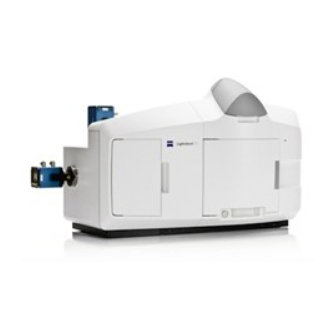
Summary of Contents for Zeiss Lightsheet Z.1
- Page 1 February 2013 ZEN 2012 (black edition)
- Page 2 Violations will entail an obligation to pay compensation. All rights reserved in the event of granting of patents or registration of a utility model. Issued by Carl Zeiss Microscopy GmbH Carl-Zeiss-Promenade 10 07745 Jena, Germany microscopy@zeiss.com www.zeiss.com/microscopy...
- Page 3 INTRODUCTION Lightsheet Z.1 Carl Zeiss Contents of this Manual Chapter 1 Hardware Chapter 2 Sample Preparation Chapter 3 Quick Guide Chapter 4 Software Operation Annex Lightsheet Z.1 - Overview 02/2013 000000-1790-528...
-
Page 5: Table Of Contents
Inserting and Removing the Sample Holder .............. 35 Installation of the Incubation Modules ..............37 3.7.1 Heating Components ..................... 37 3.7.2 -Module Lightsheet Z.1 ..................... 39 3.7.3 Heating Device Humidity S1 .................... 41 3.7.4 Registration of the incubation modules ................41 Switch Incubation ON and OFF ................... - Page 6 CHAPTER 1 - HARDWARE Carl Zeiss Content Lightsheet Z.1 Illumination and Detection Optics ................43 3.9.1 Removing and Inserting the Illumination Optics Unit ............45 3.9.2 Removing and Inserting the Detection Optics Unit............46 3.10 Removing and Inserting the Reflector Turret for Emission Selection ...... 47 3.11...
-
Page 7: Maintenance And Cleaning
CHAPTER 1 - HARDWARE Lightsheet Z.1 Maintenance and Cleaning Carl Zeiss Maintenance and Cleaning The maintenance to be carried out by the customer is limited to cleaning the painted surfaces, replacing glass windows and rubber seals on the sample chamber, cleaning and disinfecting the system cavity and... - Page 8 To clean and disinfect the temperature sensor proceed as follows: • Remove the temperature sensor from the sample chamber and loosen the connections to the main system module Lightsheet Z.1. • Once removed, thoroughly rinse the temperature sensor with distilled water and then suspend it for one hour in a 70 % ethanol solution.
-
Page 9: Maintenance Of The Liquid Cooling System
Observe the directions of the cooling system manufacturer in the supplied operating manual. If you have signed a service agreement with Carl Zeiss, our service staff will perform the check as part of the maintenance procedure. -
Page 10: Ergodrive Operating Panel
CHAPTER 1 - HARDWARE Carl Zeiss ErgoDrive Operating Panel Lightsheet Z.1 ErgoDrive Operating Panel Z-axis control Rotation button (switches 6 to rotation drive) Mode button (fine / coarse) Axis button (switches 6 to y-axis drive) X-axis control Y-axis / Rotation control Fig. - Page 11 CHAPTER 1 - HARDWARE Lightsheet Z.1 ErgoDrive Operating Panel Carl Zeiss With the ErgoDrive operating panel the movement of the sample holder and therefore the sample can be controlled. It is the manual equivalent of sliders in the Specimen Navigator tool in the ZEN software (Fig.
- Page 12 CHAPTER 1 - HARDWARE Carl Zeiss ErgoDrive Operating Panel Lightsheet Z.1 The following control elements are part of the ErgoDrive operation panel: Buttons: The Mode button (Fig. 1/3) changes the speed of the drive from coarse to fine and back.
-
Page 13: User Interfaces
− Reflector turret. Customized Sample Chamber The sample chamber delivered with the Lightsheet Z.1 can be used with a multitude of media (e.g. PBS, cell culture medium, artificial sea water) and is carefully designed to produce optimal results with the Lightsheet Z.1 system. -
Page 14: Installation And Deinstallation Of The Detection Modules
A rotation guard plate (Fig. 3/7) is mounted on the contact ring of each detection module. When mounting on both ports this must point toward the front side of the Lightsheet Z.1. It prevents rotation of the detection module and carries the laser protection notice. - Page 15 CHAPTER 1 - HARDWARE Lightsheet Z.1 User Interfaces Carl Zeiss Detector port 1 (reflection, Cam1) Detector port 2 (transmission, Cam2) Securing screw Sensors (2×) in sensor ring Contact pins (2×) on blind cap Blind cap Rotation guard plate with laser protection notice...
- Page 16 − Now disconnect all cables from the Lightsheet Z.1 system and the PC for system control. Even if the detection modules are regularly switched no cable should remain on the system.
- Page 17 − Turn on the system (see CHAPTER 4, SYSTEM OPERATION). 2) Registration and Alignment of the detection modules − After the Lightsheet Z.1 and the PC for system control are turned on and the operation system has booted, start the software ZEN Configuration Tool .
- Page 18 Carl Zeiss User Interfaces Lightsheet Z.1 − On the Configuration tab (Fig. 6) you will find a Camera field with a drop – down menu for both detector ports (Cam1, Cam2). Select the detection module(s) you are planning to use. When you use the PCO.Edge, the checkbox liquid cooling will be marked automatically.
-
Page 19: Adjustment – Detector Recognition
Detector Alignment). Adjustment – Detector Recognition When the detection modules are exchanged or attached to the Lightsheet Z.1 system for the first time, the system must identify which detection module is present for which channel. Pressing the button Detector recognition will start an identification routine to do this job. A progress bar is visible during the process. - Page 20 CHAPTER 1 - HARDWARE Carl Zeiss User Interfaces Lightsheet Z.1 After pressing the Automatic Detector Alignment button, a wizard window (Fig. 8) will open that guides you through the process. Press the Next button after each step to continue or the Previous button to move one step back.
-
Page 21: Adjustment – Manual Detector Alignment
CHAPTER 1 - HARDWARE Lightsheet Z.1 User Interfaces Carl Zeiss Step 4 is the final step in which the alignment for all filters and detection module channels is performed. A progress bar is present at the bottom of the window. When the procedure is finished the Finish button becomes available, press it to save all adjustments and leave the wizard. - Page 22 CHAPTER 1 - HARDWARE Carl Zeiss User Interfaces Lightsheet Z.1 No sample should be present when performing this task. When pressing the Manual Detector Alignment button, a window will open (Fig. 10). A grating is automatically inserted into the light path and a live image of it is shown in a new image container within the ZEN software.
-
Page 23: Adjust The Grating Focus For The Automatic Or Manual Detector Alignment Tool
CHAPTER 1 - HARDWARE Lightsheet Z.1 User Interfaces Carl Zeiss Use the sliders or the input box with arrows of Cam 1 X and Cam 1 Y (for Channel 1, Cam1) or Cam 2 X and Cam 2 Y (for Channel 2, Cam 2) to move the previously chosen structure of the grating to overlay with the cross. - Page 24 • Use the Zoom tools for the Manual Detector Alignment to zoom into the center region of the image. • While the imaging continues remove the black screw cap (Fig. 15/1) on the back of the main system module Lightsheet Z.1. This opening gives way to a screw that can be turned with an Allen wrench (Fig. 15/2).
- Page 25 CHAPTER 1 - HARDWARE Lightsheet Z.1 User Interfaces Carl Zeiss • Monitor the live image of the grating while you turn the screw in any direction and find the position of the screw that results in the sharpest image. • Stop the continuous scan and place the black screw cap back on the system.
-
Page 26: Cable Connections For The Detection Module "Pco.edge
The detection module "PCO.Edge" is equipped with a liquid cooling system. Please read the safety notes of the Lightsheet Z.1 system, as well as the separate safety data sheet for the liquid cooling system. The power supply unit (PSU) for the cooling is connected to the system's power strip at the rear of the laser module. -
Page 27: Cable Connections For The Detection Module "Standard
− The trigger cable is plugged into the respective detection module and the three ends on the rear side of the main system module Lightsheet Z.1 to [Camera 1] or [Camera 2], as well as [Camera 1/IN] and [Camera 1/OUT] (reflection port, Cam1) or [Camera 2/IN] and [Camera 2/OUT] (transmission port, Cam2). -
Page 28: Assembly Of The Sample Chamber
CHAPTER 1 - HARDWARE Carl Zeiss User Interfaces Lightsheet Z.1 Assembly of the Sample Chamber − Wear powder-free gloves to avoid fingerprints on the cover slips as well as to protect your hands from sharp edges. 3.3.1 Assembly of the Sample Chamber Windows −... - Page 29 CHAPTER 1 - HARDWARE Lightsheet Z.1 User Interfaces Carl Zeiss Repeat step 1 - 6 for each of the three windows. − Assemble the sample chamber window for the detection optic. Turn the sample chamber body (Fig. 19/1) to the largest opening window for the detection optics.
- Page 30 CHAPTER 1 - HARDWARE Carl Zeiss User Interfaces Lightsheet Z.1 Sample chamber body O-ring (blue) For the detection optic 5x/0.16 − 3 Detection optic adapter 5x/0.16 − 4 Cover slip − 5 O-ring (18mm O-ring, black) − 6 Retaining ring For the detection optic 20x/1.0, 40x/1.0, 63x/1.0...
-
Page 31: Assembly Of The Sample Chamber Body And The Sample Chamber Dove Tail Slide
CHAPTER 1 - HARDWARE Lightsheet Z.1 User Interfaces Carl Zeiss 3.3.2 Assembly of the Sample Chamber Body and the Sample Chamber Dove Tail Slide Mesh screen Sample chamber body Fig. 20 Placing the mesh screen − Place the mesh screen (Fig. 20/2) into the middle of the sample chamber body (Fig. 20/1) to reduce turbulence stress on the specimen when the chamber is filled. - Page 32 CHAPTER 1 - HARDWARE Carl Zeiss User Interfaces Lightsheet Z.1 Sample chamber dove tail slide Positioning pins Sample chamber body Screw-holes Screws (diameter = M3, length = 14 mm) Fig. 21 Assembly of the sample chamber body and the sample chamber dove tail slide...
-
Page 33: Insertion Of The Drain Connector, Luer-Lock Connectors And Blind Plugs
CHAPTER 1 - HARDWARE Lightsheet Z.1 User Interfaces Carl Zeiss 3.3.3 Insertion of the Drain Connector, Luer-Lock Connectors and Blind Plugs − Turn the assembled sample chamber body with dove tail slide so that the dove tail slide is on the table with the grip facing you (Fig. -
Page 34: Insertion Of Accessories For Incubation
− Place a cover on top of the sample chamber body (Fig. 24/3). This will minimize evaporation of medium or contamination of the sample chamber. If CO module Lightsheet Z.1 is used (configuration dependent, chapter 2.7.2) the cover is recommended to maintain the CO concentrations. -
Page 35: Removing And Inserting The Sample Chamber
The sample chamber can be removed from the system. For removal, proceed as follows: − Open the front system cavity door (Fig. 25/1) of the main system module Lightsheet Z.1. − Remove the liquid from the sample chamber (Fig. 25/6) using the hose with the corresponding syringe (Fig. -
Page 36: Assembly Of The Sample Holder
10x Teflon tips of each as well as matching Teflon tip tools are provided in the Chamber & Sample Holder Starter Kit Lightsheet Z.1 (Tab. 1). The Teflon tips can be assembled as a replacement onto the matching tip-less plungers. - Page 37 Blue Tab. 1 Capillary components in the Chamber & Sample Holder Starter Kit Lightsheet Z.1 The sleeves are tube-shaped with four slits at one end. Insert the sleeves into the capillary sample holder stem (Fig. 26/2) so that the two slit endings point outwards and the non-slit endings face toward the center.
-
Page 38: Assembly Of Sample Holder For Syringes
CHAPTER 1 - HARDWARE Carl Zeiss User Interfaces Lightsheet Z.1 Take the capillary by the glass portion (avoiding the plunger) with the agarose embedded specimen inside and carefully place it into the center of the clamp screw sample holder top. -
Page 39: Inserting And Removing The Sample Holder
Assembly of sample holder for syringes Inserting and Removing the Sample Holder • To insert the sample holder move the stage of the Lightsheet Z.1 system to the Load position via the Specimen Navigator tool (Fig. 28) in Fig. 28 Load Position in the Specimen... - Page 40 CHAPTER 1 - HARDWARE Carl Zeiss User Interfaces Lightsheet Z.1 Upper system cavity door Guide rails Capillary Hole for disc ball bearings Fig. 29 Inserting the Sample Holder − Before taking the sample holder out, retract the sample back inside the capillary glass via using plunger.
-
Page 41: Installation Of The Incubation Modules
Carl Zeiss Installation of the Incubation Modules The incubation of the Lightsheet Z.1 is configuration dependent. It is used to control and monitor temperature and CO supply to the sample environment. The TempModule S1 is required for all of the various configurations. - Page 42 (section 3.3.4, Fig. 23, Fig. 24). − Connect the Peltier Block (Fig. 32/6) to the Lightsheet Z.1 by the two provided silicone tubes (Fig. 32/7-8) and plug in the 8 pin connector of the connection cable into the right side of the front system cavity / sample room (Fig.
-
Page 43: Co -Module Lightsheet Z.1
PeCon manual. − After CO calibration, the Lightsheet Z.1 system must be completely turned off before work with the system can continue. − Position the Luer-Lock connector for CO incubation at the top right corner of the sample chamber. - Page 44 Luer-Lock connector for perfusion pump-out with tubing Drain connector with tubing Drain tray Fig. 32 Incubation connections for sample chamber body -Module Lightsheet Z.1 can be extended with Heating Device Humidity S1 to improve thermal equilibrium in the sample chamber. 000000-1790-528 02/2013...
-
Page 45: Heating Device Humidity S1
Registration of the incubation modules • After the Lightsheet Z.1 and the PC for system control are turned on and the operation system has booted start the software ZEN Configuration Tool. You find it either as a shortcut on your desktop or in this directory : C:\ZEN\HWT as Configuration Tool 2012.exe. - Page 46 • Do not check the check boxes for external Sensor as well as an Humidity Sensor. These components are not part of the Lightsheet Z.1 incubation modules. • Leave the Configuration Tool with the Store all Changes button. This will write all changes into the database of your system.
-
Page 47: Switch Incubation On And Off
ZEN stops unexpected during an experiment. Illumination and Detection Optics In the Lightsheet Z.1, one or a pair of illumination optics plus one detection optic are always required. We recommend the following combinations: Lightsheet Z.1 illumination... - Page 48 Tabelle 3 Detection module Standard Typical values for Lightsheet Z.1 systems Be aware, a homogenous light sheet illumination with full intensity is limited to about 2800 µm in the y direction. Above and below this center, the illumination intensity will fade towards the edges in the y direction.
-
Page 49: Removing And Inserting The Illumination Optics Unit
CHAPTER 1 - HARDWARE Lightsheet Z.1 User Interfaces Carl Zeiss 3.9.1 Removing and Inserting the Illumination Optics Unit The illumination optics unit (Fig. 35/2) can easily be removed and inserted after the removal of the sample chamber (see section 3.4 Removing and Inserting the Sample Chamber). -
Page 50: Removing And Inserting The Detection Optics Unit
The calibration file is created by a Carl Zeiss service personnel representative during system installation, or when a detection optic is newly purchased. Ensure that the correct detection optic unit is chosen in the ZEN software interface, based on its name and the specific serial number of the detection optic in use. -
Page 51: Removing And Inserting The Reflector Turret For Emission Selection
CHAPTER 1 - HARDWARE Lightsheet Z.1 User Interfaces Carl Zeiss 3.10 Removing and Inserting the Reflector Turret for Emission Selection The reflector turret (Fig. 37/5) can be accessed by opening the left-hand-side front system door (Fig. 37/1). After the reflector turret for emission selection has been exchanged, the Automatic Detector Alignment should be performed. - Page 52 CHAPTER 1 - HARDWARE Carl Zeiss User Interfaces Lightsheet Z.1 The emission filters can be exchanged on the filter turret (Fig. 37/4). − Use the provided tool to loosen the retaining ring around the emission filter you wish to exchange and remove the ring.
-
Page 53: Removing And Inserting The Reflector Turret For Laser Blocking Filter
CHAPTER 1 - HARDWARE Lightsheet Z.1 User Interfaces Carl Zeiss If you plan to exchange the reflector turret for emission selection on a regular basis, it can be helpful to create two databases, one for each reflector turret. To do so, copy the... - Page 54 CHAPTER 1 - HARDWARE Carl Zeiss User Interfaces Lightsheet Z.1 You can remove and replace filter modules by push and click. How to uninstall a module: − Disengage the filter module from the upper spring elements of the filter insert by tilting it forward, then lift it off the lower spring elements and remove the module.
- Page 55 CHAPTER 1 - HARDWARE Lightsheet Z.1 User Interfaces Carl Zeiss Fig. 41 Configuration Tool, Filters tab Fig. 42 ZEN software, Maintain tab, Filters tool window 02/2013 000000-1790-528...
-
Page 56: Index
CHAPTER 1 - HARDWARE Carl Zeiss Index Lightsheet Z.1 Index Automatic Detector Alignment ...... 15 Manual Detector Alignment ......17 Capillary color coding ........33 Plunger reference number ......33 Capillary size ..........33 Customized Sample Chamber ......9 Reflector Turret for Emission Selection Database configuration .......49... - Page 57 CHAPTER 2 - SAMPLE PREPARATION Lightsheet Z.1 Content Carl Zeiss CHAPTER 2 SAMPLE PREPARATION Flood P.M., Kelly R., Gutiérrez-Heredia L. and E.G. Reynaud School of Biology and Environmental Science, University College Dublin, Belfield, Dublin 4, Dublin, Ireland CONTENT Page INTRODUCTION ....................3 SAMPLE MOUNTING FOR LSFM ................
- Page 58 CHAPTER 2 - SAMPLE PREPARATION Carl Zeiss Content Lightsheet Z.1 TIPS, TROUBLESHOOTING AND ADDITIONAL INFORMATION ......37 Tips ..........................37 Troubleshooting ......................37 Suggested Additional Sources of Information ............40 References and Further Reading ................41 INDEX ......................... 44 000000-1790-528...
- Page 59 Samples must be carefully considered when using LSFMs such as the Lightsheet Z.1 as well as the label or dye used must be carefully chosen. In planning an experiment, it should be kept in mind that most labelling and imaging protocols have been developed for thin specimens and therefore many aspects are not adapted to imaging larger samples such as embryos, tissue slices or complete mosquitoes.
- Page 60 CHAPTER 2 - SAMPLE PREPARATION Carl Zeiss Introduction Lightsheet Z.1 Table 1 Topic Subtopic Sample/Model Technique/ LSFM Reference Organism implementation Technical set up of MISERB Fluorescent beads MISERB Fahrback et al, 2010 Structured illumination Mouse cochlea sTSLIM Schroter et al, 2011...
- Page 61 CHAPTER 2 - SAMPLE PREPARATION Lightsheet Z.1 Introduction Carl Zeiss Topic Subtopic Sample/Model Technique/ LSFM Reference Organism implementation Whole organism 3D Ormia ochracea; Huber et al, 2001 reconstruction Emblemasoma auditrix Large Whole organism 3D Drosophila melanogaster Ultramicroscope Jahrling et al, 2010...
- Page 62 (refractive index of 1.33) at all times, to ensure optimal image quality. The Lightsheet Z.1 is not designed for the use with clearing reagents. The Lightsheet Z.1 is built for aqueous media. Also the sample chamber and the system cavity for the sample chamber and sample holder are not compatible with aggressive chemicals.
- Page 63 CHAPTER 2 - SAMPLE PREPARATION Lightsheet Z.1 Sample Mounting for LSFM Carl Zeiss Fig. 1 Relations between the sample and the objective in microscopy. Samples are traditionally isolated from the objective by a glass coverslip (A and B) limiting access to one side only.
- Page 64 (Dodt et al., 2007), alternatively the sample can be embedded in a gel rod that can be rotated. In a horizontal configuration, like the Lightsheet Z.1, the sample can be either embedded in a stiff gel (Fig. 3/A) (Huisken et al., 2004) hooked and positioned in front of the objective (Fig.
- Page 65 In this case the sample must be mounted to support the required positioning. One approach used in Lightsheet Z.1 to support this experimental paradigm is to place the object in a gel rod that can be rotated (Fig. 3/A and E) in front of the objective. The hydrogel cylinder must be sufficiently stable to avoid movement during rotation.
- Page 66 The Lightsheet Z.1 package provides four capillaries sizes adapted to the sample holder to embed objects of various sizes. The special sample holder of the Lightsheet Z.1 adapts to hold these capillaries for precise positioning (translation and rotation) of the cylinder-shaped object for observation through the detection optics.
- Page 67 An example of how to tailor a syringe for Lightsheet Z.1 sample preparation is illustrated in Fig. 5. The tip of the syringe is cut off to create an even cylinder, and the gel solution is pumped in using the plunger.
- Page 68 Preparing a sample embedding container with a capillary. A glass capillary can be used. For the Lightsheet Z.1 they come from the manufacturer (Brand) in just the right length (A). Other capillaries can be cut to an appropriate length (B), the agarose with the object can be easily pumped in using the suitable plungers with Teflon tips (C).
- Page 69 CHAPTER 2 - SAMPLE PREPARATION Lightsheet Z.1 Sample Mounting for LSFM Carl Zeiss There are two principal methods of embedding an object. The first is to directly mix the object with the agarose then pump it into the sample embedding cylinder. This is a convenient way of embedding very small objects such as pollen grains (Swoger et al., 2007);...
- Page 70 2.2.2 Hanging Samples The Lightsheet Z.1 is optimized for gel embedding samples The sample chamber must be filled with a watery solution (refractive index of 1.33) at all times, to ensure optimal image quality. This mounting technique can also be used, but will require some initial adaptations to the sample holder.
- Page 71 Sample Mounting for LSFM Carl Zeiss The Lightsheet Z.1 has a maximum Field of View of approx. 2.5 mm (depending on zoom settings). This and the dimensions of the sample chamber might limit the size of the sample. Fig. 9 Different ways of hanging a sample.
- Page 72 CHAPTER 2 - SAMPLE PREPARATION Carl Zeiss Sample Mounting for LSFM Lightsheet Z.1 Fig. 10 Enclosed chambers for LSFM. Incubation chambers can be made by molding an agarose beaker that can be mounted on a simple plastic holder and loaded with the sample prior to imaging (A). Another solution is to create a...
- Page 73 Anna Kaufmann, Michaela Mickoleit, Michael Weber and Jan Huisken in Development 139, 3242-3247 (2012) ("Multilayer mounting enables long-term imaging of zebrafish development in a light sheet microscope") and emphasize the fact that the mounting method described in this article is fully compatible with the Lightsheet Z.1. 02/2013 000000-1790-528...
- Page 74 Carl Zeiss Microscopy GmbH (hereinafter "we") hereby informs you that we will warrant the specified and agreed performance of the Lightsheet Z.1 system only if sample chambers are applied and used that either are delivered or explicitly approved by us.
- Page 75 CHAPTER 2 - SAMPLE PREPARATION Lightsheet Z.1 Sample Mounting for LSFM Carl Zeiss Front system cavity door Sample chamber Upper sample opening Guide rails / sample chamber mount Upper system cavity door Connections for incubation Securing screw Hose and syringe Sample chamber grip Fig.
- Page 76 In the case of Lightsheet Z.1, the sample kit is provided with four types of color coded capillaries with matching plungers (Fig. 14/A and B) and color coded sleeves to fit perfectly each type of capillary to the sample holder (Fig.
- Page 77 E. Sample holder disc for capillaries F. Sample Holder diagram showing the capillary, the stem and disc of the sample holder. G and H. Sample holder handling and insertion in the Lightsheet Z.1. I. Sample holder disc for syringes, adapter ring. K. syringe (1 ml). 02/2013...
- Page 78 CHAPTER 2 - SAMPLE PREPARATION Carl Zeiss Sample Mounting for LSFM Lightsheet Z.1 A few points must be taken into account when choosing a particular mount: − Compatibility. This is a crucial issue. The mount must be compatible with the object you want to image (chemistry, temperature etc.), but it must also be compatible with the stage holder.
- Page 79 CHAPTER 2 - SAMPLE PREPARATION Lightsheet Z.1 Sample Mounting for LSFM Carl Zeiss 2.3.4 Gels and Polymers Gelling agents are commonly used for preparing semi-solid or solid tissue culture media. Gels provide support to tissues growing in static conditions. The gelling agent usually has several properties. In particular, it does not react with media constituents, is not digested by enzymes, and remains stable at all incubation temperatures.
- Page 80 CHAPTER 2 - SAMPLE PREPARATION Carl Zeiss Sample Mounting for LSFM Lightsheet Z.1 2.3.5 Hydrogel Preparation Every gelling agent is prepared following specific protocols that vary widely from supplier to supplier, and from laboratory to laboratory depending on the final application. We will not try to cover every single one of these but rather give a simple protocol that we have been using in our laboratory to prepare embedded samples in agarose as a gelling agent.
- Page 81 CHAPTER 2 - SAMPLE PREPARATION Lightsheet Z.1 Sample Mounting for LSFM Carl Zeiss Fixation and Fixatives Many experimental samples will require fixation prior to imaging. The goal of fixation is to maintain cellular structure as close as possible to the native state. Proper fixation typically facilitates immunohistochemical analyses if desired, and is an important step prior to further processing.
- Page 82 CHAPTER 2 - SAMPLE PREPARATION Carl Zeiss Sample Mounting for LSFM Lightsheet Z.1 2.5.1 Choosing a Fluorescent Label The choice of label depends upon the available equipment on your LSFM set-up (lasers, filters) and the availability of certain fluorescent protein variants, fluorochromes conjugated to required antibodies for use in multiple labeling schemes.
- Page 83 CHAPTER 2 - SAMPLE PREPARATION Lightsheet Z.1 Sample Mounting for LSFM Carl Zeiss Fig. 15 Supports for sample embedding containers. Support are used to hold three dimensional objects that cannot be held flat easily. Moreover, embedded samples need to be kept in buffer to avoid gel shrinkage or sample damage. A simple system is to use a beaker filled with PBS and place plasticine on the upper border to support the sample embedding container (A).
- Page 84 CHAPTER 2 - SAMPLE PREPARATION Carl Zeiss Specific Examples of Sample Preparation Lightsheet Z.1 Specific Examples of Sample Preparation In order to make this sample preparation section as useful as possible the following pages describe mounting techniques for specific samples, in particular describing the equipment needed, step by step protocols and illustrations based on our own laboratory experiences.
- Page 85 Beads should be fluorescent in the part of the spectrum you would like to analyze for 1 channel systems. With the Lightsheet Z.1, a two channel system, one can use one channel for the beads (e.g., red) and one channel for the specimen label (e.g, GFP.) Fluorescent beads covering whole visible spectrum are nowadays easily available from various different suppliers (e.g.
- Page 86 CHAPTER 2 - SAMPLE PREPARATION Carl Zeiss Specific Examples of Sample Preparation Lightsheet Z.1 Mounting an Oryza latypes embryo. (A) The embryo is prepared (labelling, drug treatment, Fig. 16 dissected...) (B) The embryo is deposited on the side of the Eppendorf tube and the excess of water is removed with a pipette.
- Page 87 CHAPTER 2 - SAMPLE PREPARATION Lightsheet Z.1 Specific Examples of Sample Preparation Carl Zeiss Preparation of a Fly Pupa (Drosophila melanogaster) Some type of embryos are hydrophobic once dissected and cannot be mounted using the technique described above as they will float on the agarose and will be impossible to embed. The following protocol is suitable for this type of embryo such as fly embryos or pupas.
- Page 88 CHAPTER 2 - SAMPLE PREPARATION Carl Zeiss Specific Examples of Sample Preparation Lightsheet Z.1 Preparation of a Plant Root (Arabidopsis thaliana) Plant research is an important field of investigation using plants as model systems. They are three- dimensional objects that are difficult to image fully and are usually dissected and sliced before being imaged and analyzed.
- Page 89 CHAPTER 2 - SAMPLE PREPARATION Lightsheet Z.1 Specific Examples of Sample Preparation Carl Zeiss Once the root is visible within the bottom part of the beaker, a normal plunger is inserted in the top part of the syringe, where the open part of the beaker is present.
- Page 90 Carl Zeiss Specific Examples of Sample Preparation Lightsheet Z.1 If cells are grown in a different manner it is possible to mix them with a supporting gel prior to loading into a chamber. They can also be grown within the gel already present in an incubation chamber.
- Page 91 CHAPTER 2 - SAMPLE PREPARATION Lightsheet Z.1 Specific Examples of Sample Preparation Carl Zeiss The pelleted cysts are permeabilized with PBS/1 % Triton X-100 for 10 minutes on a wheel or rocker to efficiently mix the gel pellet. The gel pellet is washed twice with PBS (500-1000 g, 5 minutes).
- Page 92 CHAPTER 2 - SAMPLE PREPARATION Carl Zeiss Specific Examples of Sample Preparation Lightsheet Z.1 Fig. 19 Mounting a complete adult Anopheles gambiae. (A) The insect is paralyzed. (B) The chitin surface is treated using 70 % ethanol. (C) The insect is positioned within the melted agarose.
-
Page 93: Troubleshooting
For optimal performance of the Lightsheet Z.1, please check the Setup Requirements information delivered with the system. For information about maintenance and cleaning any part of the Lightsheet Z.1, including the optics, please refer to the according CHAPTER in this manual. - Page 94 − This can be a simple optical problem: objectives or filters are dirty. Clean them accordingly. You can also check that all the components are well in place and aligned. − Your light sheet might not be properly aligned. For Lightsheet Z.1, please realign the light sheet using the Light sheet auto-adjust function of the ZEN software.
- Page 95 Dual Side illumination and/or Pivot scanning of the light sheet can often eliminate these effects (available in the Lightsheet Z.1) Second, the sample could be orientated differently during mounting or partially dissected to limit obstacles. The concentration of the objects can also be a problem especially with samples with optical properties (beads, tubes, glass capillaries…).
- Page 96 CHAPTER 2 - SAMPLE PREPARATION Carl Zeiss Tips, Troubleshooting and Additional Information Lightsheet Z.1 Suggested Additional Sources of Information Chemicals • Agarose: − Molecular biology grade, for routine use, SIGMA, Ref. A-9539 − Type VII, low gelling temperature, SIGMA, Ref. A-4018-50G −...
- Page 97 CHAPTER 2 - SAMPLE PREPARATION Lightsheet Z.1 Tips, Troubleshooting and Additional Information Carl Zeiss • Pipettes − 2 ml serological pipette, FALCON, BD Labware, Ref.35 7507 − 1 ml serological pipette, FALCON, BD Labware, Ref.35 7521 Equipments For the following, please refer to your local lab supply companies •...
- Page 98 CHAPTER 2 - SAMPLE PREPARATION Carl Zeiss Tips, Troubleshooting and Additional Information Lightsheet Z.1 Fuchs, E. et al., 2002. Thin laser light sheet microscope for microbial oceanography. Optics Express, 10(2), p.145. Hama H, Kurokawa H, Kawano H, Ando R, Shimogori T, Noda H, Fukami K, Sakaue-Sawano A, Miyawaki A.
- Page 99 CHAPTER 2 - SAMPLE PREPARATION Lightsheet Z.1 Tips, Troubleshooting and Additional Information Carl Zeiss Maizel, A. et al., 2011. High-resolution live imaging of plant growth in near physiological bright conditions using light sheet fluorescence microscopy. The Plant Journal, 68(2), pp.377–385.
- Page 100 CHAPTER 2 - SAMPLE PREPARATION Carl Zeiss Index Lightsheet Z.1 Index Antifading Agents ......... 26 Sample Holding ........... 8 Sample Preparation FEP Tubing ............ 17 Preparation of Fluorescent Beads .....28 Fixation and Fixatives ........25 Preparation of a Medaka Fish Embryo ..29 Preparation of a Fly Pupa ......31...
- Page 101 CHAPTER 3 - QUICK GUIDE Lightsheet Z.1 Content Carl Zeiss CHAPTER 3 QUICK GUIDE CONTENT Page LOCATING CAPILLARY AND SAMPLE ..............2 Locate Capillary ......................2 Locating Sample ......................8 CONFIGURATION OF THE LIGHT PATH ............. 15 Adjust Acquisition Parameters ................... 23 Define Channels and Tracks and adjust Detectors and Illumination Settings ..
- Page 102 CHAPTER 3 - QUICK GUIDE Carl Zeiss Locating Capillary and Sample Lightsheet Z.1 Locating Capillary and Sample Locate Capillary • To locate the capillary or similar device which is holding the sample, press the Locate Capillary button. The white LED illumination is turned ON and the overview camera acquires a live image from the inside of the sample chamber.
- Page 103 CHAPTER 3 - QUICK GUIDE Lightsheet Z.1 Locating Capillary and Sample Carl Zeiss Seeing the liquid level just at the very top of the displayed sample chamber image is a good way to monitor and be assured that there is no leakage within the sample chamber.
- Page 104 CHAPTER 3 - QUICK GUIDE Carl Zeiss Locating Capillary and Sample Lightsheet Z.1 • Use the Specimen Navigator to position the capillary from the Load position into the Work position. • Click Specimen Navigator The Specimen Navigator is a graphical tool for accessing the relative positions of the specimen embedded in agarose (light-grey cylinder), the illumination light-sheet (light-blue), as well as the X-axis (red),Y-axis (green), Z-axis (blue) and the Rotation of the sample holder.
- Page 105 CHAPTER 3 - QUICK GUIDE Lightsheet Z.1 Locating Capillary and Sample Carl Zeiss • Click Y-axis The capillary becomes now apparent in the displayed image. • Please bring the capillary in focus via the Z-axis slider. • Click Z-axis Moving the light-grey cylinder, representing the embedding medium, into the light sheet graphic (light-blue) can be used as a focus finding guide.
- Page 106 CHAPTER 3 - QUICK GUIDE Carl Zeiss Locating Capillary and Sample Lightsheet Z.1 • Please do the fine adjustment for the capillary in the X direction. • Click X-Axis The capillary is placed correctly when it is centered just above the detection optic lens.
- Page 107 CHAPTER 3 - QUICK GUIDE Lightsheet Z.1 Locating Capillary and Sample Carl Zeiss • Press the plunger of the capillary (or syringe) until the embedded specimen is roughly centered in front of the detection optics lens. The Specimen is now properly centered in front of the detection optic lens.
- Page 108 CHAPTER 3 - QUICK GUIDE Carl Zeiss Locating Capillary and Sample Lightsheet Z.1 Locating Sample • To locate the sample within the sample chamber, the Locate Sample button activates an infrared light source and shows a live transmitted light image in the Center Screen Area. This image is taken using Channel 1/ Cam1.
- Page 109 CHAPTER 3 - QUICK GUIDE Lightsheet Z.1 Locating Capillary and Sample Carl Zeiss • Please use the Zoom slider to zoom into the image. • Click Zoom • Please use the Light Intensity slider to increase the illumination on the sample.
- Page 110 CHAPTER 3 - QUICK GUIDE Carl Zeiss Locating Capillary and Sample Lightsheet Z.1 • Click on the Arrow down button to access the lower tool tabs. • The distance between the Load Position and the y-position for imaging is often quite large. This results in very large numbers for the y-coordinate which are not so convenient for use.
- Page 111 CHAPTER 3 - QUICK GUIDE Lightsheet Z.1 Locating Capillary and Sample Carl Zeiss • You can define a position you might want to return to during the course of imaging by moving the sample to the desired point and pressing the Set Home Position button.
- Page 112 CHAPTER 3 - QUICK GUIDE Carl Zeiss Locating Capillary and Sample Lightsheet Z.1 • Please rotate the capillary until the specimens back side is in front of the detection optic lens. • Click Rotation axis • Please fine adjust the X-position of the capillary and bring the specimen into the center of the image.
- Page 113 CHAPTER 3 - QUICK GUIDE Lightsheet Z.1 Locating Capillary and Sample Carl Zeiss • Please re-focus in Z until you found the desired plane. • Click Z-axis • Press the Stop button to end the live image acquisition and turn off the infrared light source.
- Page 114 CHAPTER 3 - QUICK GUIDE Carl Zeiss Locating Capillary and Sample Lightsheet Z.1 Your specimen is now properly orientated! You can now proceed with adjusting your image settings. The Zebrafish, for illustrative purposes due to its larger size, was shown for positioning in this chapter.
- Page 115 CHAPTER 3 - QUICK GUIDE Lightsheet Z.1 Configuration of the Light Path Carl Zeiss Configuration of the Light Path • Please click Acquisition to access all functions necessary for setting up and controlling light sheet fluorescent imaging and multidimensional data acquisition.
- Page 116 CHAPTER 3 - QUICK GUIDE Carl Zeiss Configuration of the Light Path Lightsheet Z.1 • Pressing the Laser icon displays the laser control submenu. • Click Laser Lines • Activate the laser(s) necessary for excitation by checking the corresponding boxes.
- Page 117 CHAPTER 3 - QUICK GUIDE Lightsheet Z.1 Configuration of the Light Path Carl Zeiss • A dialog box appears for switching ON the selected laser(s). • Click YES The Incubator button accesses all possible attached equipment (Temperature [°C] control and / or Atmosphere [%] control).
- Page 118 CHAPTER 3 - QUICK GUIDE Carl Zeiss Configuration of the Light Path Lightsheet Z.1 Detection Optics Display • Select a suitable Laser Blocking Filter for blocking the laser light. • Click Laser Blocking 000000-1790-528 02/2013...
- Page 119 CHAPTER 3 - QUICK GUIDE Lightsheet Z.1 Configuration of the Light Path Carl Zeiss • According to the chosen laser lines, an appropriate Laser Blocking Filter can be selected. • Click LBF 405/488/561 • The Beam Splitter button opens a drop-down menu for choosing the Beam Splitter to direct the emission light towards the desired detector module, Cam1 or Cam2.
- Page 120 CHAPTER 3 - QUICK GUIDE Carl Zeiss Configuration of the Light Path Lightsheet Z.1 • Click SBS LP 560 • The activated Secondary Beam Splitter (SBS) will move into position and its name is displayed in the light path. 000000-1790-528...
- Page 121 CHAPTER 3 - QUICK GUIDE Lightsheet Z.1 Configuration of the Light Path Carl Zeiss Since Beam Splitter and Emission filters form one unit, the corresponding Emission Filter 1 (in front of Cam1) and Emission Filter 2 (in front of Cam 2) will change as well in the light path.
- Page 122 CHAPTER 3 - QUICK GUIDE Carl Zeiss Configuration of the Light Path Lightsheet Z.1 • Assign a specific color to the channel image by clicking on the detection module icon "Channel Camera 1". A drop down menu opens for selecting a color or look-up-table (LUT).
- Page 123 CHAPTER 3 - QUICK GUIDE Lightsheet Z.1 Configuration of the Light Path Carl Zeiss Adjust Acquisition Parameters • In the Acquisition Mode tool tab, acquisition parameters can be set. • Click Acquisition Mode Here the actual selected Bit Depth is displayed.
- Page 124 CHAPTER 3 - QUICK GUIDE Carl Zeiss Configuration of the Light Path Lightsheet Z.1 You can change the imaged area using the Zoom function. This zoom utilizes hardware zoom optics within the system. Within the Zoom range from 0.36 to 0.7, the image quality towards the edges can decrease (Zoom numbers are displayed in red;...
- Page 125 CHAPTER 3 - QUICK GUIDE Lightsheet Z.1 Configuration of the Light Path Carl Zeiss The Light Sheet Thickness is displayed in µm. • The Pivot Scan (configuration dependent) is used to move the light sheet within the focal plane of the detection optics in a wavelike motion.
- Page 126 CHAPTER 3 - QUICK GUIDE Carl Zeiss Configuration of the Light Path Lightsheet Z.1 • Continuous produces images from all active channels and tracks. It thereby ignores the settings for multidimensional acquisition, but takes into account the imaging settings within the Acquisition Mode Tool and the Channels Tool.
- Page 127 CHAPTER 3 - QUICK GUIDE Lightsheet Z.1 Configuration of the Light Path Carl Zeiss Define Channels and Tracks and adjust Detectors and Illumination Settings • The Channels tool provides definition of channels and tracks, adjustments for Detectors, Illumination settings, as well as Display options.
- Page 128 CHAPTER 3 - QUICK GUIDE Carl Zeiss Configuration of the Light Path Lightsheet Z.1 • Using the Display characteristic curve in the histogram, you can set the limit for the white value (right). This influences the contrast and brightness of the displayed image.
- Page 129 CHAPTER 3 - QUICK GUIDE Lightsheet Z.1 Configuration of the Light Path Carl Zeiss • To end Continuous acquisition, press the Stop button. • Click Stop Lightsheet Auto-Adjust Wizard • Please adjust the light sheet right before you start the acquisition by using the Lightsheet Auto-Adjust wizard.
- Page 130 CHAPTER 3 - QUICK GUIDE Carl Zeiss Configuration of the Light Path Lightsheet Z.1 • The initial window of the Lightsheet Auto-Adjust wizard shows a live image of your sample on the left hand side. The beam path and illumination settings are taken from the Channels Tool and Light Path Tool, so no additional corrections are necessary.
- Page 131 CHAPTER 3 - QUICK GUIDE Lightsheet Z.1 Configuration of the Light Path Carl Zeiss Only the Y-drive should be used during this step since the conditions for the light sheet adjustment should be as close to the imaging conditions as possible.
- Page 132 Carl Zeiss Configuration of the Light Path Lightsheet Z.1 • The light path has been changed automatically, removing the laser blocking filter from the beam path, so stray light from the activated laser can be seen as a live image on the left hand side. Hence any bright or blocking structures, like the sample holder or the sample, are visible.
- Page 133 CHAPTER 3 - QUICK GUIDE Lightsheet Z.1 Configuration of the Light Path Carl Zeiss • Click Finish • The original position of your specimen will be restored after the wizard is finished. The resulting settings from the light sheet alignment are automatically written into the input boxes Left and Right (or only one of them if Single side illumination is chosen in the Acquisition Tool window).
- Page 134 CHAPTER 3 - QUICK GUIDE Carl Zeiss Configuration of the Light Path Lightsheet Z.1 • Snap produces one single image using all active channels and tracks. It thereby ignores the parameters activated in the multidimensional acquisition area, but takes into account the imaging settings within the Acquisition Mode Tool and Channels Tool.
- Page 135 CHAPTER 3 - QUICK GUIDE Lightsheet Z.1 Quick Steps to a Z-Stack Carl Zeiss Quick Steps to a Z-Stack Defining the First Slice and the Last Slice • Please activate the Z-Stack checkbox in the Multidimensional acquisition field to acquire an image series in z.
- Page 136 CHAPTER 3 - QUICK GUIDE Carl Zeiss Quick Steps to a Z-Stack Lightsheet Z.1 • Click on the Arrow down button to access the lower tool tabs. • If the Z-Stack tool bar is expanded, the Z-Stack control panel becomes available.
- Page 137 CHAPTER 3 - QUICK GUIDE Lightsheet Z.1 Quick Steps to a Z-Stack Carl Zeiss • Click Continuous to start a live image acquisition using the currently active imaging parameters for defining the First Slice and the Last Slice within the Z-Stack control panel.
- Page 138 CHAPTER 3 - QUICK GUIDE Carl Zeiss Quick Steps to a Z-Stack Lightsheet Z.1 Now move the focus in the other direction until the end position within the stack is reached, then press Set Last. • Click Set Last Be aware that you might need a wider range for the z-stack which includes enough fiducials or other landmarks for the landmark alignment processing later.
- Page 139 CHAPTER 3 - QUICK GUIDE Lightsheet Z.1 Quick Steps to a Z-Stack Carl Zeiss Adjustment of Z-Stack Settings The Range display box shows the total depth of the stack in micrometers. • Press the Optimal button to set the number of slices to match the optimal Z-interval. This adjusts the number of Slices to keep within the Range set from marking Set First / Set Last.
- Page 140 CHAPTER 3 - QUICK GUIDE Carl Zeiss Quick Steps to a Z-Stack Lightsheet Z.1 The Slices input box displays the number of slices for the stack. Please use the Continuous Drive to speed up the Z-Stack acquisition significantly. When the Continuous Drive checkbox is ticked, the Z-Stack is performed by continuously moving the z-drive.
- Page 141 CHAPTER 3 - QUICK GUIDE Lightsheet Z.1 Quick Steps to a Z-Stack Carl Zeiss Start Z-Stack Experiment and evaluate Data • Click Start Experiment to begin recording the Z-Stack. • Click Start Experiment You can follow the process by the displayed progress bar.
- Page 142 CHAPTER 3 - QUICK GUIDE Carl Zeiss Quick Steps to a Z-Stack Lightsheet Z.1 • The Gallery view displays images from the Z-Stack. • Click Gallery • Click on the Hide button to hide the Left Tool Area and expand the Center Screen Area.
- Page 143 CHAPTER 3 - QUICK GUIDE Lightsheet Z.1 Quick Steps to a Z-Stack Carl Zeiss All the slices from the Z-Stack are represented in the Gallery view side-by-side in a tiled fashion. • The maximum intensity projection shows an output image whose pixels contain the maximum value over all images in the stack at the particular pixel location.
- Page 144 CHAPTER 3 - QUICK GUIDE Carl Zeiss Quick Steps to a Z-Stack Lightsheet Z.1 You have successfully acquired a Z - Stack! You may now proceed with either: 1. View tabs in the Center Screen Area • Ortho View •...
- Page 145 CHAPTER 3 - QUICK GUIDE Lightsheet Z.1 Z-Stacks in Combination with Time Series Carl Zeiss Z-Stacks in Combination with Time Series Defining the Time Series • Please activate in addition to the Z-Stack, the Time Series check box in the Multidimensional acquisition field to acquire an image series in Z in combination with a time series.
- Page 146 CHAPTER 3 - QUICK GUIDE Carl Zeiss Z-Stacks in Combination with Time Series Lightsheet Z.1 • Click on the Arrow down button to access the lower tool tabs. • Click Arrow down • If the Time Series tool bar is expanded, the Time Series control panel becomes available.
- Page 147 CHAPTER 3 - QUICK GUIDE Lightsheet Z.1 Z-Stacks in Combination with Time Series Carl Zeiss • To set up a time series acquisition, first define the Duration it should acquire the images by using the slider, or the text field, or the arrow buttons.
- Page 148 CHAPTER 3 - QUICK GUIDE Carl Zeiss Z-Stacks in Combination with Time Series Lightsheet Z.1 • Click on the Hide button to hide the Right Tool Area and expand the Center Screen Area. • Click Hide Start Z-Stacks Experiment in Combination with Time Series and evaluate Data •...
- Page 149 CHAPTER 3 - QUICK GUIDE Lightsheet Z.1 Z-Stacks in Combination with Time Series Carl Zeiss • You can follow the process by the displayed progress bar. • The Gallery view displays images from the Z-Stack in combination with the Time Series.
- Page 150 CHAPTER 3 - QUICK GUIDE Carl Zeiss Z-Stacks in Combination with Time Series Lightsheet Z.1 • Click on the Hide button to hide the Left Tool Area and expand the Center Screen Area. • Click Hide The Gallery view displays images from the Z-Stack within the Time Series.
- Page 151 CHAPTER 3 - QUICK GUIDE Lightsheet Z.1 Z-Stacks in Combination with Time Series Carl Zeiss You successfully acquired a Z – Stack over Time! You may now proceed with either: 1. View tabs in the Center Screen Area • Ortho View •...
- Page 152 CHAPTER 3 - QUICK GUIDE Carl Zeiss See more from different Angles using Multiview Quick Setup Lightsheet Z.1 See more from different Angles using Multiview Quick Setup Multiview Quick Setup • Activate the Multiview checkbox in the Multidimensional acquisition field to acquire diversely sized Z-stacks using different Views.
- Page 153 CHAPTER 3 - QUICK GUIDE Lightsheet Z.1 See more from different Angles using Multiview Quick Setup Carl Zeiss • Click on the Arrow down button to access the lower tool tabs. • Click Arrow down • When the Multiview-Setup tool tab is expanded, the Multiview-Setup control panel is available.
- Page 154 CHAPTER 3 - QUICK GUIDE Carl Zeiss See more from different Angles using Multiview Quick Setup Lightsheet Z.1 The Multiview-Setup tool window is divided into three regions. (1) The upper region is the organization region and provides access to the Quick Setup wizard.
- Page 155 CHAPTER 3 - QUICK GUIDE Lightsheet Z.1 See more from different Angles using Multiview Quick Setup Carl Zeiss (3) In the lower part is an illustration showing the angle from which the detection optic (eye symbol) is facing the sample (middle cylinder). The zero angle is indicated by the dot at the bottom of the outer circle.
- Page 156 CHAPTER 3 - QUICK GUIDE Carl Zeiss See more from different Angles using Multiview Quick Setup Lightsheet Z.1 • The Quick Setup button opens a new window with a software interface that guides you through the process of setting up a multiview experiment.
- Page 157 CHAPTER 3 - QUICK GUIDE Lightsheet Z.1 See more from different Angles using Multiview Quick Setup Carl Zeiss On the right-hand-side is the Image settings tool tab. Here you have easy access to the Light Intensity slider for adjusting the infrared illumination.
- Page 158 Carl Zeiss See more from different Angles using Multiview Quick Setup Lightsheet Z.1 A short description of what to do during this step is given at the bottom of the window. • Move the Light Intensity slider to adjust the infrared illumination.
- Page 159 CHAPTER 3 - QUICK GUIDE Lightsheet Z.1 See more from different Angles using Multiview Quick Setup Carl Zeiss • If you have finished the image brightness and position adjustment, please press Next. • Click Next • To select the acquisition volume (view x-y), a red rectangle is displayed in the live image window.
- Page 160 CHAPTER 3 - QUICK GUIDE Carl Zeiss See more from different Angles using Multiview Quick Setup Lightsheet Z.1 • To change its size, grab it with the left mouse button on one of the corners and adjust the length of the adjacent sides.
- Page 161 CHAPTER 3 - QUICK GUIDE Lightsheet Z.1 See more from different Angles using Multiview Quick Setup Carl Zeiss Check, by moving the Z-drive, to see if the red rectangle encircles the whole sample along its Z-axis. • After pressing the Next button, the sample is turned by 90° before proceeding to the next step.
- Page 162 Carl Zeiss See more from different Angles using Multiview Quick Setup Lightsheet Z.1 • (1) Re-adjust the sample focus by using the Specimen Navigator tool tab if necessary. It might be necessary to reposition the specimen using the X- and Y-drives as well.
- Page 163 CHAPTER 3 - QUICK GUIDE Lightsheet Z.1 See more from different Angles using Multiview Quick Setup Carl Zeiss • To move the whole rectangle, grab it by holding the left mouse button on one of the sides and drag it to the wanted position.
- Page 164 CHAPTER 3 - QUICK GUIDE Carl Zeiss See more from different Angles using Multiview Quick Setup Lightsheet Z.1 This interface defines the number of views by entering the number of Rotations within the input box or by using the arrows.
- Page 165 CHAPTER 3 - QUICK GUIDE Lightsheet Z.1 See more from different Angles using Multiview Quick Setup Carl Zeiss • Please press the Finish button to complete the Quick Setup and close the window. • Click Finish The resulting Views are now displayed in the Multiview Setup tool tab View list.
- Page 166 Carl Zeiss See more from different Angles using Multiview Quick Setup Lightsheet Z.1 When the Zoom setting is changed during the Multiview Quick Setup, the alignment of the light sheet must be checked and adapted. Following the rule, the light sheet adjustment is the last step before image acquisition.
- Page 167 CHAPTER 3 - QUICK GUIDE Lightsheet Z.1 See more from different Angles using Multiview Quick Setup Carl Zeiss • The Gallery view displays all Views and corresponding slices from the Z-Stacks. • Click Gallery All the Views and corresponding slices from the Z-Stacks can be represented in the Gallery view side-by-side.
- Page 168 CHAPTER 3 - QUICK GUIDE Carl Zeiss See more from different Angles using Multiview Quick Setup Lightsheet Z.1 You successfully acquired a Multiview experiment! You may now proceed with either: 1. View tabs in the Center Screen Area • Ortho View •...
- Page 169 CHAPTER 3 - QUICK GUIDE Lightsheet Z.1 See more from different Angles using Multiview Quick Setup Carl Zeiss Index Acquisition ............ 15 Multidimensional acquisition ......52 Acquisition Mode .......... 23 Pivot Scan ............. 25 Beam Splitter ..........19 Rotary controls ..........4 Camera Tool ............
- Page 171 CHAPTER 4 - SYSTEM OPERATION Lightsheet Z.1 Contents Carl Zeiss CHAPTER 4 SYSTEM OPERATION CONTENTS Page STARTUP AND SHUTDOWN OF THE SYSTEM ............ 6 Startup of the System ....................6 1.1.1 Switching on the Laser Module ..................6 1.1.2 The Power Remote Switch ....................6 1.1.3...
- Page 172 CHAPTER 4 - SYSTEM OPERATION Carl Zeiss Contents Lightsheet Z.1 LEFT TOOL AREA AND HARDWARE CONTROL TOOLS ........37 Locate Tab ........................37 3.1.1 Locate Capillary ......................38 3.1.2 Locate Sample ....................... 38 3.1.3 Specimen Navigator Tool ....................39 3.1.4 Incubator Tool .......................
- Page 173 CHAPTER 4 - SYSTEM OPERATION Lightsheet Z.1 Contents Carl Zeiss 3.3.8 Processing – Ion Concentration ..................99 3.3.8.1 Single Wavelength Dyes – Offline Calibration ..............101 3.3.8.2 Ratiometric Dyes ......................102 3.3.9 Processing – Correlation ....................105 3.3.10 Processing – Modify Series .................... 106 3.3.11...
- Page 174 CHAPTER 4 - SYSTEM OPERATION Carl Zeiss Contents Lightsheet Z.1 3.4.4 Maintain – Streaming ....................171 3.4.5 Maintain – Adjustment....................172 3.4.5.1 Adjustment – Detector Recognition ................172 3.4.5.2 Adjustment – Automatic Detector Alignment ..............173 3.4.5.3 Adjustment – Manual Detector Alignment ..............175 CENTER SCREEN AREA / IMAGE CONTAINERS - DISPLAY AND IMAGE ANALYSIS ......................
- Page 175 CHAPTER 4 - SYSTEM OPERATION Lightsheet Z.1 Contents Carl Zeiss 4.10.2 Quantitative Colocalization Parameters Shown in the Data Table ........228 4.10.3 Colocalization Coefficients ................... 228 4.10.4 Overlap Coefficient after Manders ................229 4.11 Profile View ....................... 229 4.12 Mean of ROI: Additional View Type for Time Series ..........232 4.13...
- Page 176 Startup and Shutdown of the System Startup of the System The Lightsheet Z.1 system is equipped with a Laser key-operated switch on LB Rack Lightsheet and a remote control to switch on the system, the PC for system control, and the incubation. "System", "PC"...
- Page 177 Starts the ZEN software for operating the Lightsheet Z.1 system. Configuration Tool icon The Configuration Tool permits alterations to the database of the Lightsheet Z.1 that is used by the ZEN software program. This function should preferably be performed by trained personnel.
- Page 178 (Fig. 2). It displays the ZEN symbol in the upper part, the designation of the current ZEN software version (lower left) and the Zeiss logo (lower right). Once the program is loaded, the Login ZEN dialog will appear (Fig. 3) in front of the ZEN interface. The number after ZEN designates the current version.
- Page 179 CHAPTER 4 - SYSTEM OPERATION Lightsheet Z.1 Startup and Shutdown of the System Carl Zeiss When ZEN software is started for the first time, the positioning units needs to be initialized. All doors of the system must be closed and no sample can be present in the system.
-
Page 180: Shutdown Procedure
Carl Zeiss Startup and Shutdown of the System Lightsheet Z.1 − If Recent… is pressed the last couple of databases that were in use will be displayed (Fig. 9). Click on the one you want to select. By pressing the Choose… button, Windows Explorer will open. - Page 181 CHAPTER 4 - SYSTEM OPERATION Lightsheet Z.1 Startup and Shutdown of the System Carl Zeiss 1.2.1 Exiting ZEN Software The Software can be shut down from File menu of the Main toolbar by selecting Exit (Fig. 10) or by pressing the Window Close button (symbolized by a cross) in the ZEN window.
- Page 182 CHAPTER 4 - SYSTEM OPERATION Carl Zeiss Startup and Shutdown of the System Lightsheet Z.1 In the Maintain tab under the System Options dialog window, the box for activating the Lasers off on Exit is located under the Hardware tab (Fig.
- Page 183 CHAPTER 4 - SYSTEM OPERATION Lightsheet Z.1 Introduction to the Software Application Layout Carl Zeiss Introduction to the Software Application Layout Overview on the Screen Layout Fig. 13 ZEN Main Application window after Startup with empty image container Fig. 14...
- Page 184 Lightsheet Z.1 Introduction to ZEN ZEN - Efficient Navigation - is the software from Carl Zeiss which sets new standards in application- friendly usage for microscopy systems. The ZEN Interface is clearly structured and follows the typical workflow of the experiments performed with Light Sheet Fluorescence Microscope Systems (LSFM): On the Left Tool Area (Fig.
- Page 185 CHAPTER 4 - SYSTEM OPERATION Lightsheet Z.1 Introduction to the Software Application Layout Carl Zeiss Fig. 16 ZEN window Layout configuration More features of ZEN include: − The user can add more columns to the Left Tool Area or detach individual tools to position them anywhere on the monitor.
- Page 186 CHAPTER 4 - SYSTEM OPERATION Carl Zeiss Introduction to the Software Application Layout Lightsheet Z.1 Function Elements Function element Description / Explanation Tool group Tool (closed) or tool window (opened) Panel (e.g.: Zoom panel) Field with a subset of tools of a tool window List box or selection box −...
- Page 187 CHAPTER 4 - SYSTEM OPERATION Lightsheet Z.1 Introduction to the Software Application Layout Carl Zeiss Slider with input ("spin") box and arrows − Setting of numbers in the relevant input box by moving the slider or clicking on the arrow buttons or clicking on the slider and moving via the arrow keys of the keyboard.
- Page 188 CHAPTER 4 - SYSTEM OPERATION Carl Zeiss Introduction to the Software Application Layout Lightsheet Z.1 Application Bar The application bar includes the following control elements in the ZEN software application window: starts the online help minimizes the application window closes the window...
- Page 189 CHAPTER 4 - SYSTEM OPERATION Lightsheet Z.1 Introduction to the Software Application Layout Carl Zeiss Menu overview File View Acquisition Maintain Macro Tools Window New Acquisition Text View New Acquisition Hardware Macro Paste Image Close Document Displays the Document Administrator...
- Page 190 − This opens the maintain list (Fig. 20). The maintain list is bipartite. The upper two sections list user accessible tools for LSM but not for Lightsheet Z.1 systems, the lower section lists tools reserved for service personnel which are password protected.
- Page 191 This function allows the user to adjust the white balance and color balance of a connected camera of LSM systems. It is not used for any calibration tasks of the Lightsheet Z.1 detection modules. • Click on Camera in the Maintain list. − The Camera Color Adjustment window appears on the screen (see Fig.
- Page 192 CHAPTER 4 - SYSTEM OPERATION Carl Zeiss Introduction to the Software Application Layout Lightsheet Z.1 2.5.3 Help – About • The About window can be accessed via the Help item in the menu bar (Fig. 22). • Clicking About opens the About window (Fig. 23).
- Page 193 CHAPTER 4 - SYSTEM OPERATION Lightsheet Z.1 Introduction to the Software Application Layout Carl Zeiss Main Toolbar Fig. 24 Main toolbar, left side Fig. 25 Main toolbar, right side Workspace Configuration This function allows loading, saving or deleting a workspace configuration. These workspace...
- Page 194 CHAPTER 4 - SYSTEM OPERATION Carl Zeiss Introduction to the Software Application Layout Lightsheet Z.1 • To load a workspace configuration, click on the button. The following list box will appear: Selecting one of the list entries will load the respective configuration.
- Page 195 CHAPTER 4 - SYSTEM OPERATION Lightsheet Z.1 Introduction to the Software Application Layout Carl Zeiss Status Bar Fig. 26 Status bar System information − Frames per second (FPS) − Position and pixel intensity System information Shows information of the currently system tasks (state). The possible states are: −...
- Page 196 CHAPTER 4 - SYSTEM OPERATION Carl Zeiss Introduction to the Software Application Layout Lightsheet Z.1 System messages Next to the progress bar you find a green upside down exclamation mark and a white arrowhead When you click on the arrowhead a window opens in which the history of system messages is shown (Fig.
- Page 197 CHAPTER 4 - SYSTEM OPERATION Lightsheet Z.1 Introduction to the Software Application Layout Carl Zeiss Left Tool Area Main Tool tabs Switches between the Locate, Acquisition, Processing, and Maintain main tools to operate the included tool Action buttons Action buttons...
- Page 198 CHAPTER 4 - SYSTEM OPERATION Carl Zeiss Introduction to the Software Application Layout Lightsheet Z.1 Multidimensional Acquisition Tools for all multidimensional imaging The tools in this tool group will only appear if selected in the selection panel below the action buttons.
- Page 199 CHAPTER 4 - SYSTEM OPERATION Lightsheet Z.1 Introduction to the Software Application Layout Carl Zeiss To add, edit or delete configurations, click on the button next to the configuration drop-down list. 2.8.1 Tool Groups and Tools Multiple Columns Layout of tool groups This function allows moving one or several tool groups into a second or third column in the Left Tool Area.
- Page 200 CHAPTER 4 - SYSTEM OPERATION Carl Zeiss Introduction to the Software Application Layout Lightsheet Z.1 Show all functions within the tool window Clicking on the button shows or hides additional functions within the tool. Only the basic functions are shown; this functionality is adequate for most applications.
- Page 201 CHAPTER 4 - SYSTEM OPERATION Lightsheet Z.1 Introduction to the Software Application Layout Carl Zeiss 2.8.2 Context Menu for the Left Tool Area There are two different context menus in the Left Tool Area with different functionalities available. The first one (Fig. 30) is opened by clicking with the right mouse button next to the action buttons or in the area of the Experiment Manager.
- Page 202 CHAPTER 4 - SYSTEM OPERATION Carl Zeiss Introduction to the Software Application Layout Lightsheet Z.1 Center Screen Area The Center Screen Area is used for displaying acquired images or to show images in the available image views. Use the context menu for the Center Screen Area by clicking the background of a container, the view of this area can be varied individually.
- Page 203 CHAPTER 4 - SYSTEM OPERATION Lightsheet Z.1 Introduction to the Software Application Layout Carl Zeiss Image tabs For each opened image, one image tab is shown in Fig. 33 Image tabs the header of the actual container. Three modes can be selected using the context menu of the Center Screen Area (see also section 2.9.2 Context Menu for the Center Screen...
- Page 204 CHAPTER 4 - SYSTEM OPERATION Carl Zeiss Introduction to the Software Application Layout Lightsheet Z.1 Expose Mode Changes the single image display to the Expose display, showing thumbnails of all open images. Clicking on one of the image headers changes back to the single image display.
- Page 205 CHAPTER 4 - SYSTEM OPERATION Lightsheet Z.1 Introduction to the Software Application Layout Carl Zeiss 2.10 Right Tool Area The Right Tool Area is used for displaying and handling the opened images, e.g. save or close. The view of this area can be varied individually, e.g.
- Page 206 CHAPTER 4 - SYSTEM OPERATION Carl Zeiss Introduction to the Software Application Layout Lightsheet Z.1 Big View button Shows images with the textual characters and a big image preview. Save Status icon This icon appears in the image that is not saved yet.
-
Page 207: Left Tool Area And Hardware Control Tools
Acquisition Acquiring images with the Lightsheet Z.1. Processing Processing images and multidimensional (multiview) images taken with the Lightsheet Z.1. Maintain Setting imaging, system, and firmware parameters. By pressing the arrow left symbol on the Expand tab at the right-hand-side border, the left tool area will collapse to the left side. -
Page 208: Locate Capillary
CHAPTER 4 - SYSTEM OPERATION Carl Zeiss Left Tool Area and Hardware Control Tools Lightsheet Z.1 3.1.1 Locate Capillary To locate the capillary or similar device which contains the sample, press the Locate Capillary button. The white LED illumination is turned on and a live image is acquired showing the inside of the sample chamber. -
Page 209: Specimen Navigator Tool
CHAPTER 4 - SYSTEM OPERATION Lightsheet Z.1 Left Tool Area and Hardware Control Tools Carl Zeiss 3.1.3 Specimen Navigator Tool To position the sample holder for imaging while using the Locate Capillary or Locate Sample function, the Specimen Navigator tool window is available in the Locate tool tab (Fig. -
Page 210: Incubator Tool
Incubator panel by pressing the triangle in the Incubator tool tab. All incubation components you wish to use during the imaging session must be connected to the Lightsheet Z.1 system and turned on before starting the ZEN software. Once ZEN is running you can detach and reattach components. -
Page 211: Acquisition Tab
2 independent heating channels that are linked to certain devices (Peltierblock Sample Chamber Lightsheet Z.1, Heatingblock Sample Chamber Lightsheet Z.1) that are assigned to different channels. Enter the desired nominal temperature via the input boxes by typing in the Fig. -
Page 212: Experiment Manager
CHAPTER 4 - SYSTEM OPERATION Carl Zeiss Left Tool Area and Hardware Control Tools Lightsheet Z.1 3.2.1 Experiment Manager You can load a predefined experiment from the Experiment Manager field by pressing the arrow to access the list box (Fig. 46). -
Page 213: Action Buttons
CHAPTER 4 - SYSTEM OPERATION Lightsheet Z.1 Left Tool Area and Hardware Control Tools Carl Zeiss 3.2.2 Action Buttons Action buttons provide control for how the image is acquired (Fig. 49). Fig. 49 Action buttons They have the following functions, regardless of the imaging mode: The New button opens a new Image window in the Image Container into which the new image will be displayed. -
Page 214: Multidimensional Acquisition Settings
CHAPTER 4 - SYSTEM OPERATION Carl Zeiss Left Tool Area and Hardware Control Tools Lightsheet Z.1 All actions buttons (with the exception of New) turn into Stop buttons once pressed. They can be used to stop the current process or experiment. - Page 215 CHAPTER 4 - SYSTEM OPERATION Lightsheet Z.1 Left Tool Area and Hardware Control Tools Carl Zeiss The settings for multidimensional acquisition are accomplished within the tool group Multidimensional Acquisition for all functions. If available, the settings are displayed beside the function, e.g. how many images are taken for the time series (Fig.
-
Page 216: Setup Manager - Laser
(nm) and the power in mW. All lasers of the Lightsheet Z.1 are solid state lasers and are controlled by the software. To turn them on, select the power state from the power state switch box. -
Page 217: Setup Manager - Light Path
CHAPTER 4 - SYSTEM OPERATION Lightsheet Z.1 Left Tool Area and Hardware Control Tools Carl Zeiss 3.2.5 Setup Manager – Light Path The Light Path panel allows new / existing light path configurations to be created / edited and saved. -
Page 218: Track Definition
CHAPTER 4 - SYSTEM OPERATION Carl Zeiss Left Tool Area and Hardware Control Tools Lightsheet Z.1 3.2.5.1 Track Definition In this tool one can create a configuration with up to 4 tracks (Fig. 58). The sequential imaging of tracks permits several tracks to be defined as one configuration for the imaging procedure, can be Fig. -
Page 219: Transmitted Light Lamp Control
2 independent heating channels that are linked to certain devices (Peltierblock Sample Chamber Lightsheet Z.1, Heatingblock Sample Chamber Lightsheet Z.1) that are assigned to different channels. Enter the desired nominal temperature via the input boxes by typing in the numbers or by using the arrows to set the parameters. -
Page 220: Detection Optics Display
CHAPTER 4 - SYSTEM OPERATION Carl Zeiss Left Tool Area and Hardware Control Tools Lightsheet Z.1 3.2.5.5 Detection Optics Display The Detection Optic previously selected in the Maintain tab (see section 3.4.1.1 Detection Optics) is displayed as the Detection Optics icon (Fig. -
Page 221: Channel Selection
CHAPTER 4 - SYSTEM OPERATION Lightsheet Z.1 Left Tool Area and Hardware Control Tools Carl Zeiss 3.2.5.8 Channel Selection Activation/Deactivation of Channels • To activate a channel for imaging, check the check box next to the detection module icon, either Cam 1 or Cam 2. To deactivate the channel uncheck this box (Fig. -
Page 222: Specimen Navigator Tool
CHAPTER 4 - SYSTEM OPERATION Carl Zeiss Left Tool Area and Hardware Control Tools Lightsheet Z.1 3.2.6 Specimen Navigator Tool To position the sample holder for imaging the Specimen Navigator tool window is available (Fig. 69). Here the relative positions of the sample (light-grey... -
Page 223: Acquisition Parameter - Acquisition Mode
CHAPTER 4 - SYSTEM OPERATION Lightsheet Z.1 Left Tool Area and Hardware Control Tools Carl Zeiss To define the size of the sample embedding cylinder, you can adjust its diameter (Fig. 71). Use the drop-down list at the top of the Specimen Navigator tool window next to Specimen diameter. - Page 224 CHAPTER 4 - SYSTEM OPERATION Carl Zeiss Left Tool Area and Hardware Control Tools Lightsheet Z.1 Format Selection The total number of pixels of the currently used detection module is displayed in the Format drop down menu and is labeled as standard. Binning of...
- Page 225 CHAPTER 4 - SYSTEM OPERATION Lightsheet Z.1 Left Tool Area and Hardware Control Tools Carl Zeiss Information about the thickness of the light sheet with the selected zoom is available when pressing the Advanced.. button. A graphical display (Fig. 73) informs you about the thickness in the center and the border of the created light sheet in µm within the field of view.
- Page 226 CHAPTER 4 - SYSTEM OPERATION Carl Zeiss Left Tool Area and Hardware Control Tools Lightsheet Z.1 Light Sheet Illumination Settings The first choice to make regarding the Light Sheet illumination is given in the drop down menu and determines if the light sheet should only illuminate the sample from one side (Single Side) or if the light sheet is sequentially generated from both sides (Dual Side) (configuration dependent).
-
Page 227: Acquisition Parameter - Channel
CHAPTER 4 - SYSTEM OPERATION Lightsheet Z.1 Left Tool Area and Hardware Control Tools Carl Zeiss 3.2.8 Acquisition Parameter – Channel In the Channels tools, the following options are available: − Defining channels and tracks − Defining Detector and Illumination settings −... - Page 228 CHAPTER 4 - SYSTEM OPERATION Carl Zeiss Left Tool Area and Hardware Control Tools Lightsheet Z.1 If the triangle of the Look Up Table (LUT) drop down menu is pressed, a color can be assigned to a channel by highlighting it (Fig. 79).
- Page 229 CHAPTER 4 - SYSTEM OPERATION Lightsheet Z.1 Left Tool Area and Hardware Control Tools Carl Zeiss All saved track configurations will be listed in the Load configuration… panel (Fig. 82). Select the appropriate one by highlighting it in the Recent section, which lists the recently used track...
- Page 230 CHAPTER 4 - SYSTEM OPERATION Carl Zeiss Left Tool Area and Hardware Control Tools Lightsheet Z.1 The Info window pops up (Fig. 84). Press Yes to delete the configuration, No to keep the configuration. To save a configuration press the Save button (symbolized by the disc icon).
-
Page 231: Defining Detector And Illumination Settings
CHAPTER 4 - SYSTEM OPERATION Lightsheet Z.1 Left Tool Area and Hardware Control Tools Carl Zeiss 3.2.8.2 Defining Detector and Illumination Settings To define the detector and illumination settings, you can either highlight the track or channel in the Track selection display by clicking on the track or channel bar next to the track name or the colored square of the channel. - Page 232 Carl Zeiss Left Tool Area and Hardware Control Tools Lightsheet Z.1 The initial window (Fig. 89) of the Light sheet Auto-Adjust wizard shows a live image of your sample on the left hand side. The initial beam path and illumination settings are taken from the Channels Tool and Light Path Tool, so no additional corrections are necessary.
- Page 233 CHAPTER 4 - SYSTEM OPERATION Lightsheet Z.1 Left Tool Area and Hardware Control Tools Carl Zeiss The second window of the Auto-Adjust wizard displays a live image of the sample and it describes which action is necessary during this step (Please verify that no bright structures, like parts of the sample holder or the sample are visible in the image.
-
Page 234: Defining Display Options
CHAPTER 4 - SYSTEM OPERATION Carl Zeiss Left Tool Area and Hardware Control Tools Lightsheet Z.1 The detection module controls are marked as Cam1 or Cam2 and are with a vertical line in the chosen channel color. The Exposure Time [ms] is regulated with the slider, input box, and arrows (Fig. 88). The exposure times need to be synchronized with the scanning of the light sheet to ensure an even illumination of the whole image field. -
Page 235: Acquisition Parameter - Incubator
Incubator panel by pressing the triangle in the Incubator tool tab. All incubation components you wish to use during the imaging session must be attached to the Lightsheet Z.1 system before starting ZEN software. Once ZEN is running, you can detach and reattach components. -
Page 236: Multidimensional Acquisition - Z-Stack
CHAPTER 4 - SYSTEM OPERATION Carl Zeiss Left Tool Area and Hardware Control Tools Lightsheet Z.1 3.2.10 Multidimensional Acquisition – Z-Stack The Z-Stack tool tab is only available if the Z-Stack checkbox is ticked in the Multidimensional acquisition field. If the tool bar is expanded, the Z-Stack control panel becomes available (Fig. 94). - Page 237 CHAPTER 4 - SYSTEM OPERATION Lightsheet Z.1 Left Tool Area and Hardware Control Tools Carl Zeiss Pressing the Optimal button sets Interval to a value of half the optical slice thickness. Two adjacent slices have therefore always a 50% overlap. The value in Slices is adjusted accordingly to keep the Range within the limits defined by Set First and Set Last.
-
Page 238: Performing A Z-Stack Using Center Mode
CHAPTER 4 - SYSTEM OPERATION Carl Zeiss Left Tool Area and Hardware Control Tools Lightsheet Z.1 3.2.10.2 Performing a Z-Stack Using Center Mode Press the Center mode button to open the Center definition panel (Fig. 95). This mode allows setting the Range of the Z-Stack in relation to the current Focus Position which is indicated as Offset. -
Page 239: Multidimensional Acquisition - Time Series
When time series acquired with Lightsheet Z.1 a master file is created, with the following appearance: Name.czi (e.g. FishEmbryogenesis.czi). For each time point, a Fig. 96 Time Series tool... - Page 240 Show all mode) including experiment time − Interval Time − Marker − Start − End For the Lightsheet Z.1 system, Trigger In or Trigger Out signals are not supported. All settings within these advanced tools must be activated and changed manually. 000000-1790-528 02/2013...
-
Page 241: Interval Time
CHAPTER 4 - SYSTEM OPERATION Lightsheet Z.1 Left Tool Area and Hardware Control Tools Carl Zeiss 3.2.11.1 Interval Time Expand the Interval field by clicking the triangle (Fig. 99). An Interval Time is a potential time interval between the end of one cycle and the beginning of the next cycle. -
Page 242: Marker
CHAPTER 4 - SYSTEM OPERATION Carl Zeiss Left Tool Area and Hardware Control Tools Lightsheet Z.1 Select timer configuration from Configurations list by highlighting it or search for it by typing its name into the Search field. The Info window pops up (Fig. 103). - Page 243 CHAPTER 4 - SYSTEM OPERATION Lightsheet Z.1 Left Tool Area and Hardware Control Tools Carl Zeiss All saved marker configurations will be listed in the Load configuration… panel (Fig. 107). Select the appropriate one by highlighting it in the Recent section, which lists the recently used marker configurations or from the Configurations section where all saved marker configurations are displayed.
-
Page 244: Start
CHAPTER 4 - SYSTEM OPERATION Carl Zeiss Left Tool Area and Hardware Control Tools Lightsheet Z.1 The Info window pops up (Fig. 109). Press Yes to delete the configuration, No to keep the configuration. To save a marker configuration press the Save button (symbolized by the disc icon). -
Page 245: End
CHAPTER 4 - SYSTEM OPERATION Lightsheet Z.1 Left Tool Area and Hardware Control Tools Carl Zeiss 3.2.11.4 Expand the End field by clicking the triangle (Fig. 114). Fig. 114 End field In the Mode drop down menu (Fig. 115), you can select the way the acquisition is triggered. -
Page 246: Multidimensional Acquisition - Multiview-Setup
CHAPTER 4 - SYSTEM OPERATION Carl Zeiss Left Tool Area and Hardware Control Tools Lightsheet Z.1 3.2.12 Multidimensional Acquisition – Multiview-Setup The Multiview-Setup tool tab is only available, if Multiview checkbox ticked Multidimensional acquisition field. The Z-Stack checkbox will be automatically activated along... - Page 247 CHAPTER 4 - SYSTEM OPERATION Lightsheet Z.1 Left Tool Area and Hardware Control Tools Carl Zeiss By pressing Remove, the highlighted View will be deleted. Pressing Remove All will delete, after a short information about this, all Views in the list.
- Page 248 Carl Zeiss Left Tool Area and Hardware Control Tools Lightsheet Z.1 With the Range slider you can control which part of the dynamic range should be used by moving the restriction sliders on both sides. The Range slider represents simplified low grey values on the left side and high grey values on the right side.
- Page 249 CHAPTER 4 - SYSTEM OPERATION Lightsheet Z.1 Left Tool Area and Hardware Control Tools Carl Zeiss Step 2: Select the acquisition volume (view x-y) A red rectangle is displayed in the live image window (Fig. 121). The red rectangle must be adjusted to surround the sample completely.
- Page 250 CHAPTER 4 - SYSTEM OPERATION Carl Zeiss Left Tool Area and Hardware Control Tools Lightsheet Z.1 Step 3: Re-focus and select the acquisition volume (view z-y) Adjust the focus for the sample with the Specimen Navigator tool tab or the Ergo Drive control panel (Fig.
-
Page 251: Multidimensional Acquisition - Information On Experiment
CHAPTER 4 - SYSTEM OPERATION Lightsheet Z.1 Left Tool Area and Hardware Control Tools Carl Zeiss Step 4: Select number of views and z-step interval and click "finish" In this interface you define the number of views by entering the number of Rotations within the input box or using its arrows (Fig. -
Page 252: Processing Tab
CHAPTER 4 - SYSTEM OPERATION Carl Zeiss Left Tool Area and Hardware Control Tools Lightsheet Z.1 Processing Tab 3.3.1 General Structure of the Processing Tab Click on the Processing tab to have access to the processing functions. The list of available Processing tool will be displayed (Fig. - Page 253 CHAPTER 4 - SYSTEM OPERATION Lightsheet Z.1 Left Tool Area and Hardware Control Tools Carl Zeiss All processing tools can be used with stored and reloaded images or with newly acquired images (that need not be stored before processing). Please note that for PALM images, although stored in a .czi format, only the PALM and Channel Alignment Processing tools are available, which can recognize tables.
- Page 254 CHAPTER 4 - SYSTEM OPERATION Carl Zeiss Left Tool Area and Hardware Control Tools Lightsheet Z.1 The Processing tool tab (Fig. 126) is structured into different fields. The top field or header (just beneath the Main tool tab area) contains the Apply button. The button is only active when an image is selected.
- Page 255 CHAPTER 4 - SYSTEM OPERATION Lightsheet Z.1 Left Tool Area and Hardware Control Tools Carl Zeiss It is the responsibility of the user to check if an operation is suited for an image. Once an image is selected by pressing the Select button, the Output Image name field with the name of the processed image will appear.
-
Page 256: Processing - Maximum Intensity Projection
standard optional LSM ELYRA Lightsheet Z.1 Tool accepts Z-Stacks and time series (incl. combinations with several other dimensions). See full description of tool for details. The Maximum Intensity Projection function generates a maximum intensity projection image along the z-, time- or channel dimension of a multidimensional image data set (Fig. -
Page 257: Processing - Color-Coded Projection
standard optional LSM ELYRA Lightsheet Z.1 Tool accepts Z-stacks and time series (incl. combinations with several other dimensions). See full description of tool for details. The Color-coded projection function generates a maximum intensity projection image along the z-, or time- dimension of a multidimensional data set (Fig. -
Page 258: Processing - Image Calculator
standard optional LSM ELYRA Lightsheet Z.1 Tool accepts all types of image data recorded with the listed instruments. See full description of tool for details. The Image calculator tool provides a calculator- style interface to apply arithmetic operators to the selected image. - Page 259 CHAPTER 4 - SYSTEM OPERATION Lightsheet Z.1 Left Tool Area and Hardware Control Tools Carl Zeiss For an overview of the available more advanced operators, please refer to the formula panel that is shown in a pop-up window after clicking the Operators…...
-
Page 260: Processing - Average
standard optional LSM ELYRA Lightsheet Z.1 Tool accepts all types of image data recorded with the listed instruments. See full description of tool for details. The Average processing function will calculate an image that can be averaged in pixel and time. -
Page 261: Processing - Filter
LSM ELYRA Lightsheet Z.1 Tool accepts all types of image data recorded with the listed instruments. It is generally advisable to perform certain processing steps (e.g. linear unmixing or SIM processing) before applying filters. See full description of tool for details. - Page 262 CHAPTER 4 - SYSTEM OPERATION Carl Zeiss Left Tool Area and Hardware Control Tools Lightsheet Z.1 The effect of the filter increases with the matrix size. This matrix / kernel size can be set for each dimension with the three sliders Kernel size X,...
- Page 263 CHAPTER 4 - SYSTEM OPERATION Lightsheet Z.1 Left Tool Area and Hardware Control Tools Carl Zeiss Band Filter With the band filter (Fig. 138), the original image is filtered with a "band blocker" filter. It filters out a certain frequency band with the position set by Center, width set by Band Width and a steepness of the edges set by Strength.
-
Page 264: Processing - Linear Unmixing
CHAPTER 4 - SYSTEM OPERATION Carl Zeiss Left Tool Area and Hardware Control Tools Lightsheet Z.1 3.3.7 Processing – Linear Unmixing Licensing Instrument standard optional LSM ELYRA Lightsheet Z.1 Tool accepts lambda stacks and multi-channel image data (single images and multi- dimensional image data). - Page 265 CHAPTER 4 - SYSTEM OPERATION Lightsheet Z.1 Left Tool Area and Hardware Control Tools Carl Zeiss • Pressing the Select button loads the active image from the Image Display and displays it as Input Image. Only Lambda stacks (only available in LSM imaging modes) or multi-channel images can be loaded.
- Page 266 CHAPTER 4 - SYSTEM OPERATION Carl Zeiss Left Tool Area and Hardware Control Tools Lightsheet Z.1 • Select the sample-specific fluorescence dye reference spectra from the Spectra Database (Fig. 142). • Assign appropriate colors fluorescence channels by clicking on the color flag and choose from the available lookup tables (LUTs) •...
- Page 267 CHAPTER 4 - SYSTEM OPERATION Lightsheet Z.1 Left Tool Area and Hardware Control Tools Carl Zeiss Weighted unmixing: When this option is checked, spectral channels with high noise contribute less to the unmixing result. This option includes a statistical analysis of the signal-related (Poisson-) noise and weighs the respective contribution for the fitting with the combination of reference spectra to the experimental data.
- Page 268 CHAPTER 4 - SYSTEM OPERATION Carl Zeiss Left Tool Area and Hardware Control Tools Lightsheet Z.1 Multichannel Unmixing: When this option is chosen. the unmixing algorithm is applied to a multi-channel image (up to 10 channels) without the use of reference spectra.
-
Page 269: Processing - Ion Concentration
CHAPTER 4 - SYSTEM OPERATION Lightsheet Z.1 Left Tool Area and Hardware Control Tools Carl Zeiss 3.3.8 Processing – Ion Concentration Licensing Instrument standard optional LSM ELYRA Lightsheet Z.1 Tool accepts single-channel or multi-channel, multi-dimensional data (typically time series data) of samples labeled with fluorescent ion indicators. - Page 270 CHAPTER 4 - SYSTEM OPERATION Carl Zeiss Left Tool Area and Hardware Control Tools Lightsheet Z.1 Spin boxes to set the Min and Max in the Output Image Image scaling Pull down menus for To set three different calibration options, according to the dyes (single wavelength, vs ratiometric), the calibration method and if the calibration is carried out in vitro/situ.
-
Page 271: Single Wavelength Dyes - Offline Calibration
CHAPTER 4 - SYSTEM OPERATION Lightsheet Z.1 Left Tool Area and Hardware Control Tools Carl Zeiss 3.3.8.1 Single Wavelength Dyes – Offline Calibration • Subtract background/ autofluorescence image from raw images to obtain better raw data to start with (Fig. 148). -
Page 272: Ratiometric Dyes
CHAPTER 4 - SYSTEM OPERATION Carl Zeiss Left Tool Area and Hardware Control Tools Lightsheet Z.1 3.3.8.2 Ratiometric Dyes • Fura-2, Indo-, SNARF, Cameleon, Ratiometric Pericam, Phluorin. • Display fluorescence ratio R over time • Display fluorescence ratio R corrected for background/autofluorescence over time •... - Page 273 CHAPTER 4 - SYSTEM OPERATION Lightsheet Z.1 Left Tool Area and Hardware Control Tools Carl Zeiss Ratiometric Dyes - Calibration (Fig. 150) • Subtract background/autofluorescence images from raw images to obtain )/(F korr 1Background 2Background when calibration reference is not obtained with the experimental sample (in situ).
- Page 274 CHAPTER 4 - SYSTEM OPERATION Carl Zeiss Left Tool Area and Hardware Control Tools Lightsheet Z.1 Ratiometric Dyes Equation Calibration (Grynkiewicz) (Fig. 151) Fura-2, Indo-1,.. (dissociation constant) taken from literature : derived from ion-free state of the dye (e.g. 0 R max: derived from ion-bound state of the dye (e.g.
-
Page 275: Processing - Correlation
standard optional LSM ELYRA Lightsheet Z.1 Tool accepts potentially all types of image files. See full description of tool for details. • Activate Correlation in the Processing tool. The Correlation menu will appear (Fig. 152). The Correlation processing function will display... -
Page 276: Processing - Modify Series
LSM ELYRA Lightsheet Z.1 Tool accepts single image files (for concatenation) and multi-dimensional image series (typically time series and Z-stacks). See full description of tool for details. This function allows modifying dimensions of images or image series (Fig. 153). -
Page 277: Processing - Hdr-Imaging
CHAPTER 4 - SYSTEM OPERATION Lightsheet Z.1 Left Tool Area and Hardware Control Tools Carl Zeiss 3.3.11 Processing – HDR-imaging Licensing Instrument standard optional LSM ELYRA Lightsheet Z.1 " " Tool accepts image data recorded with the optional feature of the acquisition tool Acquisition Mode. - Page 278 CHAPTER 4 - SYSTEM OPERATION Carl Zeiss Left Tool Area and Hardware Control Tools Lightsheet Z.1 Fig. 157 HDR image display (left side without, right with HDR) The Sampling mode is a meant to improve the image display as it equalizes the differences between the very high and the very low intensities in the image.
- Page 279 CHAPTER 4 - SYSTEM OPERATION Lightsheet Z.1 Left Tool Area and Hardware Control Tools Carl Zeiss After acquisition, the grey dynamics of the image are automatically linearized (see description above) to match the range of the output display. This step visibly intensifies weak signals (Fig. 158).
- Page 280 CHAPTER 4 - SYSTEM OPERATION Carl Zeiss Left Tool Area and Hardware Control Tools Lightsheet Z.1 During data recording with the Illumination method, masks are applied to protect the sample from laser irradiation. The respective ROIs are displayed in an additional image (Fig. 159).
-
Page 281: Processing - Stitch
CHAPTER 4 - SYSTEM OPERATION Lightsheet Z.1 Left Tool Area and Hardware Control Tools Carl Zeiss 3.3.12 Processing – Stitch Licensing Instrument standard optional LSM ELYRA Lightsheet Z.1 Tool accepts image data recorded data recorded with the acquisition tool Tile Scan (incl. - Page 282 CHAPTER 4 - SYSTEM OPERATION Carl Zeiss Left Tool Area and Hardware Control Tools Lightsheet Z.1 First / Last threshold • Select First threshold to calculate the topography surface by using the first slice coming from the top, where the intensity reaches the value defined by the lower intensity threshold.
-
Page 283: Processing - Palm
CHAPTER 4 - SYSTEM OPERATION Lightsheet Z.1 Left Tool Area and Hardware Control Tools Carl Zeiss 3.3.13 Processing – PALM Licensing Instrument standard optional ELYRA Lightsheet Z.1 Tool exclusively accepts PALM data. See full description of tool set for details. -
Page 284: Palm - Palm
CHAPTER 4 - SYSTEM OPERATION Carl Zeiss Left Tool Area and Hardware Control Tools Lightsheet Z.1 3.3.13.1 PALM – PALM The PALM function allows setting filters for peak finding and choosing a fit model for the localization calculations. Select the PALM tab to select the PALM Processing tool (Fig. - Page 285 CHAPTER 4 - SYSTEM OPERATION Lightsheet Z.1 Left Tool Area and Hardware Control Tools Carl Zeiss The Peak mask size filter defines the radius in pixel units for a detected peak intensity that will be used to cut out the peak and put the peak into a new coordinate system. The size will also be used when fitting a 2D Gauss function to the intensity distribution of the peak.
- Page 286 CHAPTER 4 - SYSTEM OPERATION Carl Zeiss Left Tool Area and Hardware Control Tools Lightsheet Z.1 If the discard overlapping molecules was selected from the Settings drop down menu, all identified peaks with overlapping circles will be indicated in red and discarded (Fig. 168). If ignore...
- Page 287 CHAPTER 4 - SYSTEM OPERATION Lightsheet Z.1 Left Tool Area and Hardware Control Tools Carl Zeiss The Peak intensity to noise filter defines the threshold of the signal strength of an identified peak over background, at which the peak is regarded to be genuine. The filter works as follows: −...
- Page 288 CHAPTER 4 - SYSTEM OPERATION Carl Zeiss Left Tool Area and Hardware Control Tools Lightsheet Z.1 Expand the Localizer field to have access to the fit model used to localize peaks (Fig. 171). Two fit models are available that can be selected by the Fit Model drop down menu (Fig.
- Page 289 CHAPTER 4 - SYSTEM OPERATION Lightsheet Z.1 Left Tool Area and Hardware Control Tools Carl Zeiss There will be two filter settings when one performs grouping before localization: − Filters for Drift Correction based on fiducial markers (like gold particles).
- Page 290 Carl Zeiss Left Tool Area and Hardware Control Tools Lightsheet Z.1 If all parameters and filters are set press the Apply button. This will create the PALM image in the image container and the PALM view tab becomes available that allows further processing and analysis of the image.
-
Page 291: Palm - Remove Palm Outliers
CHAPTER 4 - SYSTEM OPERATION Lightsheet Z.1 Left Tool Area and Hardware Control Tools Carl Zeiss 3.3.13.2 PALM – Remove PALM Outliers The Remove PALM outliers function allows removing all molecules that within a defined radius will have less than the specified numbers of Fig. -
Page 292: Palm - Import Palm Molecules
CHAPTER 4 - SYSTEM OPERATION Carl Zeiss Left Tool Area and Hardware Control Tools Lightsheet Z.1 3.3.13.3 PALM – Import PALM Molecules The Import PALM molecules function allows importing PALM molecule tables saved as text files (extension .txt) and reconstruct a PALM image from the table. -
Page 293: Palm - Convert To Image
CHAPTER 4 - SYSTEM OPERATION Lightsheet Z.1 Left Tool Area and Hardware Control Tools Carl Zeiss 3.3.13.4 PALM – Convert to Image The Convert to Image function allows converting the vector map of centroids to be transformed to a bona fide image. -
Page 294: Processing - Particle Tracking
CHAPTER 4 - SYSTEM OPERATION Carl Zeiss Left Tool Area and Hardware Control Tools Lightsheet Z.1 3.3.14 Processing – Particle Tracking Licensing Instrument standard optional ELYRA Lightsheet Z.1 Tool was exclusively developed for tracking molecules or particles having a dot-like appearance in the image data. -
Page 295: Processing - Structured Illumination
CHAPTER 4 - SYSTEM OPERATION Lightsheet Z.1 Left Tool Area and Hardware Control Tools Carl Zeiss Once the settings are complete press Apply to obtain the trajectories of the single molecules (Fig. 189). Fig. 189 Image with trajectories of single molecules. -
Page 296: Structured Illumination - Structured Illumination
CHAPTER 4 - SYSTEM OPERATION Carl Zeiss Left Tool Area and Hardware Control Tools Lightsheet Z.1 3.3.15.1 Structured Illumination – Structured Illumination The Structured Illumination function allows automatic or manual processing of a set of structured images obtained Fig. 190 Processing –... - Page 297 CHAPTER 4 - SYSTEM OPERATION Lightsheet Z.1 Left Tool Area and Hardware Control Tools Carl Zeiss The Run Batch button will turn into a Stop Batch button (Fig. 192) once pressed. If the button is pressed, batch processing will be stopped, no matter if Apply was pressed or not and the button turns back to Run Batch.
- Page 298 CHAPTER 4 - SYSTEM OPERATION Carl Zeiss Left Tool Area and Hardware Control Tools Lightsheet Z.1 If the stack is large, available memory might be limited. In this case a Warning is issued, recommending the usage of the 3D large mode (Fig.
- Page 299 CHAPTER 4 - SYSTEM OPERATION Lightsheet Z.1 Left Tool Area and Hardware Control Tools Carl Zeiss If the Max.Isotrop checkbox is ticked, the software will compute the high resolution image with the same resolution in every direction that is it...
- Page 300 Carl Zeiss Left Tool Area and Hardware Control Tools Lightsheet Z.1 All parameters will influence the Optical Transfer Function (OTF) used for reconstruction. The shape of the OTF in x, y is displayed in the OTF display window below the Manual selection field.
-
Page 301: Structured Illumination - Tirf-Filtered
CHAPTER 4 - SYSTEM OPERATION Lightsheet Z.1 Left Tool Area and Hardware Control Tools Carl Zeiss 3.3.15.2 Structured Illumination – TIRF- Filtered This function works for SIM images (stack) in combination with a TIRF image. The lowest frame (first frame) of the stack, preferentially one taken Fig. - Page 302 CHAPTER 4 - SYSTEM OPERATION Carl Zeiss Left Tool Area and Hardware Control Tools Lightsheet Z.1 The Link drop down menu available for each channel displays all channels of the SIM Input Image (Fig. 211). You can now link the TIRF channel to one of the SIM channels by highlighting the latter.
-
Page 303: Structured Illumination - Experimental Psf
CHAPTER 4 - SYSTEM OPERATION Lightsheet Z.1 Left Tool Area and Hardware Control Tools Carl Zeiss 3.3.15.3 Structured Illumination – Experimental PSF This function allows determining the popint-spread function (PSF) of sub-resolution beads and using this experimentally determined PSF as input for Structured Illumination processing. - Page 304 Carl Zeiss Left Tool Area and Hardware Control Tools Lightsheet Z.1 Feature size sets the mask size that is used to cut out the bead and to create the Experimental PSF. The extensions are displayed in the Preview tab of the image container as circles (Fig. 215).
- Page 305 CHAPTER 4 - SYSTEM OPERATION Lightsheet Z.1 Left Tool Area and Hardware Control Tools Carl Zeiss All sub-resolution beads that will be taken into account will be circled by a white line. Those that do overlap with their circles or are considered unsuitable (beads beyond the quality thresholds) are outlined in red and will not be taken into account (Fig.
-
Page 306: Structured Illumination - Fourier Transform
CHAPTER 4 - SYSTEM OPERATION Carl Zeiss Left Tool Area and Hardware Control Tools Lightsheet Z.1 3.3.15.4 Structured Illumination – Fourier Transform The Fourier Transform tool computes and displays the Fourier transform of the image (frequency space). Fig. 217 Structured illumination – Fourier... -
Page 307: Processing - Channel Alignment
standard optional LSM ELYRA Lightsheet Z.1 Tool accepts multi-color image data (including multi-dimensional data sets). See full description of tool set for details. The Processing Channel Alignment tool allows for correction of lateral drift and chromatic aberration between different channels of an image or between images. - Page 308 CHAPTER 4 - SYSTEM OPERATION Carl Zeiss Left Tool Area and Hardware Control Tools Lightsheet Z.1 If the Fit check box is ticked and the Input 2 checkbox inactive, than the software will align all channels of Input image having the same format to the first listed channel of the same format.
- Page 309 CHAPTER 4 - SYSTEM OPERATION Lightsheet Z.1 Left Tool Area and Hardware Control Tools Carl Zeiss Lateral (Fig. 222) is used to correct for linear lateral and axial drifts, that is in all directions. Linear alignments are performed in the x, y and z direction, when the Applied button is pressed.
- Page 310 CHAPTER 4 - SYSTEM OPERATION Carl Zeiss Left Tool Area and Hardware Control Tools Lightsheet Z.1 When the Save Default button is pressed, the present coefficients will be stored and can be recalled by pressing the Restore default button, which becomes active once the Save default button was pressed once.
- Page 311 CHAPTER 4 - SYSTEM OPERATION Lightsheet Z.1 Left Tool Area and Hardware Control Tools Carl Zeiss In either case, the other loaded image does not play a role. In the coefficient table the channel (Ch) assignment column is replaced by an identity (Id) column that numbers the channels in their ascending order starting with 0.
-
Page 312: Processing - Lightsheet Processing
The Lightsheet processing module allows one to process images that were acquired with the Lightsheet Z.1 using Dual Side illumination and/or with the Multiview tool tab. Files need to be saved prior to any Lightsheet Processing task. If this is not the case the software will ask to save the data before proceeding. -
Page 313: Multiview Processing
Carl Zeiss 3.3.17.2 Multiview Processing Deconvolution For the multiview datasets acquired with the Lightsheet Z.1, Deconvolution is an available option (Fig. 230). The following datasets are available for this processing option: a) Multiview experiments before registration and fusion (single or dual side illumination) In this case, deconvolution is performed for each frame from all Z-Stacks and all Views. - Page 314 Carl Zeiss Left Tool Area and Hardware Control Tools Lightsheet Z.1 Fast Iterative: The "Fast Iterative" method is an iterative restoration method that uses only one iteration per convolution step (see Meinel, E.S.: Origins of linear and nonlinear recursive restoration algorithms. J.
- Page 315 CHAPTER 4 - SYSTEM OPERATION Lightsheet Z.1 Left Tool Area and Hardware Control Tools Carl Zeiss Adjust Strength checkbox: If you have selected the Nearest Neighbor algorithm or Fast Iterative, the set strength manually checkbox is always activated. With the other Algorithm options, you can activate or deactivate this option using the check box next to Set strength manually.
- Page 316 CHAPTER 4 - SYSTEM OPERATION Carl Zeiss Left Tool Area and Hardware Control Tools Lightsheet Z.1 To edit any of the input boxes, press the Override button. To get back to the settings provided by the software, press the Master Reset button.
- Page 317 CHAPTER 4 - SYSTEM OPERATION Lightsheet Z.1 Left Tool Area and Hardware Control Tools Carl Zeiss Multiview Processing The check-box next to Multiview Processing opens the interface to define the settings for the Registration Parameters, the Input settings, and the Fusion options for the data (Fig. 233). Each...
- Page 318 CHAPTER 4 - SYSTEM OPERATION Carl Zeiss Left Tool Area and Hardware Control Tools Lightsheet Z.1 Registration Options: Landmark Alignment For the Landmark Alignment (Fig. 234), the sample must have been embedded with appropriate fluorescent beads (fiducials) which can be recognized by the software. The beads can be present in the same channel as the image information.
- Page 319 CHAPTER 4 - SYSTEM OPERATION Lightsheet Z.1 Left Tool Area and Hardware Control Tools Carl Zeiss Expand To Maximum Volume: This checkbox is activated as default. In a multiview experiment not all views might include all the information. When Expand To Maximum Volume is activated, the volume of the resulting dataset after registration and fusion is determined and additional black pixel (x,y) and z- slices are added to the views during registration.
- Page 320 CHAPTER 4 - SYSTEM OPERATION Carl Zeiss Left Tool Area and Hardware Control Tools Lightsheet Z.1 You can select the following Pre-Registration Methods from the drop down menu: None: no Pre-Registration is performed. Registration is then only done with the settings chosen in the Registration Method menu.
- Page 321 CHAPTER 4 - SYSTEM OPERATION Lightsheet Z.1 Left Tool Area and Hardware Control Tools Carl Zeiss Expand To Maximum Volume: This checkbox is activated as default. In a multiview experiment not all views might include all the information. When Expand To Maximum Volume is activated, the volume of the resulting dataset after registration and fusion is determined and additional black pixel (x,y) and z- slices are added to the views during registration.
-
Page 322: Online Multiview Processing
(*.czi) of the experiment can be opened. When time series are acquired with the Lightsheet Z.1, a master file is created with the following appearance: Name.czi (e.g. FishEmbryogenesis.czi). For each time point, a separate file is additionally formed in the same folder with the nomenclature: Name(time point).czi (e.g. -
Page 323: Processing - Copy
Instrument standard optional LSM ELYRA Lightsheet Z.1 Tool accepts all types of image data as input. See full description of tool set for details. The Copy function provides means to copy or duplicate images. Copy Channel (Fig. 238) produces a copy of the selected image. - Page 324 1/3. The scaling is calculated to remain correct in the resulting image. Fig. 239 Subset tool with loaded Multiview (Lightsheet Z.1) image Subset The Subset function is a set of tools to truncate a multidimensional data set in all available coordinates to the desired size (Fig.
-
Page 325: Processing - Ics
CHAPTER 4 - SYSTEM OPERATION Lightsheet Z.1 Left Tool Area and Hardware Control Tools Carl Zeiss 3.3.19 Processing – ICS Licensing Instrument standard optional LSM ELYRA Lightsheet Z.1 Tool accepts exclusively input acquired with LSM 710 and LSM 780. See full description of the tool set and section on RICS (Raster Scanning Image Correlation Spectroscopy) for more details on requirements to be met. -
Page 326: Remove Structure
CHAPTER 4 - SYSTEM OPERATION Carl Zeiss Left Tool Area and Hardware Control Tools Lightsheet Z.1 3.3.19.1 Remove Structure • Activate Remove structures in the ICS tool. The Remove structures menu will appear. • To select an image, highlight the image in the image container and press the Select button. -
Page 327: Ics Correlation
CHAPTER 4 - SYSTEM OPERATION Lightsheet Z.1 Left Tool Area and Hardware Control Tools Carl Zeiss 3.3.19.2 ICS Correlation • Activate ICS correlation in the ICS tool. The ICS correlation menu will appear (Fig. 244). • Highlight the image in the image container and press the Select button. -
Page 328: Map Filter
CHAPTER 4 - SYSTEM OPERATION Carl Zeiss Left Tool Area and Hardware Control Tools Lightsheet Z.1 3.3.19.3 Map Filter • Activate Map filter in the ICS tool. The Map filter menu will appear (Fig. 247). • To select an image highlight the image in the Image container and press the Select button. -
Page 329: Processing - Adjust
Licensing Instrument standard optional LSM ELYRA Lightsheet Z.1* Tool accepts all types of image data as input. See full description of tool set for details and potential restrictions. * Except Auto Adjust Shading Correction This tool... -
Page 330: Interpolate Brightness And Contrast
CHAPTER 4 - SYSTEM OPERATION Carl Zeiss Left Tool Area and Hardware Control Tools Lightsheet Z.1 3.3.20.2 Interpolate Brightness and Contrast This function permits a continuous contrast and brightness adjustment in a Z-Stack or Z-Stacks over time by interpolation between the starting and end values of the respective image series. -
Page 331: Channel Shift
CHAPTER 4 - SYSTEM OPERATION Lightsheet Z.1 Left Tool Area and Hardware Control Tools Carl Zeiss 3.3.20.3 Channel Shift Channel shift is used to produce a congruent image with relation to the pixels of the various channels (Fig. 251). This pixel correction function is particularly important in UV applications. -
Page 332: Shading Correction
CHAPTER 4 - SYSTEM OPERATION Carl Zeiss Left Tool Area and Hardware Control Tools Lightsheet Z.1 3.3.20.4 Shading Correction Shading Correction is a tool to manipulate images which are unevenly illuminated over the field of view. This works with single images, time series, Z- Stacks, or tiled images. -
Page 333: Maintain Tab
Expand the Objectives tab to have access to the objective definition and selection. With this function one can assign a detection optic or illumination optics, e.g. when an detection optic or both illumination optics were exchanged within the Lightsheet Z.1 system without having to leave the ZEN software. -
Page 334: Detection Optics
CHAPTER 4 - SYSTEM OPERATION Carl Zeiss Left Tool Area and Hardware Control Tools Lightsheet Z.1 3.4.1.1 Detection Optics Press the Detection Optic tab to have access to the Detection Optics definition window (Fig. 254). Press the Objective icon to have access to the... -
Page 335: Add Objective
Defined Objective directory. Fig. 256 Adding a new Objective – Editing field If the Non Zeiss check box is ticked, objectives from other manufacturers can also be included in the database. If the name of the objective already exists, a warning is displayed telling the user to rename Fig. -
Page 336: Remove Objective
CHAPTER 4 - SYSTEM OPERATION Carl Zeiss Left Tool Area and Hardware Control Tools Lightsheet Z.1 3.4.1.4 Remove Objective This function allows removing objectives from the User Defined Objective database (Fig. 258). Expand the User Defined Objective directory and highlight the objective you want to delete. -
Page 337: Maintain - Filters
Expand the Filters tool to define the filters present in the Reflector Turret for Laser Blocking Filters as well as in the Reflector Turret for Emission Selection for your Lightsheet Z.1 system as will be shown in the ZEN software. Laser blocking filters are easily exchangeable push and click filter cubes. -
Page 338: Maintain - System Options
CHAPTER 4 - SYSTEM OPERATION Carl Zeiss Left Tool Area and Hardware Control Tools Lightsheet Z.1 3.4.3 Maintain – System Options The System Options tool allows for setting hardware parameters. Expand the tool to have access to the System Options settings window. -
Page 339: System Options - Reuse
CHAPTER 4 - SYSTEM OPERATION Lightsheet Z.1 Left Tool Area and Hardware Control Tools Carl Zeiss 3.4.3.2 System Options – Reuse Press the Reuse tab to open the Reuse panel (Fig. 265). The Reuse function can be found in the Dimensions View control and the File Browser window. -
Page 340: System Options - Hardware
CHAPTER 4 - SYSTEM OPERATION Carl Zeiss Left Tool Area and Hardware Control Tools Lightsheet Z.1 3.4.3.3 System Options – Hardware Press the Hardware tab to open the Hardware panel (Fig. 266). Hardware lists parameters that, when their respective check box is ticked, will set several hardware defaults. -
Page 341: Maintain - Streaming
This is advisable and should be activated by default since large datasets can be acquired with the Lightsheet Z.1 system. If you have a storage and analysis PC you wish to stream onto, make sure that the directory is mapped. -
Page 342: Maintain - Adjustment
3.4.5.1 Adjustment – Detector Recognition When the detection modules are exchanged or attached to the Lightsheet Z.1 system for the first time, the system must identify which detection module is present for which channel. Pressing the button Detector recognition will start an identification routine to do this job. A progress bar is visible during the process. -
Page 343: Adjustment - Automatic Detector Alignment
CHAPTER 4 - SYSTEM OPERATION Lightsheet Z.1 Left Tool Area and Hardware Control Tools Carl Zeiss 3.4.5.2 Adjustment – Automatic Detector Alignment This adjustment is necessary for one or two detection modules, when: − detection modules have been exchanged − reflector turrets have been removed and inserted (please restart ZEN before the alignment task) −... - Page 344 CHAPTER 4 - SYSTEM OPERATION Carl Zeiss Left Tool Area and Hardware Control Tools Lightsheet Z.1 In step 3 the zoom center is determined. A graphic of the grating is displayed on the left hand side of the window (Fig. 271). Two squares are labeled with white crosses in this image.
-
Page 345: Adjustment - Manual Detector Alignment
CHAPTER 4 - SYSTEM OPERATION Lightsheet Z.1 Left Tool Area and Hardware Control Tools Carl Zeiss 3.4.5.3 Adjustment – Manual Detector Alignment This adjustment is necessary for one or two detection modules when you recognize a pixel shift between channels or when the zoom is not centered. - Page 346 CHAPTER 4 - SYSTEM OPERATION Carl Zeiss Left Tool Area and Hardware Control Tools Lightsheet Z.1 The activated (blue) Automatic button loads the settings which have been previously defined with the Automatic Detector Alignment for each light path. After pressing the Continuous button in the...
- Page 347 CHAPTER 4 - SYSTEM OPERATION Lightsheet Z.1 Left Tool Area and Hardware Control Tools Carl Zeiss It is helpful, especially after an Automatic Detector Alignment has been performed, to use the Profile View tab in order to evaluate if the image of Cam 1 and Cam 2 overlay. While the grating is continuously imaged, press the Profile View tab and draw a line or an arrow poly-line on the grating (Fig.
-
Page 348: Center Screen Area / Image Containers - Display And Image Analysis
CHAPTER 4 - SYSTEM OPERATION Carl Zeiss Center Screen Area / Image Containers - Display and Image Analysis Lightsheet Z.1 Center Screen Area / Image Containers - Display and Image Analysis Structure and Functional Concept of the Center Screen Area and the Image Display Container 4.1.1... - Page 349 CHAPTER 4 - SYSTEM OPERATION Lightsheet Z.1 Center Screen Area / Image Containers - Display and Image Analysis Carl Zeiss Fig. 277 Image Display window – image tabs show options View tabs: The View tabs make all viewing options and image analysis functions directly available from the main view.
- Page 350 CHAPTER 4 - SYSTEM OPERATION Carl Zeiss Center Screen Area / Image Containers - Display and Image Analysis Lightsheet Z.1 Image tabs: The Image tabs show every open window so they are easy to find and access even when the Open Images panel in the Right Tool Area is hidden/minimized.
- Page 351 CHAPTER 4 - SYSTEM OPERATION Lightsheet Z.1 Center Screen Area / Image Containers - Display and Image Analysis Carl Zeiss Fig. 279 General View Options control blocks In order to reset the tab layout – use the entry Reset Tab Layout...
- Page 352 CHAPTER 4 - SYSTEM OPERATION Carl Zeiss Center Screen Area / Image Containers - Display and Image Analysis Lightsheet Z.1 Image Handling Container Layout Configuration: Fig. 281 Control area of the top of the image container The control area (Fig. 281) on the top right corner...
-
Page 353: Container Configuration With The Container Context Menu
CHAPTER 4 - SYSTEM OPERATION Lightsheet Z.1 Center Screen Area / Image Containers - Display and Image Analysis Carl Zeiss 4.1.2 Container Configuration with Container Context Menu Clicking on the background of the control area on top of the image container (Fig. 284) with the right-mouse button opens a context menu for: −... -
Page 354: View
CHAPTER 4 - SYSTEM OPERATION Carl Zeiss Center Screen Area / Image Containers - Display and Image Analysis Lightsheet Z.1 2D View This function allows to − display a single image in frame mode, − display multiple channel images in superimposed mode. - Page 355 CHAPTER 4 - SYSTEM OPERATION Lightsheet Z.1 Center Screen Area / Image Containers - Display and Image Analysis Carl Zeiss Dimensions – Zoom In the Dimensions View Options control block, there are the Zoom and Tools function lines related to changing the zoom factor of a displayed image.
- Page 356 CHAPTER 4 - SYSTEM OPERATION Carl Zeiss Center Screen Area / Image Containers - Display and Image Analysis Lightsheet Z.1 The Single Channel check box activates / deactivates single mode operation, and only one channel will be displayed. The Range Indicator check box activates / deactivates the Range Indicator function.
- Page 357 Only when the maximum Zoom is reached does the reduction of the Crop Rectangle lead to a reduction of pixels. Rotation The Crop Rectangle cannot be rotated in a Lightsheet Z.1 systems. Side ratio Click on any of the intersection points between the crossline and crop rectangle, keep the left mouse button pressed and change the side ratio as required.
-
Page 358: Display
CHAPTER 4 - SYSTEM OPERATION Carl Zeiss Center Screen Area / Image Containers - Display and Image Analysis Lightsheet Z.1 4.2.2 Display In the Display View Option control block, the intensity minimum can be set for the Black value and the intensity maximum for the White value of the image using the button. -
Page 359: Player
CHAPTER 4 - SYSTEM OPERATION Lightsheet Z.1 Center Screen Area / Image Containers - Display and Image Analysis Carl Zeiss 4.2.3 Player This function allows one to − animate frames of a Z-Stack or a time series − specify animation parameters such as range and animation speed Fig. -
Page 360: Graphics
CHAPTER 4 - SYSTEM OPERATION Carl Zeiss Center Screen Area / Image Containers - Display and Image Analysis Lightsheet Z.1 4.2.4 Graphics This function allows one to − select from a set of drawing functions such as rectangles and arrows, −... - Page 361 CHAPTER 4 - SYSTEM OPERATION Lightsheet Z.1 Center Screen Area / Image Containers - Display and Image Analysis Carl Zeiss The following list describes the most common functions in the Graphics view control block. More can be found under the pull down menu.
- Page 362 Carl Zeiss Center Screen Area / Image Containers - Display and Image Analysis Lightsheet Z.1 Color selection pull down: In this pull down, different colors can be assigned to the graphic elements with a click of the mouse. A selected color is automatically assigned to the currently selected graphic element and to all elements created afterwards.
-
Page 363: Preview View
CHAPTER 4 - SYSTEM OPERATION Lightsheet Z.1 Center Screen Area / Image Containers - Display and Image Analysis Carl Zeiss To insert a Coordinate Label into the image display, button in the More … dialog. The click the coordinate settings dialog (Fig. 295) offers the... -
Page 364: Split View
CHAPTER 4 - SYSTEM OPERATION Carl Zeiss Center Screen Area / Image Containers - Display and Image Analysis Lightsheet Z.1 Split View This function allows one to − display the individual channels of a multi-channel image as well as the superimposed image. -
Page 365: Gallery View
CHAPTER 4 - SYSTEM OPERATION Lightsheet Z.1 Center Screen Area / Image Containers - Display and Image Analysis Carl Zeiss Gallery View This function allows to: − display images (Z-Stack, Time Series, View, Illumination, and the combinations Z-Stack-View, Time- View, Z-Stack-Illumination, Time-Illumination, and View-Illumination) side by side in a tiled fashion. -
Page 366: Ortho View
CHAPTER 4 - SYSTEM OPERATION Carl Zeiss Center Screen Area / Image Containers - Display and Image Analysis Lightsheet Z.1 Ortho View This function allows one to − display a Z-Stack of images in an orthogonal view − measure distances in three dimensions The settings of the Dimensions, Display, Player, and Graphics view options control blocks apply. -
Page 367: Ortho - Select Function
CHAPTER 4 - SYSTEM OPERATION Lightsheet Z.1 Center Screen Area / Image Containers - Display and Image Analysis Carl Zeiss 4.5.1 Ortho – Select Function • By changing the parameters X, Y, and Z in the Ortho View Option control block, the section plane can be positioned to any XYZ coordinate of the Z-Stack. -
Page 368: Ortho - Distance Function
CHAPTER 4 - SYSTEM OPERATION Carl Zeiss Center Screen Area / Image Containers - Display and Image Analysis Lightsheet Z.1 4.5.2 Ortho – Distance Function • Activating the tick box permits length measurements in 3D space. • Click on the button to set the first XYZ-point for the measurement of the spatial distance. -
Page 369: Cut View
CHAPTER 4 - SYSTEM OPERATION Lightsheet Z.1 Center Screen Area / Image Containers - Display and Image Analysis Carl Zeiss Cut View This function allows to display a user defined section plane (= cut plane) of a Z-Stack. It automatically improves the image of the section plane by trilinear interpolation. -
Page 370: D View
CHAPTER 4 - SYSTEM OPERATION Carl Zeiss Center Screen Area / Image Containers - Display and Image Analysis Lightsheet Z.1 2.5 D View This function allows one to − display the two-dimensional intensity distribution of an image in a pseudo 3D mode, −... - Page 371 CHAPTER 4 - SYSTEM OPERATION Lightsheet Z.1 Center Screen Area / Image Containers - Display and Image Analysis Carl Zeiss Fig. 303 2.5 D Image Display The 2.5 D view control tab contains the following functional elements: Profile button Profile display (vertical polygon display). Setting of the Profile Distance between 1 and 20 using the slider.
-
Page 372: View (3D Visart)
Lightsheet Z.1 3D View (3D VisArt) With 3D VisArt, the Z-Stack of images generated by using the Lightsheet Z.1 system can be displayed in three dimensions. Display and rendering of Z-Stacks with up to eight color channels are supported and the data can be a time lapse series or a multiview experiment after registration and fusion. - Page 373 CHAPTER 4 - SYSTEM OPERATION Lightsheet Z.1 Center Screen Area / Image Containers - Display and Image Analysis Carl Zeiss On the 3D View control panel, the different render modes are accessible: Shadow, Transparent, Maximum, Surface, and Mixed. The rendering quality can be adjusted by using the slider.
-
Page 374: Shadow Projection
CHAPTER 4 - SYSTEM OPERATION Carl Zeiss Center Screen Area / Image Containers - Display and Image Analysis Lightsheet Z.1 Series: 3D Data can be visualized online by simply grabbing and turning the data stack with the mouse. In addition to this interactive way of interacting with the data, 3D VisArt is able to perform render series where the dataset is turned around one of the axes, along a freely defined position list, or a freely definable start and end point. - Page 375 CHAPTER 4 - SYSTEM OPERATION Lightsheet Z.1 Center Screen Area / Image Containers - Display and Image Analysis Carl Zeiss Fig. 307 Image Display window, 3D display, Shadow projection, Front view The zoom wheel to the left of the Image Display window allows continuous zooming of the 3D reconstructed image.
- Page 376 CHAPTER 4 - SYSTEM OPERATION Carl Zeiss Center Screen Area / Image Containers - Display and Image Analysis Lightsheet Z.1 The following additional buttons are available in the Any View shadow projection mode: • After activation of the Frame button (below the image), a bounding box is drawn around the 3D reconstructed image.
-
Page 377: Transparency Render Mode
CHAPTER 4 - SYSTEM OPERATION Lightsheet Z.1 Center Screen Area / Image Containers - Display and Image Analysis Carl Zeiss 4.8.2 Transparency Render Mode Principle: In the Transparent mode, a three-dimensional image with a transparent effect is calculated. At least... -
Page 378: Maximum Mode
CHAPTER 4 - SYSTEM OPERATION Carl Zeiss Center Screen Area / Image Containers - Display and Image Analysis Lightsheet Z.1 By means of the Depth Coding function (Fig. 311), the Z-information contained in a sequence can be colored with the colors of the rainbow, in which case "blue"... -
Page 379: Mixed Render Mode
CHAPTER 4 - SYSTEM OPERATION Lightsheet Z.1 Center Screen Area / Image Containers - Display and Image Analysis Carl Zeiss Basic button Surface rendering (voxel based) Advanced button Surface rendering (triangle based) Full Resolution button High accuracy surface rendering (triangle based) 4.8.5... -
Page 380: Clipping Planes
CHAPTER 4 - SYSTEM OPERATION Carl Zeiss Center Screen Area / Image Containers - Display and Image Analysis Lightsheet Z.1 4.8.6 Clipping Planes Fig. 315 Image Display window, 3D display, Clipping planes 3D VisArt can display up to three so called Clipping planes. These planes can be used to cut the volume open in order to visualize structures that are within a given volume. - Page 381 CHAPTER 4 - SYSTEM OPERATION Lightsheet Z.1 Center Screen Area / Image Containers - Display and Image Analysis Carl Zeiss All these functions can be used for creating a render series in order to show them and to make movies for presentations.
- Page 382 CHAPTER 4 - SYSTEM OPERATION Carl Zeiss Center Screen Area / Image Containers - Display and Image Analysis Lightsheet Z.1 These adjustments can be made individually for each plane. The clipping planes can be chosen by selecting the respective button.
- Page 383 CHAPTER 4 - SYSTEM OPERATION Lightsheet Z.1 Center Screen Area / Image Containers - Display and Image Analysis Carl Zeiss Clipping Planes Settings Activate or deactivate the clipping functionality globally by the check box Enable clipping. displays the three clipping planes in the default mode (opaque surface, colored outlines, clipping planes active, no clipping).
-
Page 384: Flying Mode
CHAPTER 4 - SYSTEM OPERATION Carl Zeiss Center Screen Area / Image Containers - Display and Image Analysis Lightsheet Z.1 4.8.7 Flying Mode The Flying mode is an interactive way to explore three-dimensional data. By pressing the mouse wheel you can fly into and through a dataset. Moving the mouse left / right and up / down controls the flying direction. - Page 385 CHAPTER 4 - SYSTEM OPERATION Lightsheet Z.1 Center Screen Area / Image Containers - Display and Image Analysis Carl Zeiss − Roughness (for Shadow mode only): You can also influence the roughness used to display the surface of the image structures. This changes the plasticity of the display (relative scale between 0 and 1).
-
Page 386: Series
CHAPTER 4 - SYSTEM OPERATION Carl Zeiss Center Screen Area / Image Containers - Display and Image Analysis Lightsheet Z.1 4.8.9 Series Fig. 324 Series tab The Series tab (Fig. 324) displays the Render Series control panel. This panel allows settings for the axis to be used for rotation of the 3D reconstructed images. - Page 387 CHAPTER 4 - SYSTEM OPERATION Lightsheet Z.1 Center Screen Area / Image Containers - Display and Image Analysis Carl Zeiss Position List mode In the Position List mode, the image is reconstructed between any required number of interim positions to be determined individually. Just...
-
Page 388: Interactive Measurements
CHAPTER 4 - SYSTEM OPERATION Carl Zeiss Center Screen Area / Image Containers - Display and Image Analysis Lightsheet Z.1 4.8.10 Interactive Measurements 3D VisArt features three tools for interactive measurements in Euclidian space. Measurements can be performed in all graphic-card accelerated modes (Transparency, Surface, Maximum, and Mixed). The measured tools are shown as shapes in the rendered volume and can be changed here interactively. -
Page 389: Settings
CHAPTER 4 - SYSTEM OPERATION Lightsheet Z.1 Center Screen Area / Image Containers - Display and Image Analysis Carl Zeiss 4.8.11 Settings Flying mode Ticking the tick box Flying mode activates the mode, unticking deactivates it. Fig. 330 3D Renderer Settings – Flying mode To show or hide an overview image, tick the Show overlay image box. -
Page 390: Options
Renderer: OpenGL Software is rendering the 3D data with the CPU; OpenGL Hardware uses the Graphics card to do this. Especially when having trouble with non Zeiss-certified graphics cards, it is sometimes useful to switch to a pure CPU based rendering. -
Page 391: View - Basic
CHAPTER 4 - SYSTEM OPERATION Lightsheet Z.1 Center Screen Area / Image Containers - Display and Image Analysis Carl Zeiss Intensity Alpha textures: This feature optimizes the usage of the graphic ram with single channel images. Display Lists activates a batch processing of the operations the graphics card is doing. It results in faster rendering. -
Page 392: Histogram View
CHAPTER 4 - SYSTEM OPERATION Carl Zeiss Center Screen Area / Image Containers - Display and Image Analysis Lightsheet Z.1 Histogram View The Histogram function (Histo View) allows to − display a histogram (distribution of pixel intensities) of an image or Region of Interest, −... - Page 393 CHAPTER 4 - SYSTEM OPERATION Lightsheet Z.1 Center Screen Area / Image Containers - Display and Image Analysis Carl Zeiss Fig. 336 Image Display, Histogram view The Histo button can also be used online during image acquisition. 02/2013 000000-1790-528...
- Page 394 CHAPTER 4 - SYSTEM OPERATION Carl Zeiss Center Screen Area / Image Containers - Display and Image Analysis Lightsheet Z.1 Histogram functions: Skip Black and Skip White tick boxes: Ignore black pixels (gray value 0) and Ignore white pixels (gray value 255 or 4096) in the histogram.
-
Page 395: Colocalization View
CHAPTER 4 - SYSTEM OPERATION Lightsheet Z.1 Center Screen Area / Image Containers - Display and Image Analysis Carl Zeiss Histogram Threshold: When the Threshold tick box is selected, the controls to set a lower and upper threshold for the Fig. - Page 396 CHAPTER 4 - SYSTEM OPERATION Carl Zeiss Center Screen Area / Image Containers - Display and Image Analysis Lightsheet Z.1 Fig. 339 Image Display, Colocalization view Fig. 340 Scatter diagram and threshold with crosshair 000000-1790-528 02/2013...
-
Page 397: How A Scatter Diagram Is Generated
CHAPTER 4 - SYSTEM OPERATION Lightsheet Z.1 Center Screen Area / Image Containers - Display and Image Analysis Carl Zeiss 4.10.1 How a Scatter Diagram is Generated All pixels having the same positions in both channels are considered a pair. Of every pair of pixels (P1, P2) from the two source channels, the intensity level of pixel P1 is interpreted as X coordinate, and that of pixel P2 as Y coordinate of the scatter diagram. -
Page 398: Quantitative Colocalization Parameters Shown In The Data Table
CHAPTER 4 - SYSTEM OPERATION Carl Zeiss Center Screen Area / Image Containers - Display and Image Analysis Lightsheet Z.1 4.10.2 Quantitative Colocalization Parameters Shown in the Data Table Number of pixels in image ROI or scatter region − Area / relative area of image ROI or scatter region −... -
Page 399: Overlap Coefficient After Manders
CHAPTER 4 - SYSTEM OPERATION Lightsheet Z.1 Center Screen Area / Image Containers - Display and Image Analysis Carl Zeiss 4.10.4 Overlap Coefficient after Manders (Manders, Verbeek and Aten, J. Microscopy 169:375-382, 1993) ∑ ∑ ∑ Another parameter used to quantify colocalization in image pairs −... - Page 400 CHAPTER 4 - SYSTEM OPERATION Carl Zeiss Center Screen Area / Image Containers - Display and Image Analysis Lightsheet Z.1 Fig. 342 Image Display - Profile View, Line Profile with markers Fig. 343 Image Display - Profile View, Profile displayed in Image...
- Page 401 CHAPTER 4 - SYSTEM OPERATION Lightsheet Z.1 Center Screen Area / Image Containers - Display and Image Analysis Carl Zeiss Control block tools The Profile toolbar contains the following tools: Select button: Activates the mouse button for selection, resizing or movement of the profile line in the Image Display window.
-
Page 402: Mean Of Roi: Additional View Type For Time Series
CHAPTER 4 - SYSTEM OPERATION Carl Zeiss Center Screen Area / Image Containers - Display and Image Analysis Lightsheet Z.1 4.12 Mean of ROI: Additional View Type for Time Series The Mean of ROI View Type allows one to − display the intensity - time diagram (mean intensity in user defined ROIs over time), −... - Page 403 CHAPTER 4 - SYSTEM OPERATION Lightsheet Z.1 Center Screen Area / Image Containers - Display and Image Analysis Carl Zeiss Fig. 346 Image Display window, Mean ROI display for time series of Z-Stack The Mean of ROI view options control block contains the following function elements: ROIs can be managed in the Graphics View Option control block.
- Page 404 CHAPTER 4 - SYSTEM OPERATION Carl Zeiss Center Screen Area / Image Containers - Display and Image Analysis Lightsheet Z.1 Ellipse and 3D Ellipse button: Activates the ellipse drawing mode. The first click sets the center point, the displayed line permits the...
-
Page 405: Information View
CHAPTER 4 - SYSTEM OPERATION Lightsheet Z.1 Center Screen Area / Image Containers - Display and Image Analysis Carl Zeiss 4.13 Information View The Information View is a view type that shows a summary information sheet of all relevant image acquisition parameters. -
Page 406: Right Tool Area, Data Management And Storage
When time series are acquired with the Lightsheet Z.1 a master file is created, with the following appearance: Name.czi (e.g. FishEmbryogenesis.czi). For each time point, a separate file is additionally formed in the same folder with the nomenclature: Name(time point).czi (e.g. -
Page 407: Comments On Data Management
Carl Zeiss Comments on Data Management Using the Lightsheet Z.1 system which is designed to acquire fast Z-Stacks from multiple views over a period of time will easily produce large datasets. To give a rough idea, here are some typical examples:... - Page 408 PCI board and the information on the cable can be obtained from Carl Zeiss Microscopy GmbH. However, ensure that all additional storage devices are disconnected from the PC of system control or the storage and analysis PC during image acquisition to ensure the full acquisition performance of the system.
-
Page 409: Zen File Browser
CHAPTER 4 - SYSTEM OPERATION Lightsheet Z.1 Right Tool Area, Data Management and Storage Carl Zeiss ZEN File Browser The ZEN File Browser is a versatile tool to access your image data. • In the menu bar, click on File and select the New File Browser … item for opening the ZEN File Browser. -
Page 410: Gallery View Of The Zen File Browser
Carl Zeiss Right Tool Area, Data Management and Storage Lightsheet Z.1 Pressing the Reuse button loads the hardware settings of the .czi image highlighted (blue) in the browser window. You do not have to open the image to apply the Reuse function. - Page 411 CHAPTER 4 - SYSTEM OPERATION Lightsheet Z.1 Right Tool Area, Data Management and Storage Carl Zeiss Opening Images: • To open an image, double click on it. • For opening multiple images at a time, select the desired images and press Load.
- Page 412 CHAPTER 4 - SYSTEM OPERATION Carl Zeiss Right Tool Area, Data Management and Storage Lightsheet Z.1 Fig. 351 Images with information 000000-1790-528 02/2013...
-
Page 413: Form View Of The Zen File Browser
CHAPTER 4 - SYSTEM OPERATION Lightsheet Z.1 Right Tool Area, Data Management and Storage Carl Zeiss 5.3.2 Form View of the ZEN File Browser All images in a given folder can be accessed by using the slider below the Info-Panel or by using the stepper on the right side of the slider. - Page 414 CHAPTER 4 - SYSTEM OPERATION Carl Zeiss Right Tool Area, Data Management and Storage Lightsheet Z.1 5.3.3 Table View of the ZEN File Browser The Table View is especially useful if a lot of files are present in one folder.
- Page 415 CHAPTER 4 - SYSTEM OPERATION Lightsheet Z.1 Right Tool Area, Data Management and Storage Carl Zeiss The columns displayed in the table are selectable from the Table columns button. The options shown in Fig. 354 are available. The width of the columns is editable and the order of the columns can be freely shifted by dragging the header of the columns.
-
Page 416: Images And Documents Panel
CHAPTER 4 - SYSTEM OPERATION Carl Zeiss Right Tool Area, Data Management and Storage Lightsheet Z.1 Images and Documents Panel The Images and Documents panel (Fig. 355) is located in the Right Tool Area of ZEN. It displays all images and data which are open in ZEN. Its purpose is multi-fold: −... - Page 417 CHAPTER 4 - SYSTEM OPERATION Lightsheet Z.1 Right Tool Area, Data Management and Storage Carl Zeiss The context menu of the Images and Documents section allows the following functions: Selecting one of the view options Textual − View, Thumbnail View, or Big View Selecting all opened image files by clicking −...
- Page 418 CHAPTER 4 - SYSTEM OPERATION Carl Zeiss Right Tool Area, Data Management and Storage Lightsheet Z.1 − A progress bar (Fig. 359) is shown for each image facilitating the overview if e.g. many processing function are running at the same time.
-
Page 419: Opening Of Files Via The "Open" Command In The File Menu
CHAPTER 4 - SYSTEM OPERATION Lightsheet Z.1 Right Tool Area, Data Management and Storage Carl Zeiss Opening of Files via the "Open" Command in the File Menu • Click on the File menu of the menu bar and select Open or click on the icon of the main toolbar. -
Page 420: Export Of Images
CHAPTER 4 - SYSTEM OPERATION Carl Zeiss Right Tool Area, Data Management and Storage Lightsheet Z.1 Export of Images The Export function allows the export of acquired images and images loaded • Select the image to be exported. • Click on the Export menu Item in the File menu of the menu bar −... -
Page 421: Index
CHAPTER 4 - SYSTEM OPERATION Lightsheet Z.1 Index Carl Zeiss Index Application bar ..........18 Arrow down button ........34 2D View ............184 Auto-Adjust ..........61 Automatic detector alignment ..... 173 3D distance measurement ......198 Average ............90 3D view basic .......... - Page 422 CHAPTER 4 - SYSTEM OPERATION Carl Zeiss Index Lightsheet Z.1 Copy ............153 Fourier transform ......... 136 Copy channel .......... 153 Function elements ..........16 Delete image ........... 153 Fusion ............152 Duplication ..........153 Subset ............. 154 Gamma ............188 Copy channel ..........
- Page 423 CHAPTER 4 - SYSTEM OPERATION Lightsheet Z.1 Index Carl Zeiss Initialization of positioning unit ......9 Intensity based alignment ......149 Main tool tabs ..........27 Interval time ..........71 Main toolbar ..........23 Ion concentration .......... 99 Maintain ..........20, 163 Administrator ..........
- Page 424 CHAPTER 4 - SYSTEM OPERATION Carl Zeiss Index Lightsheet Z.1 New image document button ......35 PALM ............113 Particle tracking ........124 SIM ............125 Offline calibration ........101 Stitch ............111 Online multiview processing ......152 Structured illumination ......125 Online ratio ..........
- Page 425 CHAPTER 4 - SYSTEM OPERATION Lightsheet Z.1 Index Carl Zeiss Show all function........... 30 Interval time ..........71 Shutdown ............6 Marker ............72 Shutdown procedure ........10 Master file ..........69 Single wavelength dyes ........ 101 Start ............74 Small thumbnail view ........
- Page 426 CHAPTER 4 - SYSTEM OPERATION Carl Zeiss Index Lightsheet Z.1 Workspace zoom ........15, 18 Zoom selection ..........54 Z-Stack ............66 Center mode ..........68 ZEN File Browser ......20, 236, 239 Continuous drive ........67, 69 Form view ..........243 First/Last mode ...........66 Gallery view ..........
- Page 427 Lightsheet Z.1 - Overview...