Table of Contents
Advertisement
Advertisement
Table of Contents

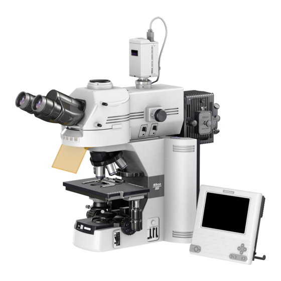
Summary of Contents for Nikon Eclipse 80i
- Page 1 M318E 03.12.CF.1(2/2) Microscope ECLIPSE 80i Instructions <Reference>...
-
Page 3: Introduction
Introduction Thank you for purchasing this Nikon product. This instruction manual, which describes basic microscope operations, is intended for users of the Nikon ECLIPSE 80i microscope. To ensure correct use, please read this manual carefully before operating the product. • This manual may not be reproduced or transmitted in whole or in part without Nikon's express consent. -
Page 4: Warning/Caution Symbols Used In This Manual
Decontaminate the contaminated part according to the standard procedure specified for your laboratory. Caution for heat This symbol found on the lamphouse of the ECLIPSE 80i indicates the following: • The lamp and surrounding areas (including the lamphouse) become very hot during and immediately after a period of illumination. -
Page 5: Safety Precautions
Abbreviations Used in The Manual The product names and abbreviations used in this manual are given below. The manual uses the following abbreviations: Name of device Abbreviation Microscope ECLIPSE 80i C-ER Eye Level Riser Eye Level Riser C-TE Ergonomic Binocular Tube Ergonomic Binocular Tube... -
Page 6: How To Use This Instruction Manual
How to use this instruction manual How to use this instruction manual This instruction manual is composed of two parts, as below: Manual 1 "Microscopy" describes basic microscope operations that you must follow. Please read this manual carefully before operating the product. Manual 2 "Reference"... -
Page 7: Table Of Contents
Contents Contents Introduction ....................... 1 Warning/Caution symbols used in this manual ..............2 Meaning of symbols used on the product ................. 2 Safety Precautions ....................3 WARNING....................3 CAUTION..................... 5 Abbreviations Used in The Manual ................... 3 How to use this instruction manual.................. 4 Contents........................ - Page 8 Contents 12.1 Adjusting the aperture diaphragm opening using the condenser scale ....22 12.2 Adjusting the aperture diaphragm opening using the centering telescope (optional)22 Selecting a Condenser ..................23 Adjusting the Field Diaphragm ................23 Oil Immersion Operation ..................24 Water Immersion ....................
- Page 9 Contents...
-
Page 10: Individual Operations
Individual Operations Item Title Operating sections Power ON/OFF Power switch, battery, AC adapter Brightness Adjustment Brightness control knob, preset switch, ND filter, color compensating filter, filter holder Optical Path Switching Optical path switching knob Vertical Stage Motions Coarse/fine adjustment knobs, coarse torque adjustment ring, refocusing lever Lateral Stage Motions X knob, Y knob, XY knob torque adjustment screws... -
Page 11: Power On/Off
Individual Operations 1 Power ON/OFF Power ON/OFF Turning on the microscope To turn on the microscope, press the power switch to the “|” position. To turn off the microscope, press the power switch to the “◯” position. Power switch DIH-M Power Supply Push in the power switch located on the side the C-Box to switch on power for the DIH-M. -
Page 12: Brightness Adjustment
Individual Operations 2 Brightness Adjustment Brightness Adjustment Image brightness can be adjusted by the following methods: Method Operating controls Explanation Transmitted Adjusting lamp voltage Brightness control knob image (The 50i is subject to shifts Preset switch in color temperature.) ND filter ND filter IN/OUT lever attachment/detachment (for 50i only) -
Page 13: Adjustment Using The Preset Switch
Individual Operations 2 Brightness Adjustment Adjustment using the preset switch Pushing in the preset switch switches the lamp voltage to 9V. Push in the preset switch and move the NCB11 filter into the optical path to achieve optimum color reproduction accuracy. Push the switch to restore it to the out position. -
Page 14: Transmitted Image In Fluorescence Observation
Individual Operations 2 Brightness Adjustment ND filter sliders ND filter sliders Epi-illumination attachment DIH-M Brightness ND16 1/16 1/32 1/64 1/128 1/512 Transmitted image in fluorescence observation For fluorescence observations, turn off the microscope power switch to cancel the transmitted image. Bright ambient lights will make it more difficult to view the image. -
Page 15: Optical Path Switching
Individual Operations 3 Optical Path Switching Optical Path Switching Optical path distribution With the ergonomic binocular tube or trinocular eyepiece tube, the optical path switching lever allows distribution of light to the binocular section and camera port. Optical path distribution (%) Position of optical path switching lever Binocular section... -
Page 16: Disabling The Clicking Of The Optical Path Switching Lever
Individual Operations 3 Optical Path Switching With the DIH-M, two optical path switching levers allows distribution of light to the binocular section, front port and rear port. Position of optical Position of optical Optical path distribution (%) path switching path switching Binocular Front Rear port... -
Page 17: Vertical Stage Motion
Individual Operations 4 Vertical Stage Motion Vertical Stage Motion Prohibited actions Avoid the following actions, which can cause equipment malfunctions. Rotating the right and left coarse/fine focus knobs in opposite directions. • • Rotating the coarse focus knob past the stopper. Knob rotation direction and stage motion direction Turn the coarse or fine focus knob to raise or lower the stage and to adjust image focus. -
Page 18: Adjusting The Rotating Torque Of The Coarse Focus Knob
Individual Operations 4 Vertical Stage Motion Adjusting the rotating torque of the coarse focus knob Adjust the rotation torque of the coarse focus knob (rotation resistance) by turning the torque adjustment ring (TORQUE) located at the base of the coarse focus knob. If the torque is too low, the stage may descend under its own weight. -
Page 19: Lateral Stage Motion
Individual Operations 5 Lateral Stage Motion Lateral Stage Motion Prohibited action Avoid the following actions, which can cause equipment malfunctions. Moving the stage or the specimen holder to the left and right by holding stage • or holder directly. Knob rotation direction and stage motion direction X direction To move the stage in the X or Y direction, rotate Y direction... -
Page 20: Stage Rotation
Individual Operations 6 Stage rotation Stage rotation Stage with Rotating Mechanism Loosen the rotation clamp screw on the bottom Rotation clamp surface of the stage to allow the stage to swivel. screw Centering the Stage 180° (1) Move the 10x objective into the optical path rotation and focus on the specimen. -
Page 21: Diopter Adjustment
Individual Operations 8 Diopter Adjustment Diopter Adjustment Diopter adjustment compensates for differences in visual acuity between the right and left eyes, improving binocular observation. It also minimizes focal deviations when switching objectives. Adjust diopter settings for both eyepiece lenses. (1) Turn the diopter adjustment ring of each Line eyepiece lens and align the end face of the diopter adjustment ring with the line. -
Page 22: Interpupillary Adjustment
Individual Operations 9 Interpupillary Adjustment Interpupillary Adjustment Interpupillary adjustment improves the ease of binocular observation. The right and left fields of view Perform steps 1) to 10) in “Bright-Field Microscopy” converge until in the "Microscopy" instruction manual and focus on they coincide. -
Page 23: Adjusting The Condenser Position
Individual Operations 11 Adjusting the Condenser Position Adjusting the Condenser Position Adjust the condenser (focusing and centering) so that the light passing through the condenser forms an image at the correct location (center of the optical path) on the specimen surface. (1) Perform steps 1) to 10) in “Bright-Field Microscopy”... -
Page 24: Adjusting The Aperture Diaphragm
Generally, aperture settings that are 70 to 80% of the maximum aperture of the Plan 40X objective will provide satisfactory images with 40x / 0.75 Nikon JAPAN suitable contrast. Plan 40x 40x / 0.75 / -WD / -WD Since an excessively small aperture diaphragm Condenser aperture diaphragm scale=... -
Page 25: Selecting A Condenser
Individual Operations 13 Selecting a Condenser Selecting a Condenser Condenser ( : Optimum, : Suitable, x: Not suitable) Objective Achromatic/aplanat Achromat Abbe condenser 1-100x condenser magnification condenser condenser Note 1 Note 1 10x to 100x Note 1: The entire field of view may not be covered if a UW eyepiece is attached. Depending on the type of objective, the indicated numerical aperture of the objective may not be achieved. -
Page 26: Oil Immersion Operation
Individual Operations 15 Oil Immersion Operation Oil Immersion Operation Objectives marked “Oil” are oil-immersion objectives. Objectives of this type are used with immersion oil applied between the specimen and the tip of the objective. For maximum performance, oil-immersion objectives with numerical apertures of 1.0 or higher should be combined with oil-immersion chromatic/aplanat condensers. -
Page 27: Water Immersion
Individual Operations 16 Water Immersion Water Immersion Objectives marked “WI” or “W” are water-immersion objectives. These objectives are used with immersion water (distilled water or physiological saline) applied between the specimen and the tip of the objective. Microscopy procedures are the same as for oil-immersion objectives. Since water evaporates readily, monitor the immersion water during observation. -
Page 28: Fluorescence Observation
Individual Operations 17 Fluorescence Observation Fluorescence Observation Warning 17.1 The mercury lamp (or xenon lamp) used with the Epi-illumination attachment or DIH-M requires careful handling. Be sure to read the warnings described in the beginning of this manual and in the operating manual provided by the manufacturers of the super high-pressure mercury light source (or high-intensity light source) and lamp. -
Page 29: Field Diaphragm Of The Epi-Illumination Attachment
Individual Operations 17 Fluorescence Observation Field diaphragm of the Epi-illumination attachment 17.4 The field diaphragm controls the illumination on the area of the specimen being viewed. Operating the field diaphragm lever changes the size of the field diaphragm. Position of field diaphragm open/close Field diaphragm setting lever Pushed in... -
Page 30: Epi-Illumination Aperture Diaphragm
Individual Operations 17 Fluorescence Observation Epi-illumination Aperture Diaphragm 17.5 The aperture diaphragm determines the numerical aperture for illumination and the optical system. In Epi-fluorescence microscopy, it is used to adjust image brightness. Use the aperture diaphragm open/close lever to adjust the size of the opening of the aperture diaphragm. Position of aperture diaphragm Size of aperture diaphragm open/close lever... -
Page 31: Switching Excitation Methods
Individual Operations 17 Fluorescence Observation (6) Stop down the aperture diaphragm. (Pull the Aperture diaphragm aperture diaphragm lever.) open/close lever (7) While observing the centering tool screen, move the center of the aperture diaphragm image to the center of the screen. (Using the hexagonal screwdriver provided with the Aperture diaphragm microscope, turn the field diaphragm... -
Page 32: Epifluorescent Nd Filters
Individual Operations 17 Fluorescence Observation Epifluorescent ND Filters 17.7 Pushing in an ND filter slider sets the respective ND filter into the optical path and darkens the fluorescent image. ND filters reduce light intensity without altering the color balance of the illumination. Higher filter numbers correspond to lower transmission rates (i.e., darker images). -
Page 33: Selecting Fluorescent Filters
Individual Operations 18 Selecting Fluorescent Filters Selecting Fluorescent Filters A filter cube is comprised of the following three optical components: an excitation filter (EX filter), a barrier filter (BA filter), and a dichroic mirror (DM). Select a combination of filter cubes appropriate for the specimen characteristics, Barrier filter fluorescent pigment, and the purpose intended. -
Page 34: Selection Of Barrier Filter (Ba Filter)
Individual Operations 18 Selecting Fluorescent Filters Narrow EX filter bandwidth Wide Brightness of fluorescent image Dark Light Generation of self-fluorescence High Degree of color fading High Selection of barrier filter (BA filter) 18.2 The barrier filter allows only fluorescent light emitted by the specimen to pass, blocking excitation light. -
Page 35: Replacing Excitation And Barrier Filters
Individual Operations 18 Selecting Fluorescent Filters BP filter (bandpass filter) The bandpass filter passes only light of a certain wavelength, blocking all other wavelengths. BP filters are used for microscopy of fluorescent BA520-560 (BP type) images involving a specific dye in multiple-dye specimens. -
Page 36: Excitation Light Balancer
Individual Operations 19 Excitation Light Balancer Excitation Light Balancer Mount the optional D-FB excitation light balancer to the Epi-illumination attachment or DIH-M to adjust the wavelength characteristic of the excitation light. The excitation light balancer is used in combination with a dual-band filter cube. Required accessaries •... - Page 37 Individual Operations 19 Excitation Light Balancer Detailed view of excitation light balancer B Effective diameter on aperture diaphragm plane A Transmission rate 100% B TRITC FITC DAPI Texas-Red Wavelength The excitation light balancer is set so that the transmission rate remains at about 100% for typical dark fluorescent FITC.
-
Page 38: Image Capture
Individual Operations 20 Image Capture Image Capture Images can be captured by mounting a camera head to the ergonomic binocular tube or trinocular eyepiece tube. For more detailed discussion of this topic, refer to the operating manual provided with the camera head or camera control software. -
Page 39: Fluorescence Photomicrography
Individual Operations 20 Image Capture Fluorescence photomicrography 20.6 The fluorescence of fluorescent specimens may fade during exposure. To prevent this, do the following: Select a brighter optical combination. Even if the overall magnification is the same on the monitor, the combination of objective and camera zoom can result in significant variations in exposure time.


Need help?
Do you have a question about the Eclipse 80i and is the answer not in the manual?
Questions and answers