Table of Contents
Advertisement
Quick Links
Advertisement
Table of Contents

Related Manuals for Mindray DC-T6


Summary of Contents for Mindray DC-T6
- Page 1 DC-T6 Diagnostic Ultrasound System Operator’s Manual [Basic Volume]...
-
Page 3: Table Of Contents
Contents Contents ..........................i Intellectual Property Statement ......................I Responsibility on the Manufacturer Party ................... I Warranty ............................II Exemptions ........................... II Customer Service Department ..................... II Company Contact ........................III Important Information ........................III About This Manual ..........................IV Notation Conventions ........................ - Page 4 3.3.2 Powering OFF the System .................... 3-5 3.3.3 Standby ......................... 3-6 Control Panel Position Adjusting ..................3-7 Monitor Adjusting ........................3-7 3.5.1 Monitor Position Adjusting .................... 3-7 3.5.2 Adjusting Brightness/Contrast on the Monitor .............. 3-8 Connecting a Probe ......................3-9 3.6.1 Connecting a Probe ......................
- Page 5 Power Mode Image Optimization ..................5-20 5.6.1 Power Mode Exam Protocol ..................5-20 5.6.2 Power Mode Image Parameters ................. 5-21 5.6.3 Power Mode Image Optimization ................5-21 PW/CW Doppler Mode Optimization .................. 5-22 5.7.1 PW / CW Mode Exam Protocol ................... 5-22 5.7.2 PW/CW Mode Image Parameters ................
- Page 6 6.7.2 Protocol Edit ........................ 6-12 Saving Stress Echo Data ....................6-14 Exiting the Stress Echo Feature ..................6-14 6.10 Measurement ........................6-14 Display & Cine Review ....................7-1 Image Display ........................7-1 7.1.1 Splitting Display......................7-1 7.1.2 Image Magnification ...................... 7-1 7.1.3 Spot ..........................
- Page 7 10.2.2 Adding Body Marks ..................... 10-5 10.2.3 Moving Body Marks ....................10-6 10.2.4 Deleting Body Marks ....................10-6 11 Patient Data Management ..................11-1 11.1 Patient Information Management ..................11-1 11.1.1 Enter Patient Information .................... 11-1 11.1.2 Patient Information Setting ..................11-1 11.2 Image File Management .....................
- Page 8 12.6 DICOM Task Management ....................12-23 13 Probes and Biopsy ....................13-1 13.1 Probe ..........................13-1 13.1.1 Name and Function of Each Part of the Probe ............13-5 13.1.2 Orientation of the Ultrasound Image and the Probe Head ......... 13-6 13.1.3 Procedures for Operating ...................
- Page 9 15.6.2 Comment Softkey Preset ..................15-19 15.7 Peripheral Preset ......................15-19 15.8 Network Preset ......................... 15-21 15.9 Manage Settings ....................... 15-22 15.9.1 Exporting Setup Data ....................15-22 15.9.2 Importing Setup Data ....................15-23 15.10 Maintenance ........................15-23 15.11 System Information ......................15-23 16 Batteries ........................
-
Page 11: Contents
©2011-2017 Shenzhen Mindray Bio-Medical Electronics Co., Ltd. All rights Reserved. For this Operator’s Manual, the issue date is 2017-04. The MindrayVNC ver. 1.0 contained in this product is revised by MINDRAY on Aug, 2009, based on the UltraVNC ver. 1.0.5.5, and it complies with GNU General Public License. -
Page 12: Warranty
FITNESS FOR ANY PARTICULAR PURPOSE. Exemptions Mindray's obligation or liability under this warranty does not include any transportation or other charges or liability for direct, indirect or consequential damages or delay resulting from the improper use or application of the product or the use of parts or accessories not approved by Mindray or repairs by people other than Mindray authorized personnel. -
Page 13: Company Contact
7. Important data must be backed up on external memory media. 8. Mindray shall not be liable for loss of data stored in the memory of this system caused by operator error or accidents. -
Page 14: About This Manual
9. This manual contains warnings regarding foreseeable potential dangers, but you shall always be alert to dangers other than those indicated as well. Mindray shall not be liable for damage or loss that results from negligence or from ignoring the precautions and operating instructions described in this operator’s manual. -
Page 15: Software Interfaces In This Manual
2. When you find that the contents of the manuals in CD are NOT consistent with the system or English manuals, please ONLY refer to the corresponding English manuals. 3. The accompanying manuals may vary depending upon the specific system you purchased. -
Page 17: Safety Precautions
Safety Precautions Safety Classification According to the type of protection against electric shock: Class I equipment powered by outer power & equipment powered by inner batteries According to the degree of protection against electric shock: Type-BF applied part According to the degree of protection against harmful ingress of water: The main unit belongs to IPX0, and the probes belong to IPX7 Footswitch: 971 SWNOM belongs to IP68 Footswitch: SP-997-350 (3-pedal) belongs to IP68... -
Page 18: Meaning Of Safety Symbols
Meaning of Safety Symbols Symbol Description Type-BF applied part. The ultrasound probes connected to this system are type-BF applied parts. The ECG module connected to this system is Type-BF applied part. General warning, caution, risk of danger. Safety Precautions Please observe the following precautions to ensure patient and operator’s safety when using this system. - Page 19 DO NOT use a probe that has a damaged, scratched surface, or exposed wiring of any kind. Immediately stop using the probe and contact Mindray Customer Service Department or sales representative. There is risk of electric shock if a damaged or scratched probe is used.
- Page 20 Accessory equipment connected to the analog and digital interfaces must comply with the relevant IEC standards (e.g., IEC 60950 information technology equipment safety standard and IEC 60601-1 medical equipment standard). Furthermore, all configurations must comply with the standard IEC 60601-1-1. It is the responsibility of the person, who connects additional equipment to the signal input or output ports and configures a medical system, to verify that the system...
- Page 21 You cannot repair the system under this circumstance and must call the Mindray Customer Service Department or sales representative. There is no risk of high-temperature burns during normal ultrasound examinations.
- Page 22 The system and its accessories are not disinfected or sterilized prior to delivery. The operator is responsible for the cleaning and disinfection of probes and sterilization of biopsy brackets according to the manuals, prior to the use. All items must be thoroughly processed to completely remove harmful residual chemicals, which will not only harmful to the human body, but also damage the accessory.
- Page 23 If the system is used in a small room, the room temperature may rise. Please provide proper ventilation and free air exchange. 10. To dispose of the system or any part, contact Mindray Customer Service Department or sales representative. Mindray is not responsible for any system content or accessories that have been discarded improperly.
- Page 24 The ultrasonic probe is only for use with the specified WARNING: ultrasonic diagnostic system. Please refer to 2.5 System Configuration to select the proper probe. Ultrasound transducers can only be used by qualified professionals. Confirm that the probe and cable are normal before and after each examination.
- Page 25 Please use the disinfection or sterilization solution that recommended in this operator’s manual; otherwise Mindray will not be liable for damage caused by other solutions. If you have any questions, please contact Mindray Customer Service Department sales representative. The probe sheath contains natural rubber that can cause allergic reactions in some individuals.
-
Page 26: Latex Alert
Latex Alert When choosing a probe sheath, it is recommended that you directly contact CIVCO for obtaining probe sheath, pricing information, samples and local distribution information. For CIVCO information, please contact the following: CIVCO Medical Instruments Tel: 1-800-445-6741 WWW.civco.com Allergic reactions in latex (natural rubber) sensitive patients WARNING: may range from mild skin reactions (irritation) to fatal anaphylactic shock, and may include difficulty in breathing... -
Page 27: Warning Labels
Warning Labels The warning labels are attached to this system in order to call your attention to potential hazards. The symbol on the warning labels indicates safety precautions. The warning labels use the same signal words as those used in the operator’s manual. Read operator’s manual carefully before using the system. -
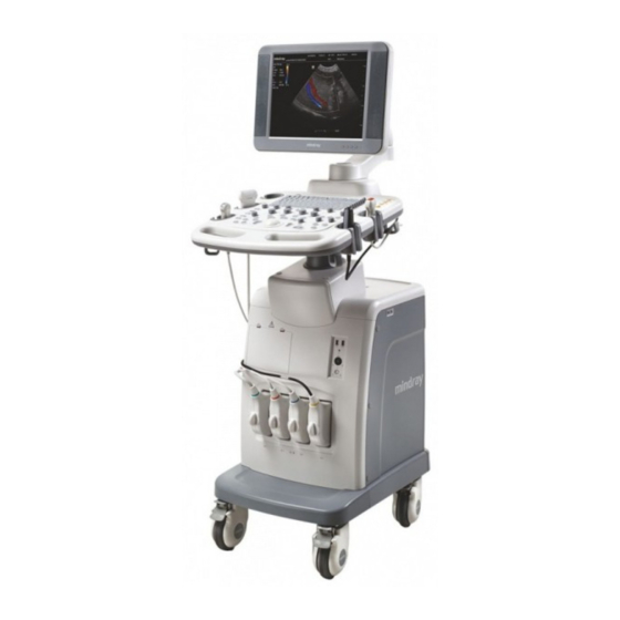
Page 29: System Overview
System Overview Intended Use The DC-T6 Diagnostic Ultrasound System is applicable for adults, pregnant women, pediatric patients and neonates in fetal, abdominal, pediatric, small organ (breast, thyroid, testes), neonatal cephalic, adult cephalic, trans-rectal, trans-vaginal, musculo-skeletal (conventional, superficial), cardiac (adult, pediatric), peripheral vascular and urology exams. -
Page 30: Product Specifications
Product Specifications 2.4.1 Imaging Modes B Mode M Mode Anatomical M: Free Xros M, Free Xros CM Color M Mode (CM) Color Free Xros M C Mode Color Power (Dirpower) D Mode PW Doppler CW Doppler Special Imaging Smart 3D Static 3D iScape 2.4.2... -
Page 31: Environmental Conditions
2.4.3 Environmental Conditions Operating conditions Ambient temperature 0℃~40℃ 10℃~40℃ (For 4CD4 only) Relative humidity 30%~85% (no condensation) Atmospheric pressure 700hPa~1060hPa Storage and transportation conditions Ambient temperature -20℃~55℃ -10℃~60℃ (For 4CD4 only) Relative humidity 30%~95% (no condensation) Atmospheric pressure 700hPa~1060hPa Do not use this system in the conditions other than those WARNING: specified. -
Page 32: System Configuration
System Configuration 2.5.1 Standard Configuration Main unit Accessories Operator's manuals Ultrasound gel (made by Eco-Med Pharmaceuticals, model: Eco Gel 200 ) Ultrasound gel holder Ultrasound probe holder System power line Dust-proof cover Probe ports dust-proof cover 4D probe port dust-proof cover Grounding line Video printer remote line Multilingual controls overlay... - Page 33 Probe model Intended Use Abdominal, Pediatric, Small organ, Neonatal Cephalic, Musculo-skeletal L14-6 Conventional, Musculo-skeletal Superficial, Peripheral Vascular P4-2 Abdominal, Pediatric, Adult Cephalic, Cardiac Adult, Cardiac Pediatric 4CD4 Fetal, Abdominal Abdominal, Pediatric, Neonatal Cephalic, Adult Cephalic, Cardiac Adult, P7-3 Cardiac Pediatric Pediatric, Small organ (Small organ-breast, thyroid, testes), L14-6N Musculo-skeletal Conventional, Musculo-skeletal Superficial, Peripheral...
- Page 34 13G, 15G, NGB-011 (metal/needle P4-2/2P2 11°, 23° 16G, 18G, un-detachable) 14G, 16G, NGB-015 25°, 35°, 18G, 20G, C5-2 45° Metal/needle detachable 14G, 16G, NGB-016 30°, 40°, L14-6 18G, 20G, 50° Metal/needle detachable 2.5.2.2 Options Item iClear module CW module (requires to be configured in factory) iScape module Free Xros M module Free Xros CM (Curved Free Xros M)module...
- Page 35 Item DICOM Basic module (including: task management, DICOM storage, DICOM print, DICOM storage commitment, DICOM media storage (including DICOM DIR) and etc.) DICOM Worklist module (only can be applied with the DICOM basic function module configured) DICOM MPPS (only can be applied with the DICOM basic function module configured) DICOM OB/GYN structured report (only can be applied with the DICOM basic function module configured)
- Page 36 Item Model Sony DVO-1000MD SYMBOL LS2208 Barcode reader SYMBOL DS6707 Parts that can be used within patient environment: Main unit Probes Footswitch Printers: SONY UP-D897, MITSUBISHI P93DC, MITSUBISHI P93W, SONY UP-897MD, SONY UP-D25MD, SONY UP-D23MD, MITSUBISHI CP910E, SONY UP-D898MD, SONY UP-X898MD NOTE: If the ultrasound system can not recognize the SONY UP-X898MD and SONY UP-D898MD printers automatically, you may need to change the settings on...
-
Page 37: Introduction Of Each Unit
Introduction of Each Unit System Overview 2-9... -
Page 38: I/O Panel
Name Function Monitor Displays the images and parameters during scanning. Control Panel Operator-system interface or control. DVD drive DVD drive. Used for turning on/off the power supply (on the upper Power button left corner of control panel). Video printer room Used for placing black/white video printer. -
Page 39: Power Supply Panel
Symbol Function <1> USB port, used for connecting USB devices <2> Parallel port, connects the parallel port devices, reserved. <3> Unavailable to the user. <4> Ethernet interface (<3> is reserved for future use). <5> Remote interface for connecting video printer <6>... -
Page 40: Physiological-Signal Panel
Name Function Used for equipotential connection, balancing the protective Equipotential <1> earth potentials between the system and other electrical terminal equipment. <2> Power inlet AC power inlet <3> Power outlet Supply power for optional peripheral devices (e.g. VCR). <4> Circuit <5>... -
Page 41: Control Panel
2.10 Control Panel Name Description Function <1>. Power button It does not illuminate when the system is turned off. Press the button to turn on the system, at the moment the indicator lights on and becomes green and flashes .When the system enters work status the indicator becomes green. - Page 42 Name Description Function 2. When the battery capacity is charged to the full capacity, the indicator color turns green. 3. When the system is supplied with power by the batteries and the power capacity is more than 20%, the indicator color is green. 4.
- Page 43 Name Description Function controls 1 displayed on the bottom of the screen. Refer to the subsequent contents for specific functions. <19>. Soft menu Press or rotate to select the soft menu items controls 2 displayed on the bottom of the screen. Refer to the subsequent contents for specific functions.
- Page 44 Name Description Function make the 3D image to rotate around X axis. <36>. Pressable Press to enter PW mode, and rotate to adjust PW(Y) knob PW (in PW mode) or CW gain (in CW mode); while in 3D/4D mode, rotate the knob to make the 3D image to rotate around Y axis.
-
Page 45: Symbols
Name Description Function <56>. Pressable Multifunctional knob. knob <57>. Menu Menu Press to display or hide a mode-specific parameter menu. <58>. Freeze Freeze Press to freeze or unfreeze the image. <59>. Press to confirm an operation, same as the left-button of the mouse. <60>. - Page 46 Symbol Description USB port S-VIDEO signal interface; VIDEO signal interface Battery Status Indicator Port (reserved) Connects serial port devices Audio signal Microphone input jack Remote control port Unavailable to the user Hard disk status indicator Standby status indicator AC power supply status indicator Product serial number Manufacture date 2-18 System Overview...
-
Page 47: System Preparation
System Preparation Move/Posit the System Please read and understand the safety precautions before placing the system to ensure safety for both operator and device. 1. Switch off the power, unplug the power cord. 2. Disconnect all cables from the off-board peripheral devices (printer, recorder, etc.). 3. - Page 48 2. Push the retaining clamp downward, and lock the power cord, as shown in the figure below. 3. Plug the other end power plug into an appropriate power socket. The grounding terminal should be connected with a power grounding cable to ensure that protective grounding works normally.
-
Page 49: Powered By Batteries
2. Be sure to connect this system and its peripheral devices to wall receptacles, which meet the rated power requirement written on the nameplate. If adapters or multifunctional receptacles are used, it may cause the leakage current to exceed the safety requirement. In addition, please connect the video printer to the special power cable in the video printer room of this system or the special auxiliary power outlet of this... -
Page 50: Power On /Off
If the system begins to function improperly – immediately stop scanning. If the system continues to function improperly – fully shut down the system and contact Mindray Customer Service Department or sales representative. If you use the system in a persistent improperly functioning state –... -
Page 51: Powering Off The System
3. Press the power button in the upper left corner on the control panel 4. The system enters the work status. When the battery capacity is sufficient, you can also press the power button directly to turn on the system. To check the system after the system is turned on: Check Item <1>... -
Page 52: Standby
Shut down: To power off the system normally. Standby Click to enter the standby status. If you want to exit standby, press the button. Cancel: to cancel the operation. To shut down the system in a direct way if you cannot do it normally: Long press the power button and the system will power off without displaying the [Shutdown Confirm] screen. -
Page 53: Control Panel Position Adjusting
Control Panel Position Adjusting To adjust height of the control panel: Use one hand to pinch the button located at the left internal side of the handle. Use the other hand to move the control panel up or down. When you adjust the control panel to a desired position, loosen your hand on the button and the position of the control panel is fixed. -
Page 54: Adjusting Brightness/Contrast On The Monitor
Height adjust (1) Toggle the lever located at the monitor support arm to the unlocking position (2) Move the monitor support arm up or down to adjust the height. (3) When you adjust the monitor to a desired position, toggle the lever to the locking position and the monitor height is fixed. -
Page 55: Connecting A Probe
Hang the probe cable to the hanger located under the control panel to avoid excessively bending and damaging the cable. Only use the probes provided by Mindray. Aftermarket probes may result in damage or cause a fire. NOTE: If a probe port is not used for a long period of time, please use the dustproof cover to protect the probe port from dust;... -
Page 56: Disconnecting A Probe
1. Keep the cable end of the probe to the right side of the system, and insert the connector into the port of the system, and then press in fully (Shown as the left figure). 2. Turn the lock handle 90°clockwise to lock it securely (Shown as the right figure). 3. -
Page 57: Connecting Peripheral Devices
Connecting Peripheral Devices 3.7.1 Connecting the USB Devices DO NOT directly remove a USB memory device; otherwise, the USB memory device and/or the system may be WARNING: damaged. Available USB ports on the system: 1. Two USB ports on the physiological-signal panel. 2. -
Page 58: Graph /Text Printer
Function setting The function of the footswitch can be set, for details, please refer to “15.1.7 Key Config”. 3.7.3 Graph /Text printer Connecting a local printer As shown in the figure below, a graph /text printer has a power cord and data cable. The power cord shall be directly connected to a wall receptacle as required. - Page 59 4. Select "Add Local Printer" and click [Next] to enter the screen of browsing driver; select the desired driver and click [OK] to install the driver. Printers listed in "2.5 System Configuration" have drivers installed already. Click [Printer Attribute] to see the printer attribute. 5.
-
Page 60: Video Printer
Please refer to the accompanying manuals of the printers for more details. 3.7.4 Video Printer The video printers supported by the system include analog video printers and digital video printers. The video printers, whether analog or digital, consist of B / W printers and color printers. - Page 61 4. Place the B/W video printer into the printer room, make the foot mats clip in the groove to fix the printer. 5. Collect the window cover to a proper place. 6. Load a paper roll, and turn on the system and printer. 7.
- Page 62 Select the image to be printed on the iStation or Review screen, and click [Send To] to select the printer to print. To print by the user-defined key (<Print> key as an example): a) Enter "[Setup] → [System Preset] → [Key Config]". b) Select "Print"...
-
Page 63: Installing The Barcode Reader
5. If it is a digital color video printer, connect the power cable and USB cable to the system. Operations of adding/modifying print service is same as the operations of the B/W video printer, please refer to it. Please refer to the accompanying manuals of the printers for more details. 3.7.5 Installing the Barcode Reader Directly insert the USB port of the barcode reader to the system applicable USB ports. - Page 64 To preset whether gender, age or operator is displayed: Enter "[Setup]→[System Preset]→[General Preset]" and check "Gender", "Age" or "Operator" in the [Patient Info] box in the upper left corner of the screen. Logo Manufacture logo, displayed in the upper left corner of the screen. Hospital name Display the hospital name.
- Page 65 Menu title Page-turning button Drop-down list button Items Page-turning button Shared items Menu title Displays the menu name. Divide the menu items according to measurement conditions. Page-turning button When there are too many items in a menu, the items will be divided into more than one page.
- Page 66 For an item with sub-menu: press the knob to extend the sub-menu, and the cursor navigates to the first sub-item. Operate the sub-menu through the multifunctional knob. At the same time, you can exit from the sub-menu to the previous item by clicking “Exit” in the sub-menu. For tab items in the menu: e.g.
- Page 67 The soft menu controls are located at the top of the control panel, shown in the following figure. Page-turning: Use the up/down keys in directional soft menu controls <21> to turn pages up or down; you can operate the items only when they are highlighted. Mode switching: Left / right keys in directional soft menu controls <21>...
-
Page 68: Basic Operations Of Screens
3.8.2 Basic Operations of Screens A screen consists of title, page tabs, contents and buttons, as shown in the following figure: Title Content Control button Composition Description The title bar is used to give a description for the content and Title Bar function of the screen. -
Page 69: Exam Preparation
Exam Preparation Before examining a new patient, press <End Exam> to end the CAUTION: exam of the previous patient, update the patient ID and information, to avoid mixing data of the next new patient. To Start an Exam You can start a patient exam in the following situations: New patient information: Enter the patient information, if it is a new patient, refer to "4.2.1 New Patient Information"... - Page 70 You can also change the cursor position by <Tab>, <Enter> or direction keys. Detailed information is described as follows: 1. General information Patient ID Once you enter the ID and confirm it, you are not allowed to change it. There are 2 ways to generate the patient ID.
- Page 71 You can either enter the birth date of a patient manually according to the format displayed in the field, or click to select the date. In the table, you can select the desired year (or enter it manually); month and day, then click [OK] to finish it. Age: Auto generated age: once the DOB is entered, the system can display an auto-generated age in the field box;...
- Page 72 Exam Type Information Description According to the entered index (can be selected in the drop-down list); including LMP (last menstrual period), IVF (in vitro fertilization), PRV (previous exam date), BBT (basic body temperature)), the system can automatically calculate GA and EDD (estimated delivery date); or, calculates GA and LMP according to the EDD and entered date.
-
Page 73: Retrieve Patient Information
Exam Type Information Description SMP (Small None Parts) None (Pediatrics) 3. Operating Information Accession #: exam number used in DICOM. It should be entered within 16 letters or characters; “\” is not permitted. Diagnostician: people who is responsible for the exam. "\", "^","=" and "," are not permitted. - Page 74 2. Select the data source: Select the data source in the drop-down list of "Data Source". The occupying space percentage of the selected data source will be displayed. 3. Set the searching condition Define the searching item: Name, ID, BOD or Exam Date; and then enter the related keyword.
-
Page 75: Select An Exam Mode And Probe
1. Select data source: choose a worklist server in the drop-down list of “Worklist Server”, and then all the patient exam records in the server are listed out. 2. Set the searching condition: Enter the period which includes the exam date, and click [Query] to search. Enter patient ID, patient name, accession #, the system provides the result in real-time. -
Page 76: Select The Imaging Mode
(2) Roll the trackball and press <Set> to select the exam mode. To save the image parameters for the current exam mode quickly: (1) Press <Probe> to open the dialog box. (2) Click [Save], to save the image parameters of the current image mode as setup data. -
Page 77: Continue An Exam
The system can automatically load the patient information and exam data to continue the exam. If you want to activate an exam with data in an external memory database, you have to first import the patient data to the system’s patient database. 4.5.2 Continue an Exam Select an exam that is paused within 24 hours, click [Continue Exam] in “iStation”... -
Page 78: Anonymous Patient Exam
4. Select the reason and click [OK]. 5. After the above operations being taken, the exam is cancelled, and images, measurements and report are saved with the status of the exam to be "Canceled". If the system has been configured with a MPPS server, it will send the status information "Cancelled"... -
Page 79: Image Optimization
Image Optimization 1. The images displayed in this system are only reference WARNING: for diagnosis. Mindray is not responsible for the correctness of diagnostic results. It is the responsibility of the clinician, who performs the exam, to capture the correct diagnostic results. -
Page 80: Switching Between Imaging Modes
Switching Between Imaging Modes Description B mode knob: press to enter B mode. M mode knob: press to enter M mode. PW mode knob: press to enter PW mode. CW mode key: press to enter CW mode. Color mode knob: press to enter Color mode. Power mode key: press to enter Power mode. -
Page 81: Image Adjustment
Image Adjustment Before optimizing the image by adjusting image parameters, adjust the brightness and contrast of the monitor to the best. Requirement Available Operations Adjust gain Adjust TGC To modify the brightness Adjust [A. power] (do try to adjust gain first before increasing the acoustic power) Adjust [Dyn Ra.] Adjust [Gray Map]... -
Page 82: B Mode Image Optimization
Image mode switching Use left/ right controls in soft menu controls <21> to switch among the modes. The soft menu items vary depending on the mode. The menu navigation will cause the change of the menu, while the change of the menu will cause the menu navigation. - Page 83 Display F 5.0 D 17.6 G 50 FR 40 IP 4 DR 60 Parameter Frequency Depth Gain Frame Rate B IP B Dynamic Range Parameters that can be adjusted to optimize the B Mode image are indicated in the following. Adjustment Items Control Panel...
- Page 84 Operation To increase the gain compensation at an area of interest, move the TGC slider to the right. To decrease the gain compensation at the corresponding area of interest, move the TGC slider to the left. About 1.5s after the adjustment is finished, the TGC curve disappears. Effects Adjust the signal gain for the certain image area to get a balanced image.
- Page 85 Impacts The greater the number of focus, the slower the frame rate to image. Imaging Display Adjustment Description More information can be obtained without moving the probe or changing the sampling position. FOV (Field Change the scan range through the [FOV] item in the soft menu or menu. of View) The system provides four levels of scan range: W, N, M1, and M2.
- Page 86 Effects The more the dynamic range, the more specific the information, and the lower the contrast with more noise. iClear Description This function is used to increase image profile, so as to distinguish the image boundary. Operation Adjust through the [iClear] item in the soft menu or menu. The system provides 5 levels of iClear effects adjustment, off represents no iClear is turned on, and the bigger the value the stronger the effect.
- Page 87 Operation Adjust through the [iBeam] item in the soft menu or menu. The system provides 3 levels of iBeam adjustment, off represents no iBeam, and while 2 represents maximum iBeam optimization. Effects Images after iBeam processing can be optimized with less spot noise and higher resolution, so that more details for the structure are revealed.
- Page 88 Impacts The function is available in real-time imaging, freeze or cine review status. TSI (Tissue Specific Imaging) Description The TSI function is used to optimize the image by selecting acoustic speed according to tissue characteristics. Operation Select among the TSI modes through the [TSI] item in the soft menu or menu.
-
Page 89: M Mode Image Optimization
HScale Description Display or hide the width scale (horizontal scale). The scale of the horizontal scale is the same as that of vertical scale (depth), they change together in zoom mode, or when the number of the image window changes. When image is turned up/down, the HScale will also be inverted. - Page 90 During M mode imaging, menus for B mode and M mode are displayed in the soft menu at the same time; use the left/right keys of directional soft menu controls to switch between the menus of B mode and M mode. During M mode scanning, frequency and acoustic power of the probe are synchronous with that of B mode.
- Page 91 Speed Description This function is used to set the scanning speed of M mode imaging, and the real-time speed value is displayed in the image parameter area in the upper left corner of the screen. Operation Change the speed through the [Speed] item in the soft menu or menu. Or adjust it in the image parameter area.
-
Page 92: Color Mode Image Optimization
Click [γ] in the soft menu or menu to adjust. The adjusting range is 0-3. Impacts The function is available in real-time imaging, freeze or cine review status. The post process adjustment will not influence the cine review. Gray Map Description This function applies the gray correction to obtain the optimum image through adjusting 3 parameters: curve, gray rejection and γ. -
Page 93: Color Mode Exam Protocol
Generally, the color above the color bar indicates the flow towards the probe, while the color below the color bar indicates the flow away from the probe; the brighter the color, the faster the flow speed; while the darker the color, the slower the flow speed. 5.5.1 Color Mode Exam Protocol 1. - Page 94 Operation Rotate the <Color> knob clockwise to increase the gain, and anticlockwise to decrease. Or adjust it in the image parameter area. The adjusting range is 0-100. Impacts Increasing the gain will increase the flow signal presented as well as noise, while the signals may be missing when the gain is adjusted too low.
- Page 95 Impacts Frame rate will increase when the function is turned on. Dual Live Description This function is used to display B image and Color image synchronously. Operation Click [Dual Live] in the soft menu or menu to turn on or off the function. When the function is turned on, the window will be automatically switched to the dual windows (one for B image, and the other for Color image).
- Page 96 Persistence Description This function is to adjust the temporal smooth in Color mode to optimize the image. Operation Adjust through the [Persistence] item in the menu or the soft menu. The system provides 5 levels of persistence adjustment, 0 represents no persistence and the bigger the value the stronger the effect.
- Page 97 Invert Description To set the display mode of the color flow, and the color scale will be inverted when the function is turned on. Operation Turn on or off the function through the [Invert] item in the soft menu or menu.
-
Page 98: Power Mode Image Optimization
Priority Description This function is used to set levels of the flow display, to display the grayscale signal or color signal. Operation Select the value through the [Priority] item in the soft menu or menu. The adjusting range is 0-100%. Effects The higher the value, color signals are prior to be displayed;... -
Page 99: Power Mode Image Parameters
5.6.2 Power Mode Image Parameters In Power mode scanning, the image parameter area in the upper left corner of the screen displays the real-time parameter values as follows: Display F 2.5 G 38 IP 1 WF32 PRF 0.3k Parameter Frequency Power Power Power Wall Pulse Repetition... -
Page 100: Pw/Cw Doppler Mode Optimization
Description This feature indicates the display effect of power image. The maps in Power mode image are grouped into two categories: Power maps and Directional Power maps. Operation Select the map through the [Map] item in the soft menu or menu. There are 8 kinds of maps provided: P0-3 belong to Power Mode maps, while dP0-3 belong to Directional Power Mode maps. -
Page 101: Pw/Cw Mode Image Parameters
CW Sampling Line Angle Angle 0° Adjustment CW Focus Depth SVD 13.2cm 3. Set the SVD by rotating the trackball; adjust the angle and SV size according to the actual situation. 4. Press <PW>/<CW> again or <Update> to enter PW/CW Mode and perform the examination. -
Page 102: Pw/Cw Doppler Mode Optimization
During PW/ CW mode imaging, menus for B mode and PW/ CW mode are displayed in the soft menu at the same time, use the left/right keys of directional soft menu controls to switch the among menus of B mode and PW/ CW mode. In PW/CW mode, acoustic power of the probe is synchronous with that of B mode. - Page 103 Scale Description This function is used to adjust the speed range of color flow, which is adjusted through PRF in the system. The real-time PRF value will be displayed in the image parameter area in the upper left corner of the screen.
- Page 104 Invert Description This function is used to set the display manner of spectrum. Operation Turn on or off the function through the [Invert] item in the soft menu or menu. Select "Auto Invert" in the "[Setup] → [System Preset] → [Image Preset]", then the color bar can automatically invert when the color flow is steered to a certain angle to accommodate operator’s habit of distinguishing flow direction.
- Page 105 Colorize and Colorize Map Description Colorize function provides an imaging process based on color difference rather than gray distinction. Operation Turn on or off the function through the [Colorize] item in the soft menu or menu. Select the map through the [Colorize Map] item in the soft menu or menu. The system provides 10 colorize maps to be selected among.
- Page 106 Display Format Description To set the display format of PW mode image with B mode image. Operation Adjust through the [Display Format] item in the soft menu or menu. There are 4 formats to display the images: V1:1, L/R, V1:2, Full. Impacts The function is available in real-time imaging, freeze or cine review status.
- Page 107 Quick Angle Description To adjust the angle faster in increments of 60°, and the real-time value of which is displayed on the right part of the spectrum map. Operation Adjust through the [Quick Angle] item in the soft menu or menu. There are 3 angles for quickly adjustment: -60°, 0°, and 60°.
-
Page 108: Anatomical M Mode
Post Process Description Post process is used to apply modifications to the image in order to optimize overall image quality. Post process function includes 3 parameters : curve, gray rejection and γ. Gray This function is to reject image signals less than a certain gray scale. Rejection Click [Gray Rejection] in the soft menu or menu to adjust. - Page 109 5.8.1.1 Imaging Shortcut key for Free Xros M mode Assign a user-defined shortcut key for Free Xros M: [Setup] → [System Preset] → [Key Config]. Please refer to "15.1.7 Key Config" for details. Real-time Imaging 1. In real-time B mode or M mode, adjust the probe and image to obtain the desired plane.
- Page 110 During real-time Free Xros M mode imaging, menus for B mode, Free Xros M mode and other modes are displayed in the soft menu at the same time, use the left/right keys of directional soft menu controls <21> to switch the menus. While imaging in freeze mode, menus of image optimization for B mode, Free Xros M mode and other modes are displayed in the soft menu at the same time.
-
Page 111: Free Xros Cm (Curved Anatomical M Mode)
5.8.1.3 Exit In Free Xros M mode, click [Free Xros M] in the soft menu or press <B> or the user-defined key to exit. 5.8.2 Free Xros CM (Curved Anatomical M Mode) Free Xros CM images are provided for reference only, not for CAUTION: confirming a diagnosis. -
Page 112: Tdi
Delete the Sample Line Click the sample line to select it, and click [Delete] to delete the selected line and you can re-depict a new sample line. Tips: Parameters of Free Xros M mode and Free Xros CM mode are dependent from each other, but their functions are the same, parameters of Free Xros CM are not to be introduced, please refer to relevant sections of the M mode and Free Xros M mode. -
Page 113: Tdi Image Parameters
As in TDI mode, [TDI] item in the soft menu is the pathway for entering and exiting TDI mode, you can preset its performance through the path "[Setup] → [Image Preset] → [Menu Preset]". 5.9.2 TDI Image Parameters In TDI mode scanning, the image parameter area in the upper left corner of the screen will display the real-time parameter values as follows: Display F 3.3... -
Page 114: Tdi Image Optimization
5.9.3 TDI Image Optimization Parameters that can be adjusted to optimize the TDI mode image are indicated in the following. Operation Items Control Panel Gain, Depth Baseline, TVI IP, Acoustic Power, Line Density, B Display, Menu and Soft Smooth, Persistence, Focus Position, Packet Size, B/C Wide, Menu Dual Live, Map, Priority, WF, Frequency, Scale, Invert, Tissue State, Line Density... - Page 115 1. Perform image scanning on cardiac muscle, freeze the image and select a range of images for analysis; or select a desired cine loop from the stored images. Tips: Images from the current scan session (already in freeze mode) or from a saved image loop can be used for TDI QA.
- Page 116 Frame marker: a line that perpendicular to the X axis, can be moved horizontally left to right (right to left) by rolling the trackball. Click the check box beside the ROI to set if to hide or to display the TIC curve. 4---Displays ECG trace.
- Page 117 You cannot go outside the image boundary when drawing a freehand ROI. Delete ROI Press <Clear> key to clear out the last ROI; click [Delete All] on the soft menu to clear out all ROIs. The corresponding traces for the deleted ROIs are erased from the plot. Deleting an ROI causes the ROIs to be deleted from all frames in the analysis loop.
-
Page 118: Color M Mode
5.10 Color M Mode Color M mode provides information of color flow or tissue movement on the M mode images to indicate cardiac motion state. It is highly sensitive to the flow or tissue movement. The Color M mode includes Color Flow M mode and Color Tissue M mode (also known as TVM). -
Page 119: Note Before Use
Set the position of the sampling line by moving the trackball left and right, and set the ROI position by moving the trackball up and down. Set the ROI size by moving the trackball up and right. Press <Set> to switch between the cursor status between the ROI position adjustment and ROI size adjustment. -
Page 120: Overview
The region desired is isolated by amniotic fluid adequately. The region to imaging is not covered by limbs or umbilical cord. The best condition is that the fetus keeps still. If there is a fetal movement in Smart 3D or Static 3D imaging, you need a rescanning when the fetus is still. Angle of a B tangent plane The optimum tangent plane to the fetal face 3D/4D imaging is the sagittal section of the face, or the coronal section. - Page 121 Volume data: the image data set of a 3D object rendered from 2D image sequence. 3D image: the image displayed to represent the volume data. View point: a position for viewing volume data/3D image. Section image: tangent planes of the 3D image obtained by algorithm. As shown in the figure below, XY-paralleled plane is C-section, XZ-paralleled plane is B-section, and YZ-paralleled plane is A-section.
- Page 122 Roll the trackball to change the ROI size and position, press the <Set> key to toggle between setting the size (dotted line) and position (solid line, with a small box at each corner of ROI). Curved VOI adjustment Roll the trackball to change the curved VOI position, press <Set> key to switch among the state of changing ROI and curve VOI.
- Page 123 c. Left/Right d. Right/Left e. Front/Back f. Back/Front Tips: Changing the view direction only changes the 3D/4D image; images of the 3 sections do not change. Probe A 2D imaging probe can be applied for Smart 3D imaging, however, to realize static 3D imaging or 4D imaging, a volume probe should be selected.
- Page 124 A sectional plane window B sectional plane window C sectional plane window 3D image window The current window’s icon is highlighted, e.g., the icon shows that A window is the current window. A, B, C section images are illustrated as the following sections of 3D image. Section A: corresponds to the 2D image in B mode.
-
Page 125: 4D Preset
besides, a yellow plane in the wire cage presents the position of the sectional plane. See the graphic below: Wire Cage The ultrasound images are provided for reference only, not for CAUTION: confirming a diagnosis. Please use caution to avoid misdiagnosis. - Page 126 Select the Probe Select a probe model in the drop-down list General Parameters Preset Display Format: to set the image display format in 3D image viewing status. Tool: to set the edit type in image edit mode. Edit Depth: to set the edit depth in image edit mode. MPR Line: to set the display manner of MPR Line in image rendering mode.
- Page 127 1. Name: move the cursor to the Name column and enter the preset package name. 2. General parameters preset: Gray Render Type: select image rendering mode. Direction: select 3D view direction. Quick Rot.: set the angle for quick rotation. ROI Height: set ROI height. ROI Width: set ROI width.
-
Page 128: Smart 3D
5.11.4 Smart 3D 5.11.4.1 Basic Procedures for Smart 3D Imaging NOTE: In Smart 3D image scanning, if the probe orientation mark is oriented to the operator’s finger, please perform the scanning from right to left in linear scan; or rotate the probe from left to right in fan scanning; otherwise, the 3D image direction is wrong. - Page 129 Fan scanning Rotate the probe once from the left to the right side (or from the right to the left) to involve the whole region desired. See the following figure. Parameters description: Type Parameter Description Start To begin image acquisition. Acquisition Stop To stop image acquisition.
- Page 130 Type Parameter Description Function: to set the distance the probe covered from one end to the other end during a linear sweep. Distance Range: 10-200mm, in increments of 10mm. Function: to set the angle the probe covered during a fan sweep.
- Page 131 A small ROI results in an incomplete 3D image, which can be corrected by resetting ROI after acquisition. For details, please refer to “Reset ROI”. 4. Press <Update> or click [Start] to begin the acquisition. 5. Perform the acquisition according to the scanning technique stated above. (Take fan scanning as an example).
- Page 132 Image reset. Entering/Exiting Image Viewing Exit To return to 3D/4D image acquisition preparation status, you can, Click [Return] in the soft menu, Press <Freeze>, <Update> or <Esc>. B Mode Parameters Adjustment Use the left/ right keys of directional soft menu controls to switch to the B menu, and adjust the image effect by B image parameters.
- Page 133 Parameter Description Function: to set the transparency value for 3D image rendering. Range: 0%-100%, in increments of 5%. Transparency The higher the number, the more transparent the gray scale information. Only used in surface imaging. Function: to set the smoothness of 3D image. Selection: 0-20.
- Page 134 Adjust VOI Function Adjusting the VOI box size and position is to select the volume data needed to re-render the 3D image and improve the re-render effect. Procedures (1) In image viewing mode, click [Accept VOI] to be “Off”. (2) Select a desired sectional plane by clicking [Current Window X]. Then VOI on section A, B and C are adjustable.
- Page 135 When 3D image or the MPR the direction of which is the same as 3D image is active, a control point is displayed on the 3D image, roll the trackball to move the control point. Section Image Viewing The principle of 3D imaging is to render a 3D image from multiple 2D image information. You can move the MPR line to view the different position of the section image.
- Page 136 Sectional A Sectional B Sectional C E.g. select A as the current window and roll the trackball, MRP lines (indicating the position of section A) in the B and C windows move, and image in A window changes. Tips: In actual system displaying, different colors of the window box and the section lines are used to identify the section A, B and C.
- Page 137 Rotating the Image The system supports the following rotation modes: Sphere center rotation Axial rotation Auto rotation Quick rotation Sphere center rotation Rotate the 3D image around the center point of the 3D image. Procedures a) Set 3D image window as the current window. Click [Current Window] to be “3D”...
- Page 138 5. Set Start position and End position: Start position: roll the trackball to view to a certain position, click [Set Start Pos]. End position: roll the trackball to view to a certain position (after you rotate the image), click [Set End Pos]. 6.
- Page 139 In image edit status, no image parameter can be changed. There displays a cutting cursor or an eraser cursor The editing function is only available on a 3D rendered image. Editing of the 3D image does not affect the sectional plane of A, B and C window Procedures (1) Enter image editing status by clicking [Edit].
- Page 140 Type Parameters Description Function: allows you to define the image depth to be cut. 3D image changes in real time along with the depth. Full: the full depth of the selected region will be cut. Edit Depth Depth Define: allow you to define the depth to be cut. When [Full Depth] is “Off”, you can define the depth by clicking [Edit Depth].
- Page 141 5.11.4.5 Image Saving and Reviewing in Smart 3D Image saving In the 3D viewing mode, press the image-saving key (Save Image to hard drive) to save the current image to the patient information management system in the set format and image size. Save clip: in 3D viewing mode, press the cine-saving key (Save Clip (Retrospective) to hard drive) to save CIN-format clip to the hard drive.
- Page 142 Angle Function: to set the range for 4D imaging. Adjusting range: 10°-70°, in increment of 5°. Quality Function: to adjust the image quality by changing the line density. Image quality can affect the imaging speed that the better the image quality, the lower the speed, so the frame rate is lower.
-
Page 143: Static 3D
Rotate, zoom in/out and shift the 3D images, and reset the images to original status by clicking [Reset]. Set 4D image view direction by clicking [Direction]. 3D image editing (You can enter image cutting mode by clicking [Edit].). Set the parameters to render 3D image. Set the brightness, contrast and colorize map and so on for 3D image. - Page 144 Tips: if the current probe doesn’t support static 3D imaging, the [Static 3D] item in the soft menu is disabled when entered 3D/4D imaging. 2. Select the scanning probe and exam mode, and do parameter preset if necessary. 3. Obtain a 2D image. Optimize the image as usual. 4.
-
Page 145: Iscape
5.12 iScape The iScape panoramic imaging feature extends your field of view by piecing together multiple B images into a single, extended B image. Use this feature, for example, to view a complete hand or thyroid. When scanning, you move the probe linearly and acquire a series of B images, the system piece these images together into single, extended B image in real time. -
Page 146: Iscape Preset
Click [Return], or press <Esc> to return to iScape preparation; or press <B> button to return to B mode.If it is in the capture status, click [Exit] in the menu; or, press the <Esc> key; or, press <B> button on the control panel. 5.12.2 iScape Preset Shortcut key preset The system supports shortcut key to enter iScape mode. -
Page 147: Iscape Viewing
Status ROI Color Tips Appropriate Green None Speed too high Red Moving speed of the probe is too high. Guidance and precautions for uniform motion: Make sure that there is enough coupling gel along the scan path. Always move the probe slowly and steadily. Continuous contact is required throughout the length of the extended image. -
Page 148: Cine Review
5.12.4.3 Rotating the Image For the convenience of viewing the image, you can rotate the image by clicking [Rotation] in the soft menu. 5.12.4.4 Measurement, Comment, and Body Mark In iScape image viewing status, you can perform measurement, comment, and body mark. The operations are the same as that of B mode. -
Page 149: Save Image
5.12.6 Save Image Panoramic image viewing mode: Press <Save> key (with user-defined saving function) to save the extended image to the default position in the format of FRM. Click [Save Cine] item in the soft menu to save the extended image and its series of images acquired in the format of CIN. - Page 150 3. Select the exam mode and probe. Select “Adult ABD” from the “Exam Mode” drop-down list. Select “4CD4” from the “Probe” drop-down list. 4. Image parameter preset The [Adult ABD-All Probe] field on the left side of the screen displays the parameter preset for all probes in Adult ABD exam mode.
-
Page 151: Soft Menu And Menu Preset
Parameter Type Description Drop-down list To set the default FOV in B mode. Focus Position Drop-down list To set the default focus position in B mode. Focus Number Drop-down list To set the default focus number in B mode. To set the default steer angle for the linear probe in B Steer Drop-down list mode. - Page 152 5.13.2.1 Soft Menu Preset The soft menu preset screen is shown as the figure below: 1. Click [Image Params] page tab to enter image parameter setting page. 2. Select the probe type: select “Linear” in the corresponding drop-down list. 3. Select image mode: select “B” in the drop-down list. 4.
- Page 153 Blank line Click [Add Blank Line] to add a new blank line. Click [Del Blank Line] to delete an extra line. 6. Click [OK] to complete the setting. 5.13.2.2 Menu Preset On Menu Preset page, click Menu on the right side to enter the menu preset page. See the figure below: 1.
- Page 154 To change position of an item, select the item in the right side box and click [Move Up] or [Move Down]. 6. Click [OK] to confirm the setting and exit Menu Preset page. 5-76 Image Optimization...
-
Page 155: Stress Echo
Stress Echo Stress echo data are provided for reference only, not for CAUTION: confirming a diagnosis. Please compare the data with that of other machines, or make diagnosis using none-ultrasound methods. About the Stress Echo Feature The optional Stress Echo feature allows you to capture and review cardiac loops for multiple-phase (multiple-stage) Stress Echo protocols. - Page 156 corresponding view from the first stage (if the stage is non-continuous and if reference image display is enabled). To acquire Stress Echo loops: To acquire Stress Echo loops that are based on the systolic cycle, enable the ECG option. ECG must be enabled for continuous stages. 1.
-
Page 157: Navigation Toolbar
At completion of acquisition for each stage, the system advances to the next stage. If the stage is non-continuous, then the system displays the views of the stage. When image acquisition is completed for all views and continuous stages, the system shifts the red dot to the [End Acquisition] selection in the Protocol window. -
Page 158: Selecting Preferred Stress Echo Loops (Select Mode)
Toggle Play. Frame-by-frame loop review or direct access to first/last loop in series. Decrease Speed/Increase Speed: Decrease or increase playback speed. Sweeping Play: Continuous sweeping playback of loops forward-backward-forward. Labels on/off: hide/display graphics on loops. Specifies loop segment of display. Bookmark Review: turns on/off the bookmark function. - Page 159 selection is the loop acquired immediately prior to the last loop. For example, if four loops are acquired for every view, then the default loop selection is the third loop. Loops for views in non-continuous stages are labeled with the view name at acquisition. Loops for views in continuous stages are labeled with the view name when you select the “preferred”...
-
Page 160: Review Mode
Selection Description Bookmark Review For continuous acquisition, when bookmark is set “On”, only the selected loops for the current view can be displayed. Clip Length Set the clip length: Systole, User Defined. Save Selected Saves only the selected clips for the study. Save All Saves all the clips acquired for the study. -
Page 161: Playing Back Loops
The system displays all phases for the selected view. If fewer than four phases exist, then the system also displays phases for the next view. To display a loop in full-screen format 1. Select the loop to activate it. The system outlines the loop in gray to indicate activation. 2. -
Page 162: Wall Motion Scoring And Reports
systole to end systole and then reversed from end systole to beginning systole. You can change the speed of playback and you can display a specific frame. To playback the displayed loops 1. Select (highlight) the Toggle Play toolbar button at the top of the screen. The system plays the displayed loops in one direction, from beginning systole to end systole. -
Page 163: Wall Motion Scoring
Besides, you can insert text for indication: 1. Select [Indication] on soft menu to open the Indication dialog box. 2. Click [Insert Text] on the Indication dialog box to pop up the Select Text to Insert dialog box. 3. Select the available texts. Besides, you can: Edit a selected text by clicking [Edit]. -
Page 164: Designating Stages For Inclusion In Reports
To assign a normal wall motion score (WMS) to all currently displayed loops: Select the All Visible Loops Normal button on the right of the screen, or select [Set All Normal] on the soft menu. To assign a normal wall motion score (WMS) to the currently selected loop: Select the Selected Loop Normal button on the right of the screen, or select [Set Current Normal] on the soft menu.. - Page 165 Item Function Description Acquire mode Set the type of ROI: manual ROI or full-screen. Image compression Set image compression level. Overlay Select the items to be labeled on each loop. WMS type Set the methods of chamber segments division. Printer Set the printer used for report printing.
-
Page 166: Protocol Edit
6.7.2 Protocol Edit You can create, edit, and delete Stress Echo protocols using the Protocol Editor dialog box. Your changes will display in the Select Protocol to Load window the next time you activate the Stress Echo feature. Access the Protocol Editor dialog box by clicking [Protocol Editor] on the Select Protocol screen. - Page 167 1. Click [New] button on the right of the Protocol Editor dialog box. 2. Enter the protocol name in the Protocol Name box at the top. 3. For each phase in the protocol: Select “new” in the Stage list. Enter a phase name in the Description box. Select the required option from the Clip Capture drop-down list.
-
Page 168: Saving Stress Echo Data
Saving Stress Echo Data Stress Echo data consists of Stress Echo loops, wall motion scores, and all other information pertaining to the Stress Echo portion of a patient examination. You can save all acquired loops (all loops acquired for each view) or you can just save the preferred loops (the single representative loop for each view). -
Page 169: Display & Cine Review
Display & Cine Review Image Display 7.1.1 Splitting Display The system supports dual-split and quad-split display format. However, only one window is active at one time. Dual-split: press <Dual> key on the control panel to enter the dual-split mode, and using <Dual> key to switch between the two images; press <B> on the control panel to exit. -
Page 170: Pan
7.1.4 Procedures: 1. Press <Zoom> knob to directly enter the pan zooming status. Image-in-image is displayed. 2. Rotate the <Zoom> knob to change the magnification factor. The system provides a zoom range of 1-10. 3. Exit pan zooming: press <Zoom> knob; or press <Esc> key. 7.1.5 iZoom (Full-screen Zooming) Function: to magnify the image in full screen. - Page 171 7.1.6.1 Imaging Mode Switching When Frozen Imaging mode switching in freeze mode follows the following principles: In splitting display B mode, press <B> to exit splitting display mode and display the image of the currently activated window in full screen. In freeze mode, the system supports imaging mode switching between the sub-modes (only for the activated window).
-
Page 172: Cine Review
Cine Review After you press <Freeze>, the system allows you to review and edit the images prior to the image frozen. This function is known as cine review. The magnified images can also be reviewed after <Freeze> is pressed, and the operating method is the same. You can perform post process operations, measurements, comments adding and body marks on the images being reviewed. -
Page 173: Cine Review In M Or D Mode
Auto Review Reviewing all a) In the manual cine review status, click [Auto Play] in the soft menu to activate auto cine review. b) Reviewing speed: In the auto cine review status, click [Auto Play] in the soft menu to adjust the review speed. The available values are ×0, ×1/10, ×1/5, ×1/4, ×1/3, 1/2, ×1, ×2, and ×... -
Page 174: Linked Cine Review
Cine review operations are the same as those of 2D mode. 7.2.4 Linked Cine Review The linked cine review refers to review of the images captured at the same moment. Dual live mode(B+C) PW+B duplex mode PW+C+B triplex mode There displays the frame mark on the time mark of M/PW image indicating the current 2D image. -
Page 175: Frame Compare
In non-single display format, image will be saved in BMP-format; in single display format, FRM image will be saved in FRM-format, and CIN image file will be saved in CIN-format. 6. Click [Return] or press <Freeze> to exit image compare. 7.3.2 Frame Compare 1. -
Page 176: Cine Memory
There is sound feedback after a saving is completed; meanwhile, a thumbnail is showed in the Thumbnail area. If the saving fails, please pay attention to the help information. If you perform any operation that will clear the cine memory during the cine saving process, the cine saving is cancelled. -
Page 177: Cine Settings
Parameters that can result in imaging region or direction changing, such as depth, FOV, trapezoid, steer, zoom and so on. Parameters that can result in image frame changing, such as line density, focus number and so on. Change ROI position and size. Change speed in M/D mode, or change the scale in PW mode. -
Page 179: Ecg
The system can be configured with the optional physio module. In that case, the ECG signal is displayed on the image, and can be reviewed simultaneously with the image. When the system is connected with the ECG module: The Physio menu is available. The ECG icon including heart-shaped symbol and heart rate symbol are displayed on the screen. -
Page 180: Ecg Operation Basic Procedures
ECG Operation Basic Procedures 1. Connect the ECG device. Set ECG parameters if necessary. For details, please refer to “8.2 ECG Setting”. Turn off the power supply of the system, and connect the ECG cable to the port in the system (For connection, please refer to “2.9 Physiological-signal Panel”) Turn on the power supply of the system Place the ECG electrodes on the patient’s body (as shown in the following figure) Right... -
Page 181: Parameter Description
Menu Preset Click [Menu Preset] in the Image Preset screen to enter the menu preset page, and click “Physio” page tab to go for the setting. Parameter Description Display Function: to set if to display ECG trace on the screen. Function: to set the vertical position of the ECG trace on the image display. -
Page 183: Measurement
Measurement You can perform measurements on a zoomed image, cine reviewing image, real-time image, or frozen image. For measurements details, please refer to the [Advanced Volume]. 1. Be sure to measure areas of interest from the most WARNING: optimal image plane to avoid misdiagnosis from inaccurate measurement values. -
Page 184: M General Measurements
Measurement Function Tools Distance Measures the distance between two points of interest. The distance between probe surface and the probing point along Depth ultrasound beam. Angle The angle between two intersected planes. Area Measures the area and perimeter of a closed region. Volume The volume of a target. -
Page 185: Application Measurement
Measurement Function Tools N intervals (n≤8) are measured to calculate a PW mode derived HR value in Beats Per Minute (BPM). On the Doppler mode image, measure velocity, PG (pressure D Velocity gradient) and spectrum correction angle of a point on the Doppler spectrum waveform and etc. -
Page 186: Measurement Accuracy
Measurement Accuracy Table 1 Error of 2D Images Parameter Value Range Error Within ±3%; or when the measured value is Distance Full screen less than 40 mm, the error is less than1.5 mm. Within ±7%; or when the measured value is Area (Trace) Full screen less than 16 cm... -
Page 187: Comments And Body Marks
Comments and Body Marks 10.1 Comments (Annotations) Comments can be added to an ultrasound image to bring attention, notate or communicate information observed during the examination. You can add comments to: zoomed image, cine review image, real-time image, frozen image. You can type the character as comments;... -
Page 188: Adding Comments
Change Font Size/Arrow Size Click [Font Size] to change the font size of comment: Small, Mid, Big. Click [Arrow Size] to change the arrow size of comment: Small, Mid, Big. Navigating through comment libraries Press <Menu> to show the comment menu, and select the menu title to change the comment library (the available ones are those customized libraries for all the exams assigned to the current probe). -
Page 189: Moving Comments
To type the upper characters, press <Shift> on the control panel and the character key at the same time. 3. Move to a new line: In the edit status (the characters are in green color), press <Enter> to move the cursor to the new line, and the location of the cursor is aligned with that of the first line. -
Page 190: Modifying (Editing) Comments
2. Roll the trackball to move the comment to the new position. 3. Press the <Set> key to anchor the comment in the new position, and the comment-moving operation is complete. 10.1.5 Modifying (Editing) Comments Modifying (Editing) characters 1. In the comment status, move the cursor onto the comment that needs to be modified: Directly enter the character at the position that the cursor stays;... -
Page 191: Body Marks (Pictograms)
10.2 Body Marks (Pictograms) The Body Mark (Pictogram) feature is used for indicating the exam position of the patient as well as probe position and orientation. The system can be configured with body mark libraries including Abdomen, Cardiology, GYN (Gynecology), OB (Obstetrics), Urology, SMP (Small Part), Vascular, EM (Emergency) and Nerve. -
Page 192: Moving Body Marks
10.2.3 Moving Body Marks You can move the body mark graphics to any desired position within the image area. 1. Roll the trackball to move the cursor onto the body mark. The cursor changes into indicating you can move the pictogram to a new position. 2. -
Page 193: Patient Data Management
2. The system patient database space is limited, please back up or clear patient data in time. 3. Mindray is not responsible for lost data if you DO NOT follow suggested backup procedures. 11.1 Patient Information Management 11.1.1 Enter Patient Information The general patient information and exam information are entered through the Patient Info screen, for details;... -
Page 194: Image File Management
11.2 Image File Management You can store the image files either in the patient database in the system, or to external memory devices. For a saved image, you can perform operations like image reviewing, analyzing and demonstration (iVision). 11.2.1 Memory Media System supported memory media including: System hard disk USB memory devices: USB flash drive, removable USB hard disk... -
Page 195: Saving Images To The System
Set single frame export format Format You can select the image export format in the “Send To” dialogue box. You can set the JPG compression ratio via “[Setup] → [System Preset] → [General]”. NOTE: Compression in a JPEG format may result in image distortion. Set cine saving length Live Capture Clip length... -
Page 196: Quickly Saving Images To Usb Flash Drive
The thumbnail of this image will appear in the thumbnail area on the right side of the screen. When you move the cursor onto the thumbnail, its filename with suffix will be displayed. 11.2.5 Quickly Saving Images to USB Flash Drive Use user-defined keys to quickly save the single-frame or cine to USB flash drive. -
Page 197: Image Review And Analysis
11.2.8 Image Review and Analysis You can review and analyze the stored images (only refer to the images stored in the system default path). 11.2.8.1 Review an Image You can review all images stored in an exam, and send, delete or analyze the stored images. - Page 198 3. Double-click a thumbnail to view and analyze an image. Rotating the Multifunctional knob will navigate through thumbnails. The function buttons are described as follows: Exam History: You can select one certain exam from the exam directory to review the images. If entered from iStation, the screen displays the record(s) selected in the iStation.
-
Page 199: Ivision
Click [Return] on the lower right corner of the screen to exit the image analysis. For BMP-formatted image, click [Exit] at the lower right corner of the image to exit. In image analysis status, the selected image is open on the screen, and the thumbnails of the same exam are displayed on the Thumbnail area, you can click to turn pages, click to delete or click... -
Page 200: Sending Image File
demonstration list, the images in the directory and subdirectory are played one by one, and the system will automatically jump over the files that can’t be opened. Demonstration item There are two kinds of catalogs: Demo Catalog and Customize Catalog. Demo Catalog: demo catalog is the folder in hard disk (E disc), where the factory DEMO is stored. -
Page 201: Report Management
For external memory devices (e.g. USB memory devices, DVD-RW or network storage server): a) PC format transfer: JPG/ AVI, BMP/ AVI, TIFF/ AVI. Where a single-frame image is exported as JPG, TIFF or BMP, and the cine file exported as AVI. b) DCM format transfer: DCM (including single-frame DCM and multi-frame DCM). -
Page 202: Patient Data Management (Istation)
In the iStation or Review screen, click [Send To] to send patient data to an external memory device or network storage server, you can choose if reports are exported with images. See the figure below. To export the report: (1) Check “Export Report” on the screen, and select the exported format, RTF or PDF. -
Page 203: Viewing Patient Information
To Enter iStation Press <iStation> key on the control panel; or Click [iStation] in the Patient Info screen; or Click [iStation] in the Review screen. The iStation screen is shown as follows: 11.4.1 Viewing Patient Information Data Source Select the data source of patient data, the system patient database is default. Patient list Display patient information, exam mode, number of images and cines, exam state, backed up or not. -
Page 204: Searching A Patient
11.4.2 Searching a Patient (1) Select the data source. (2) Set search items of Name, ID, DOB, and Exam Date in the "Item" drop-down list. (3) Enter the keyword in accordance with the “Item” selected, and the system searches and displays the results in the patient list. (4) When you select a patient in the patient list, the images of this patient will be displayed at the bottom of the screen. -
Page 205: Examinations
(1) Select items to be recovered in the list. (2) Select operations: Click [Restore Items] to restore the item back to iStation. Click [Delete] to delete the item permanently, and the item can never be restored again. Click [Restore All Items] to restore all the items back to iStation. Click [Empty Recycle Bin] to empty the recycle bin and all items can never be restored again. -
Page 206: Print Job Management
User-defined key for network storage Send Image to Network Storage: to send image(JPG format) to the default network server. Send AVI Cine to Network Storage: to send image(AVI format) to the default network server. 11.6 Print Job Management You can manage the pending print tasks for graph/text or digital video printer. After the printer is successfully connected and print task is undergoing, click the printer icon in the lower right corner of the screen to open the Print Task Manager screen. -
Page 207: Patient Task Management
To erase data from a CD/DVD (1) Put the CD/DVD in the tray. (2) Click the symbol to pop up the Disc Option dialogue box, as shown in the figure below. (3) Click [Erase] to erase data from a CD/DVD. (4) After the erasing process is completed, click [Eject] in the Disc Option dialogue box. - Page 208 The system supports three types of task management: Storage Task: displays the DICOM storage task. Print Task: displays the DICOM print task. Media Storage Task: DICOM media storage task(including DVD-RW and USB devices) Backup task (system-relevant format): select the exam in iStation and click [Backup].
-
Page 209: Administration
11.9 Administration 11.9.1 Access Setting There are two kinds of users: the system administrator and operator. The system administrator can manage other operators, such as add/delete user, modify password as well as view all patient data, including patient information, image and report, etc. -
Page 210: Add/ Delete A User
(2) Click [Change User] to pop up the Login dialogue box. (3) Enter the user name and password in the field box. 11.9.4 Add/ Delete a User The system administrator can add and delete a user, while the operator can’t. Add a User Premise: you must log on the system as the system administrator. -
Page 211: Modify Password
3. Enter the user name (you are not allowed to enter the same name or modify the name already exist). 4. Enter the password and the confirm password (the password is consist of 6-16 characters) 5. Set the user role in the drop-down list: administrator or operator. 6. - Page 212 Session Manage page (general operator and administrator can modify the password). When the user has logged on the system, you can see at the lower right corner of the screen. (1) Click at the lower right corner to pop up the Session Manage dialogue box, on which you can see the current user’s information.
-
Page 213: Dicom
DICOM NOTE: Before using DICOM, please read the electronic file DICOM CONFORMANCE STATEMENT along with the device. This chapter is confined to the preset, connection verification and DICOM services of the DICOM-configured ultrasound machine, not including SCP configurations like PACS/ RIS/ HIS. - Page 214 3. Local TCP/ IP preset items are described as follows: Name Description Name of the ultrasound system. Station Tips: you need to restart the system to make the modification effective Name (press the power button and select ”Shut Down” to power off the system, and then wait for a while to power on the system.).
-
Page 215: Dicom Local Setting
12.1.2 DICOM Local Setting To set the DICOM server properties and log saving manner. 1. Press <Setup> to enter the Setup menu. 2. Move the cursor onto [Network Preset], select [DICOM Local] to open the screen, as shown in the figure below: 3. -
Page 216: Dicom Server Setting
Tips: AE Title should be the same with the SCU AE Title preset in the server (PACS/ RIS/ HIS), for example, if the AE Title of the server preset in the storage server is AAA, and the AE Title of the accepted SCU is preset as MMM, then in the figure above, the AE Title of Local should be MMM, and the AE Title of storage server should be AAA. -
Page 217: Dicom Service Setting
Tips: If the currently entered name has already existed, the system will pop up: “The server name exists!” Click [OK] to enter another name. 12.1.4 DICOM Service Setting When the system is configured with DICOM basic function module, and installed DICOM Worklist, MPPS, DICOM Structured Reporting, DICOM storage commitment, and Query/ Retrieve modules, the corresponding preset settings can be found in DICOM Service screen. - Page 218 Name Description After you set the server(s) in DICOM Server Setting, the name(s) Device will appear in the drop-down list, select the name of the storage server. Service Name Default is xxx-Storage, and it can be modified. Application Entity title, here, it should be consistent with that of the AE Title storage server.
- Page 219 Select an item in the service list, and click [Delete] to delete the service. Select an item in the service list, and click [Default] to set the server to be the default service. 2. Select an item in the service list, and click [Verify] to verify the connection. 3.
- Page 220 Name Description The system supports RGB (color printing) and MONOCHROME2 (black Settings and white printing). Please select the type the printer supports. Film Orientation Select between LANDSCAPE and PORTRAIT. Priority Specify printing task priority among HIGH, MED and LOW. Film Size Select film size among the selections listed in the drop-down list.
- Page 221 Select an item in the service list, and click [Delete] to delete the service. Select an item in the service list, and click [Default] to set the server to be the default service. 2. Select an item in the service list, and click [Verify] to verify the connection. 3.
- Page 222 Name Description To set the AE Title of the scheduled station. For example, if the AE Scheduled Station Title of the Worklist server is AAA, then the scheduled station AE Title AE Title here should be AAA. Click to add the Worklist service to the service list. Cancel Click to cancel the parameter setting.
- Page 223 DICOM MPPS setting items are described as follows: Name Description After you set the server(s) in DICOM Server Setting, the name(s) will Device appear in the drop-down list, select the name of the MPPS server. Service Default is xxx-MPPS, and it can be modified. Name Application Entity title, here, it should be consistent with that of the MPPS AE Title...
- Page 224 Name Description Click to verify if the two DICOM application entities are normally Verify connected. Exit Click to exit the screen. 12.1.4.5 Storage Commitment Setting 1. Enter the “Storage Commitment” page: “[Setup] → [Network Preset] → [DICOM Service]→Storage Commitment. Select device, enter the information. For device setting, please refer to “12.1.3 DICOM Server Setting”.
- Page 225 Name Description Application Entity title, Here, it should be consistent with that of the AE Title storage commitment server. DICOM communication port, 104 by default. Here, the port should be Port consistent with that of the storage commitment server port. Maximum Retries Reserved feature.
- Page 226 DICOM query/retrieve setting items are described as follows: Name Description After you set the server (s) in DICOM Server Preset screen, the Device name (s) will appear in the drop-down list, select the name of the server. Service Name Default: xxx-Store, and it can be modified AE Title Application Entity title DICOM communication port, 104 is default.
-
Page 227: Verify Connectivity
Name Description Exit Click to exit the screen. 12.2 Verify Connectivity If you want to verify connectivity (it is not a must), you can click [Verify] button on the pages of DICOM Service screen respectively. If the verification succeeds, it will prompt “xxx Verify Succeed”. Otherwise, it prompts “xxx Verify Failed”. - Page 228 (3) Select DICOM in the “Target” list; select a server in the “Storage Server” list. (4) Click [OK] to start the sending. To send images by shortcut key (1) You can save single frame image or multi-frame images to DICOM server by shortcut key: Save Image to DICOM Storage.
-
Page 229: Dicom Print
To send images for storage after an exam ends: (1) Open [Setup] → [System Preset] →[General], and then check in the Patient Management area. (2) Set a default storage server as described before. (3) After finishing the setting, you can perform image scanning, and each time you press <End Exam>, the system will send images to the default DICOM storage server. -
Page 230: Dicom Worklist
12.3.3 DICOM Worklist After successfully connected DICOM Worklist server with ultrasound system, you can query patient records from Worklist server, and then import the desired information to your system. To query patient information via Worklist server: (1) Press <Patient> to enter Patient Info screen. (2) Click [WorkList] to enter the WorkList page. -
Page 231: Mpps
Use the automatic query function via Worklist server (1) Enter DICOM Service Preset screen, and open the Worklist page: [Setup]→[Network Preset]→[DICOM Service]→Worklist. (2) Select an item in the service list, and click [Default] to set the server to be the default service. -
Page 232: Query/ Retrieve
To send storage commitment after an exam ends 1. Open [Setup] →[System Preset]→[General], then check in the Patient Management area. 2. Set the default storage server and storage commitment server (refer to “12.1 DICOM Preset”). 3. After finish the above settings, every time you press <End Exam>, the system will send images to the default DICOM storage server for storage and storage commitment to default DICOM storage commitment server. -
Page 233: Dicom Media Storage
3. Select the server in “Server and Service” area for the source and the destination. 4. Enter the query information, such as patient name, patient ID, accession number, exam time, and select keyword for searching. Click [Clear] to empty the entered query information. 5. - Page 234 3. Select destination in the drop-down list (CD/DVD/USB disk) and select “DICOM Format”. 4. Set whether to remove patient exam data or only images from the local hard disk. 5. Click [Backup] to begin storage. If the backup succeeded, there will be a tick marked in the Backup list in iStation screen; otherwise, no tick is marked.
-
Page 235: Structured Report (Sr)
12.5 Structured Report (SR) The system supports DICOM OB/ GYN structured report, Cardiac structured report and Vascular structured report, and for OB, GYN, Vascular and Cardiac exams, structured report functions are included in DICOM storage, DICOM storage commitment and DICOM media storage. -
Page 237: Probes And Biopsy
Probes and Biopsy 13.1 Probe Note: For details of storage time and condition for disinfected probes or sterilized probes and brackets, please refer to Technical standard for Disinfection of Medical and Health Structures The system supports the following probes: Probe Type Illustration Model... - Page 238 Probe Type Illustration Model 3C5A Convex Linear Phased L12-4 Linear L14-6 Linear P4-2 Phased 4CD4 Convex(4D) 13-2 Probes and Biopsy...
- Page 239 Probe Type Illustration Model 7L4A Linear P7-3 Phased L14-6N Linear Bi-plane CB10-4 (convex & convex) 7LT4 Linear CW5s Pencil probe Probes and Biopsy 13-3...
- Page 240 Probe Type Illustration Model CW2s Pencil probe 13-4 Probes and Biopsy...
-
Page 241: Name And Function Of Each Part Of The Probe
13.1.1 Name and Function of Each Part of the Probe The basic structures and corresponding functions of probes are basically the same; the following will take probe C5-2 as an example. <2> <1> <3> <4> <5> Name Function <1> Probe head It converts the electrical signal into ultrasound signal, making the sound beams focus in the given direction;... -
Page 242: Orientation Of The Ultrasound Image And The Probe Head
13.1.2 Orientation of the Ultrasound Image and the Probe Head The orientation of the ultrasound image and the probe are shown as below. The “Mark” side of the ultrasound image on the monitor corresponds to the mark side of the probe. Check the orientation before the examination (Here takes linear probe as an example). - Page 243 Procedures for operating (with biopsy function): Inspection before examination Connection to the ultrasonic diagnostic system Examinations Biopsy procedure Disconnection to the ultrasonic diagnostic system Sterilization of the needle- guided bracket Wiping off the ultrasound gel Inspection after use Washing the transducer with water Storage Draining/drying Immersion into disinfectant...
- Page 244 Procedures for operating (with no biopsy function): Inspection before examination Connection to the ultrasonic diagnostic system Examinations Disconnection to the ultrasonic diagnostic system Wiping off the ultrasound gel Washing the transducer with water Draining/drying Disinfection Immersion into disinfectant Removing the transducer from disinfectant Rinsing the transducer into sterile water Draining/drying...
-
Page 245: Wearing The Probe Sheath
13.1.4 Wearing the Probe Sheath A legally marketed probe sheath must be installed over the probe before performing intra-cavitary and intra-operative examination. Protective barriers may be required to minimize disease transmission. Probe sheaths are available for use with all clinical situations where infection is a concern. -
Page 246: Probes Cleaning And Disinfection
102.4 69.7 25.9 2260±50mm 102.7 38.1 30.4 2260±50mm V10-4 315.84 40.95 31.85 2260±50mm V10-4B 32.25 31.01 2260±50mm 33.5 24.8 2260±50mm L11-4 101.6 25.7 2260±50mm L12-4 101.6 25.7 2260±50mm L7-3 101.6 25.7 2260±50mm L14-6 91.4 47.1 22.8 2260±50mm 4CD4 122.74 51.6 2260±50mm P4-2 102.7... - Page 247 Never immerse the probe connector into liquid such as water WARNING: or disinfectant. Immersion may cause electrical shock or malfunction. When performing cleaning and CAUTION: disinfection of the probe to prevent infection, wear sterile gloves. After disinfection, rinse the probe thoroughly with sterile water to remove all chemical residues.
- Page 248 Disinfecting by Immersion 1. Wear sterile gloves to prevent infection. 2. Clean the transducer before disinfecting it.MINDRAY recommends the following solutions to disinfect the transducer. Refer to the instructions provided by the chemical manufacturer concerning concentration of the disinfectant solution, method of disinfection and dilution and cautions during use.Do not soak the transducer connector or the cable near it into...
- Page 249 Strain relief Probe handle NOTE: 1. Observe the graph here carefully to immerse the transducer. Only soak parts of the transducer below the strain relief. Compatible Disinfectants Manufacturer Trade Name Procedures Type Pharmaceutical T-Spray II Please refer to the instructions Spray* Innovations, provided by the manufacturer of the...
- Page 250 For intra-operative probes (7LT4), they have to be sterilized after completing each examination. 1. Wear sterile gloves to prevent infection. 2. Clean the probe before sterilizing it. MINDRAY recommends the following solutions to sterilize the probe. Hydrogen Peroxide and Peroxyacetic Acid -based sterilization solution...
-
Page 251: Storage And Transportation
Relative humidity: 30% ~ 95% (no condensation) Atmospheric pressure:700 hPa ~ 1060 hPa 3. When the probe is sent to MINDRAY Customer Service Department or sales representative for repair, be sure to disinfect it and keep it in the carrying case to prevent infection. - Page 252 If an abnormality is found on the needle-guided bracket, immediately stop using it and contact MINDRAY Customer Service Department or sales representative. DO NOT use a needle-guided bracket when scanning is performed.
- Page 253 13. Image of the biopsy target and the actual position of the biopsy needle: Diagnostic ultrasound systems produce tomographic plane images with information of a certain thickness in the thickness direction of the probe. (That is to say, the information shown in the images consist all the information scanned in the thickness direction of the probe.) So, even though the biopsy needle appears to have penetrated the target object in the image, it may...
-
Page 254: Basic Procedures For Biopsy Guiding
A needle-guided bracket is available for purchase as an optional accessory; it is used in combination with the probe. Some of the probes have matched needle-guided bracket and needles. To order needle-guided brackets, contact MINDRAY Customer Service Department or sales representative. - Page 255 For biopsy or treatment, ultrasound-guided biopsy procedures can be performed using the probe in combination with a needle-guided bracket (optional accessory) and a biopsy needle (provided by the user). Names of Parts This section describes the parts and corresponding functions of each needle-guided bracket.
- Page 256 NGB-005 Needle-guided bracket Probe NGB-006 Metal/needle detachable needle-guided bracket: Name Description <1> Support of needle-guided Used for installing the needle-guided bracket on the bracket probe. <2> Groove and tab of the Respectively matched with the tab and groove of the needle-guided bracket probe.
- Page 257 Name Description <7> Guiding block Used for installing biopsy needle; there are five specifications of guiding blocks for different biopsy needles. <8> Specification of guiding Matched with the corresponding biopsy needle block (14G) (14G). <9> Guiding hole of the biopsy Used for installing the biopsy needle.
- Page 258 NGB-007 Metal needle-guided bracket: No. Name Description Support of needle-guided Used for installing the needle-guided bracket on the bracket probe. Groove and tab of the Respectively matches with the tab and groove of the needle-guided bracket probe Angle adjusting base There are 3 types of angles available to be adjusted.
- Page 259 Plastic needle-guided bracket: Name Description Support of needle-guided Used for installing the needle-guided bracket on the <1> bracket probe. Used for determining the angle of the biopsy; there <2> Angle block are three specifications of blocks of angle. Used for installing biopsy needle; there are five <3>...
- Page 260 Name Description Guiding hole of the <2> Used for installing the biopsy needle. biopsy needle Support of Used for installing the needle-guided bracket on the <3> needle-guided probe. bracket Knob of fixing <4> needle-guided Used for fixing the needle-guided bracket on the probe. bracket Specification of <5>...
- Page 261 NGB-015 Guiding hole Groove V-shaped guiding block Lock pin Needle type adjusting base Needle fixing nut Needle type dial scale V-shaped cover Clamp Angle block Angle adjusting base Pinch nut Angle pinch nut Angle shift sign NGB-016 Name Description Clamp of needle-guided Used for installing the needle-guided bracket on the <1>...
-
Page 262: Biopsy Preset
Be sure to perform inspections before and after use of the needle-guided bracket. If an abnormality is found on the needle-guided bracket, immediately stop using it and contact MINDRAY Customer Service Department or sales representative. 1. Sterilize the needle-guided bracket before and after use. - Page 263 3. When the retaining clamp is turned to the right position, the locking nut will lock the retaining clamp and the needle-guided bracket is then mounted to the right position. NGB-005 (1) Inosculate the locating groove on the clamp with the two raised edges on the probe head and aligning the locating pit of the clamp to the convex point on the probe head.
- Page 264 Plastic needle-guided bracket: (1) Put on the sterile probe sheath. (2) Hold the probe by one hand, select proper needle-guided bracket, and hold it with the other hand. Align the narrow end tab of the needle-guided bracket with the groove of the probe, then push the needle-guided bracket forward, making the tabs and the grooves of the needle-guided bracket to match with the grooves and tabs of the probe.
- Page 265 d) Select a proper guiding block and push it into the groove above the angle block, and clamp it tightly. e) Screw the nut of the block to secure the block. f) Insert a biopsy needle with the same specification as that of the guiding block into the hole of the guiding block.
- Page 266 NGB-010 metal/needle detachable needle-guided bracket (1) Put on the probe cover. (2) Hold the probe by one hand, select proper needle-guided bracket, and hold it with the other hand, and align the grooves of the needle-guided bracket with the tabs of the probe, then push the needle-guided bracket forward, making the grooves of the needle-guided bracket to match with the tabs of the probe.
- Page 267 2. Turn the grip knob at the tail of the needle-guided bracket tightly. NGB-015 1. Put on the sterile transducer sheath. 2. Hold the transducer by one hand, select the proper needle-guided bracket, and hold it with the other hand. Match the groove of the bracket with the tab of the transducer. Amount the bracket onto the transducer.
-
Page 268: Biopsy Menu
Ensure that all guide parts are seated properly prior to CAUTION: performing a biopsy. 13.2.5 Biopsy Menu Move the cursor onto the image menu; navigate the cursor to [Other] item and press <Set> or the multifunctional knob. Click [Biopsy] to enter the biopsy menu. Select biopsy bracket angle If the needle-guided bracket supports more than one biopsy angle, you can select the angle from the drop-down list (the last two numerals). -
Page 269: Ineedle
13.2.6 iNeedle In the course of biopsy, the metal needle attached to the probe is punctured into the tissue with a certain angle; because of the needle acoustic impedance, the ultrasonic beam cannot penetrate the metal needle, a reflecting boundary is formed. As in figure 1, if the deflection angle is very large, the needle display is not clear. -
Page 270: Verifying The Biopsy Guide Line
13.2.7 Verifying the Biopsy Guide Line Prior to each biopsy procedure, be sure to verify the WARNING: guide line. If the needle is not consistent with the guide line, DO NOT perform the biopsy procedure. NOTE: You can perform guide line verification on a single live B/C image only, and all biopsy-irrelevant operations are forbidden. - Page 271 NGB-005 Hold the probe and the needle-guided bracket, open the grip knob of the needle-guided bracket. NGB-006 Metal needle-guided bracket: (1) Screw out the nut on the guiding block to loose the guiding block and the needle-guided bracket. (2) Take the guiding block away in the direction of the needle tail, and then separate the needle from the probe and the residual parts of the needle-guided bracket.
- Page 272 (1) Screw the nut of the guiding block and remove the guiding block slightly along the direction of the needle’s tail. (2) Separate the residual part of the needle-guided bracket and the probe from the needle. (3) Screw the pinch nut of the bracket, and remove the needle-guided bracket from the probe.
-
Page 273: Clean And Sterilize The Needle-Guided Bracket
Sterilization 1. Wear sterile gloves to prevent infection. 2. Clean the needle-guided bracket before sterilizing it. MINDRAY recommends the following solution or sterilizing system to sterilize the needle-guided bracket. 3. Follow local regulations when selecting and using the disinfectant. -
Page 274: Storage And Transportation
2. Between examinations, keep the needle-guided bracket in a sterile environment. 3. When the needle-guided bracket is sent to your MINDRAY representative for repair, be sure to disinfect or sterilize it and keep it in the carrying case to prevent infection. -
Page 275: Recording
3. Accidental exposure to strong electromagnetic fields or mishandling of the video cassette may result in image and data loss, so check if the recording is successful as soon as possible. Mindray is not responsible for any data loss. 14.1 VCR VCR is used to record exam images. - Page 276 1. Connect the DVR recorder to the ultrasound system and insert an available DVD disc or make sure there is memory in the hard disc. 2. Enter "[Setup]→[Peripheral Preset]→[Input& Display]", preset the output setting. For details, please refer to “15.7 Peripheral Preset”. 3.
-
Page 277: Setup
When the setup data is changed, be sure to save the CAUTION: preferences according to the methods described in this chapter. Mindray is not responsible for the loss of the setup data. To enter Setup: Press the <Setup> to enter Setup; or On image menu, select [Setup] in the [Other] menu to enter the Setup. -
Page 278: System Preset
[Cancel]: click to give up the setting, exit the current page and then return to the previous page. [Load Factory]: click [Load Factory], and all parameter settings will be restored to the original factory default. 15.1 System Preset Click [System Preset] on the Setup menu to open the following page: Page Description To set the hospital name, language, time zone, time format, system... -
Page 279: Region
Page Description Displays the installation of system supported options. You can install or Option uninstall the options through the page. Admin To set the user account control relevant information. Scan Code To set the parameters for barcode scanning for patient information. 15.1.1 Region Open the Region page via “[Setup] →... -
Page 280: Image Preset
Region Items Description For an anonymous patient exam, you can save Save image images without entering any patient information. If without patient you check “Auto generated ID” ([Setup]→[System info Preset]→ [General]), the system will save the images using the auto generated ID. Patient Management Check the item to generate an ID automatically... -
Page 281: Meas
Region Items Description Probe To set the default probe model for the system. Reset Reload Preset Check to set if to reload setup data when begins a new Config on New patient exam. Patient Freeze Status after To set the system status after image is frozen. Config Freeze To set the steer mode in B+ Color +PW imaging mode. - Page 282 a) Click to select the desired key in the Key Function column at the left side of the page. b) Click to select a function in Function area. You can see the function selected is displayed at the right side of the selected key. c) Click [OK] to complete function setting.
- Page 283 Type Function Description Auto Calc To enter/exit auto calculation To enter/exit TDI imaging mode Free Xros M To enter/ exit Free Xros mode To enter/exit Free Xros CM imaging Free Xros CM mode. Frame Compare To enter/exit image compare function Colorize To turn on the colorize function Save Image Para...
- Page 284 Type Function Description Send Image to Network To save single frame image to network Storage server Send AVI Cine to Network To save multi-frame image to network Storage server in AVI format. Send Image to DICOM Printer Send Image to DICOM Printer The connected or the configured printers Print display here: Digital B&W Print;...
-
Page 285: Biopsy
Type Function Description To make the cursor return to the home Home position ABC Display To display/hide the comments Erase All Text To clear all text on the screen iZoom To enter/ exit full-screen zoom status iVision iVision Cine To enter cine status Exit To exit from the current screen Other Settings... -
Page 286: Admin
15.1.10 Admin Open the Admin page via “[Setup] → [System Preset] → [Admin]”. For details of access control, please refer to “11.9 Administration”. 15.2 Exam Preset Open “[Setup][Exam Preset]” or click [Exam Preset] in the “Probe & Exam” selection screen to enter Exam Preset screen. 15.2.1 Exam Selection After the Exam Preset screen is entered, the screen will display the Exam Selection page. -
Page 287: Exam Configuration
Click [Up] and [Down] to adjust the sequence of the items in Selected Items. Click [Default] to set a selected exam mode as the default exam mode. 3. Click [OK] to confirm the modified setup; or click [Cancel] to cancel the modified setting. -
Page 288: User-Defined Exam Modes
15.2.3 User-defined Exam Modes To define exam modes for a probe: 1. Click to select a user-defined item in the [Exam Mode] list in Exam Config page, such as [User-defined 1]. Click a selection in Application list, and select an application for the exam mode in the popped up drop-down list (see the figure below). -
Page 289: Measure Preset
Imaging mode relevant parameters. Parameters pertinent to both exam mode and probe. Other image specific parameters. For details, please refer to the relevant imaging modes chapters. 15.4 Measure Preset For measurements details, please refer to the [Advanced Volume]. 15.5 Body Mark Preset In order to accommodate your workflow –... -
Page 290: User-Defined Body Marks
Click to add the item in Available Items on the left into Selected Items on the right. Click to add all items in Available Items on the left into Selected Items on the right. Click to delete the selected item in Selected Items. Click to clear the content in Selected Item. - Page 291 Copy a body mark a) Select a body mark of a certain application area. b) Click [Copy] to enter the “Body Mark Preset - Add Picto” screen, thus you can edit the copied body mark. Add new body mark a) Click [New] to enter the “Body Mark Preset - Add Picto” screen; here you can draw a body mark to your preference.
- Page 292 Available tools are described as follows: Brush Eraser Up/down flip Left/right flip Zoom in Zoom out Rotate Drag b) Check on the screen, move the cursor onto the probe mark and press <Set>, adjust the position with the trackball and direction with the multifunctional knob.
-
Page 293: Body Mark Softkey Preset
15.5.3 Body Mark Softkey Preset Click [Softkey Preset] on the Body Mark Preset screen to open the body mark softkey preset dialogue box. Exam mode: displays the exam mode selected in the Body Mark Preset screen, not editable. Apply to All Exam Modes: set whether to apply the softkey preset to all exam modes. : to add the softkey in Available Items on the left into Selected Items on the right. - Page 294 2. Select exam mode. 3. Enter Library Name: you can enter characters for the library name, or accept the default name (the same as that for the exam mode). 4. Add user-defined comments: directly enter comment texts, or select comment texts for the comment library.
-
Page 295: Comment Softkey Preset
You can only delete the user-defined items rather than the items in the system library. After a user-defined item is deleted, it will not be available. 7. After you customize comments, click [OK] to confirm and exit the [Comment Preset] screen. - Page 296 For the printers aforementioned in “2.5 System Configuration”, you do not need to install them. For printers that need to install the driver, please refer to “3.7.3 Graph /Text printer 3.7.4 Video Printer” for details. NOTE: If you use the user-defined key to print, the user-defined printer type shall be consistent with that of the default printer.
-
Page 297: Network Preset
15.8 Network Preset Please refer to DICOM chapter for details about DICOM setting. The Network Storage screen is as follows: Setup 15-21... -
Page 298: Manage Settings
Name Description Service Name The name of the network service, cannot be empty Destination Name of the device, cannot be empty Device IP Address IP address of the network service device, cannot be empty Ping Press Ping to confirm that a device is connected User name for entering the shared PC server, which is decided by User Name the server... -
Page 299: Importing Setup Data
15.10 Maintenance The [Maintenance] function is designed for you to update the system software or other special functions. If you require these functions, please contact Mindray Customer Service Department or sales representative. Through the menu, you can perform net update, remote desktop, system test, log operation, etc. -
Page 301: Batteries
The battery is inside the machine; only technical WARNING: professionals from Mindray or engineers authorized by Mindray after training can perform battery installation and uninstallation. If you need to change the battery or buy a new one, please contact your sales representative. -
Page 302: Checking Battery Performance
“Warning! Battery is out of power! Please connect to AC power or system will be shut down in one minute.” Connect to AC power to afford normal work. 16.3 Checking Battery Performance The battery performance may be degraded over time, so you need to check the battery performance periodically. -
Page 303: Acoustic Output
Acoustic Output This section of the operator’s manual applies to the overall system including the main unit, probes, accessories and peripherals. This section contains important safety information for operators of the device, pertaining to acoustic output and how to control patient exposure through use of the ALARA (as low as reasonably achievable) principle. -
Page 304: Mi/Ti Explanation
Additional information regarding the concept of ALARA and the possible bioeffects of Ultrasound is available in a document from the AIUM (American Institute of Ultrasound Medicine) title “ Medical Ultrasound Safety ”. 17.4 MI/TI Explanation 17.4.1 Basic Knowledge of MI and TI The relationship of various ultrasound output parameters (frequency, acoustic pressure and intensity, etc.) to bioeffects is not fully understood presently. -
Page 305: Mi/Ti Display
Although the output power is automatically controlled for the selected applications, high TI values should be kept to a minimum or avoided in obstetric applications. WFUMB (World Federation for Ultrasound in Medicine and Biology) guidelines: state that temperature increase of 4 degree C for 5 min or more should be considered as potentially hazardous to embryonic and fetal tissue. -
Page 306: Acoustic Power Control
NOTE: This system automatically returns to the settings whenever changes are made to the values (when you turn on the power, switch between probes, press <End Exam>, or select Return in the Setup menu).In the factory default settings, the Acoustic Output is limited below settings. Following the ALARA restriction, you are allowed to increase the acoustic power under FDA 510(k) Guidance-Track3 limits and to set it in the image preset screen. -
Page 307: Limits Of Acoustic Output
× × × - atten water Where I is the attenuated intensity, I is the intensity measured in a water tank (at atten water distance z), fc is the center frequency of the ultrasound wave (as measured in water), and z is the distance from the transducer. -
Page 308: Measurement Uncertainty
peak-rarefactional pressure will be lower. Further, the actual peak-rarefactional pressure will change depending upon the region of the body being scanned. For these reasons, the TI and MI displays should only be used to assist the operator in implementing ALARA at the time of the patient examination. 17.8 Measurement Uncertainty I spta 28.5%... -
Page 309: Guidance And Manufacturer's Declaration
Guidance and Manufacturer’s Declaration The system complies with the EMC standard IEC60601-1-2: 2007. The use of unapproved accessories may diminish system WARNING: performance. NOTE: 1 Use of accessories, probes, and cables other than those specified may result in increased emission or decreased immunity of system. 2 The system or its components should not be used adjacent to or stacked with other equipment. - Page 310 TABLE 1 GUIDANCE AND MINDRAY DECLARATION—ELECTROMAGNETIC EMISSIONS The system is intended for use in the electromagnetic environment specified below. The customer or the user of system should assure that it is used in such an environment. ELECTROMAGNETIC ENVIROMENT- EMISSIONS TEST...
- Page 311 TABLE 2 GUIDANCE AND MINDRAY DECLARATION—ELECTROMAGNETIC IMMUNITY The system is intended for use in the electromagnetic environment specified below. The customer or the user of system should assure that it is used in such an environment. IEC 60601 COMPLIANCE ELECTROMAGNETIC...
- Page 312 TABLE 3 GUIDANCE AND MINDRAY DECLARATION—ELECTROMAGNETIC IMMUNITY The system is intended for use in the electromagnetic environment specified below. The customer or the user of system should assure that it is used in such an environment. IMMUNITY IEC 60601 COMPLIANCE...
- Page 313 TABLE 4 RECOMMENDED SEPARATION DISTANCES BETWEEN PORTABLE AND MOBILE RF COMMUNICATION DEVICE AND SYSTEM The system is intended for use in an electromagnetic environment in which radiated RF disturbance are controlled. The customer or the user of system can help prevent electromagnetic interference by maintaining a minimum distance between portable and mobile RF communication equipment (transmitters) and system as recommended below, according to the maximum output power of the communication equipment.
-
Page 315: System Maintenance
System Maintenance Routine system maintenance shall be carried out by the user. Service maintenance will be provided by Mindray service engineers while the system is under warranty. System maintenance after the warranty has expired is the full responsibility of the owner/operator. - Page 316 Cleaning the main panel, cover and bracket (1) Use dry soft cloth to clean the surface of the system. If the system is dirty, moisten the soft cloth with a mild or neutral detergent and then wipe off any stains. (2) Use dry soft cloth to remove any moisture and allow all hard surfaces to completely air-dry.
-
Page 317: Checking The Probe
19.2 Maintenance Checks by Service Engineer The following checks must be performed to ensure and maintain system safety and performance. Please contact Mindray Customer Service Department or sales representative to schedule and carry out these checks. Check Category Check Item... -
Page 318: Consumables And Periodic Part Replacement
19.3 Consumables and Periodic Part Replacement This system contains some consumables and parts requiring periodic replacement. Before replacing them, please contact Mindray Customer Service Department or sales representative for instructions. 19.4 Troubleshooting 1. DO NOT spill water or other liquid into the system while you perform the cleaning. - Page 319 Failure Cause Measure The transmission power, overall gain or TGC controls are Adjust the transmission power, The monitor improperly set. gain or TGC control. displays the characters and Check if a probe Ensure proper probe menus but no is connected connection.
-
Page 321: Appendix A Wireless Lan
Appendix A Wireless LAN It is prohibited to use the wireless LAN function in WARNING airplane, as it may violate the relevant provisions in the aviation regulations. 2. Use the wireless LAN function prudently in the emergency ambulance (or other vehicles) as other devices or communication signals may be interfered with. - Page 322 To Use the W-LAN Function The system can be configured with wireless net adapter, so as to assist information communication. 1. Connect the wireless LAN adapter using the USB port. 2. Local TCP/IP setting in preset: a) Press <Setup> to enter the Setup menu. b) Move the cursor onto [Network Preset], select [Local TCP/IP] to open the screen, as shown in the figure below: c) Local TCP/ IP preset items are described as follows:...
- Page 323 The IP address of the system should not be occupied by other devices in the LAN, or failure may result. NOTE DO Not edit IP address in Network Preset page as the network task is executed, otherwise it fails. View if there is any undergoing task in task manager of the system.
- Page 324 To Install Drive for Wireless Adapter You can install drive for wireless adapters through the following path (take DWA-125 D-LINK as an example): a) Acquire the License file from the service engineer. b) Press <Setup> to enter the Setup menu. c) Move the cursor onto [Network Preset], select [Setup Wireless Driver] to open the screen, as shown in the figure below: d) Select the path for installing the drive and click [OK] to pop up the driver...
-
Page 325: Appendix B Remote Control And Net Update
Appendix B Remote Control and Net Update Remote control (remote desktop) is a function provided by Mindray with service engineer monitoring and controlling the ultrasound system in real-time so as to handle malfunctions or for instruction. Using net update, you can update the system by downloading upgrade software from the manufacturer sever. -
Page 327: Appendix C Barcode Reader
Appendix C Barcode Reader The product supports two kinds of readers for logging data as patient ID: 1-D barcode reader (SYMBOL LS2208) and 2-D barcode reader (SYMBOL DS6707). The laser transmitted by the two readers is Class 2 laser. WARNING: Class 2 laser adopts low power, visible LED. DO NOT stare into beam because of unknown hazards of transient radiation provided by class 2 laser. - Page 328 Setting The reader has factory settings; please refer to A.4 for details. The reader supports some user-defined functions as introduced below. For more details, please contact the SYMBOL reader agents or Mindray Customer Service Department. Volume setting: Scan the following barcode to set the volume parameter.
- Page 329 code 93 and codebar scanning: To enable or disable Code 93, scan the appropriate barcode below. To enable Codebar, scan the appropriate barcode below. code 39 full ASCII scanning: Code 39 Full ASCII is a variant of Code 39 which pairs characters to encode the full ASCII character set.
- Page 330 C-4 Barcode Reader...
- Page 331 C.1.4 Scanning in Hand-Held Mode 1. Ensure all connections are secure. 2. Aim the reader at the barcode. Press the trigger. Note: Ensure the scan line crosses every bar and space of the symbol, see the figure below. 3. Upon successful decode, the reader beeps and the LED turns green. Tips: Do not hold the reader directly over the barcode.
- Page 332 C.1.5 Scanning in Hands-Free Mode Assembling the Intellistand NOTE Before tightening the wingnut under the base, ensure that the flat areas on the flexible neck fit securely in the grooves in the base. Mounting the Stand (optional) You can attach the base of the reader’s stand to a flat surface using two screws or double-sided tape (not provided).
- Page 333 Screw Mount 1. Position the assembled base on a flat surface. 2. Screw one #10 wood screw into each screw-mount hole until the base of the stand is secure Tape Mount 1. Peel the paper liner off one side of each piece of tape and place the sticky surface over each of the three rectangular tape holders.
- Page 334 2-D Barcode Reader (Take DS6707 as an example) C.2.1 Overview 1. LED Green: A barcode was successfully decoded. Red: A data transmission error or reader malfunction occurred. 2. Scan window Scan the barcode. 3. Trigger Press to decode C.2.2 Setting Up the Digital Imager Reader Installing the Interface Cable 1.
- Page 335 Setting The reader has factory settings; please refer to A.4 for details. The reader supports some user-defined functions as introduced below. For more details, please contact the SYMBOL reader agents or Mindray Customer Service Department. Volume setting: Scan the following barcode to set the volume parameter.
- Page 336 code 39 full ASCII setting Code 39 Full ASCII is a variant of Code 39 which pairs characters to encode the full ASCII character set. To enable or disable Code 39 Full ASCII, scan the appropriate barcode below. I 2 of 5 symbols setting Select this option to decode only I 2 of 5 symbols containing a selected length.
- Page 337 Barcode Reader C-11...
- Page 338 C.2.4 Scanning in Hand-Held Mode 1. Ensure all connections are secure (see the appropriate host chapter.) 2. Aim the digital imager reader at the barcode. 3. When the digital imager reader senses movement, in its default Auto Aim trigger mode, it projects a red laser aiming pattern which allows positioning the barcode or object within the field of view.
- Page 339 C.2.5 Scanning in Hands-Free Mode The optional Intellistand adds greater flexibility to scanning operation. When the digital imager reader is seated in the stand’s “cup,” the digital imager reader’s built-in sensor places the digital imager reader in hands-free mode. When the digital imager reader is removed from the stand it operates in its normal hand-held mode.
- Page 340 Setting in Ultrasound System 1. Press <Setup> to enter the Setup menu. 2. Select [System Preset]- “General”, if “Scan Code” tab is invisible, click Setting items are described as follows: Parameter Description Bar Code Reader Select type: 1D or 2D. Select the mode for inputting patient data.
- Page 341 Parameter Defaults Refer to the following table for parameter defaults of LS2208 and DS6707. Parameter Defaults Parameter Defaults 1-D Symbologies Code 39 Check Digit Disable Verification UPC/EAN Transmit Code 39 Check UPC-A Enable Disable Digit UPC-E Enable Code 39 Full ASCII UPC-E1 Disable Disable...
-
Page 343: Appendix D Electrical Safety Inspection
Appendix D Electrical Safety Inspection The following electrical safety tests are recommended as part of a comprehensive preventive maintenance program. They are a proven means of detecting abnormalities that, if undetected, could prove dangerous to either the patient or the operator. Additional tests may be required according to local regulations. - Page 344 Device Enclosure and Accessories D.2.1 Visual Inspection Test Item Acceptance Criteria No physical damage to the enclosure and accessories. No physical damage to meters, switches, connectors, etc. The enclosure and accessories No residue of fluid spillage (e.g., water, coffee, chemicals, etc.). No loose or missing parts (e.g., knobs, dials, terminals, etc.).
- Page 345 normal polarity( Normal Condition), reverse polarity( Normal Condition), normal polarity with open neutral(Single Fault Condition), reverse polarity with open neutral(Single Fault Condition) LIMITS For UL60601-1, 300 μA in Normal Condition 1000 μA in Single Fault Condition For IEC60601-1, 500 μA in Normal Condition 1000 μA in Single Fault Condition Enclosure Leakage Test The following outlet conditions apply when performing the Enclosure Leakage test.
- Page 346 LIMITS For BF applied parts 100μA in Normal Condition 500μA in Single Fault Condition Mains on Applied Part Leakage The Mains on Applied Part test applies a test voltage, which is 110% of the mains voltage, through a limiting resistance, to selected applied part terminals. Current measurements are then taken between the selected applied part and earth.
- Page 347 P/N: 046-001523-01 (V1.0)
Need help?
Do you have a question about the DC-T6 and is the answer not in the manual?
Questions and answers