Sign In
Upload
Manuals
Brands
Olympus Manuals
Microscope
Fluoview FV1000
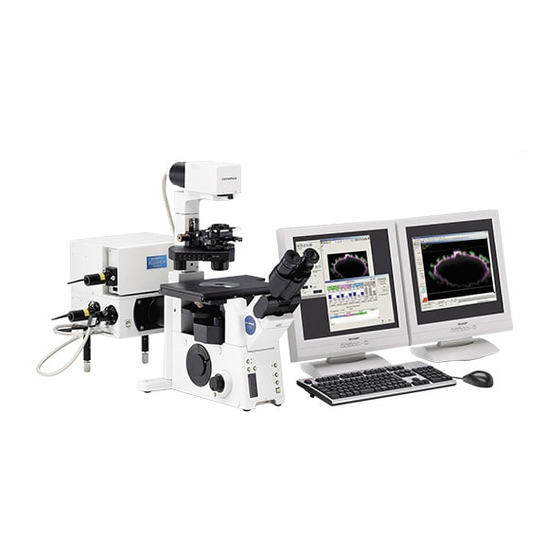
Olympus Fluoview FV1000 Manuals
Manuals and User Guides for Olympus Fluoview FV1000. We have
8
Olympus Fluoview FV1000 manuals available for free PDF download: User Manual, Overview, Short Instructions, Manuallines
Olympus Fluoview FV1000 User Manual (540 pages)
Confocal laser scanning biological microscope
Brand:
Olympus
| Category:
Microscope
| Size: 15.94 MB
Table of Contents
Fluoview Manual Configuration
4
Table of Contents
4
Getting Started Fluoview 1-1
13
Basic Operations
23
Getting Started FLUOVIEW
23
Microscope
23
General Mouse Operation Procedures
29
Names of Major Panel and Window Controls and Their Functions
30
Outline of LSM Observation Procedures
31
Turning Power on
33
Combination with BX
34
Focusing on the Specimen
34
Combination with IX81 FVF
36
Combination with Upright Microscope (BX)
38
Setting the LSM Light Path
38
Combination with Inverted Microscope (IX81 FVF)
40
Selecting the Dyeing Method
42
Selecting the Filters
45
Setting the ND Filters
48
Setting the Objective Magnification
49
Setting the Observation Condition
49
Setting the Channels
50
Setting the Zoom Ratio to 1X
50
Setting the Highest Scan Speed
51
Repeated Scanning Operation
52
Setting the XY Observation Mode
52
Setting the Cross-Section to be Observed
53
Setting a Lower Scan Speed
54
Setting the Area to be Observed
54
Acquiring Image
55
Stopping Repeated Scanning
55
Saving Image
56
Exiting from the Software, Turning Power off
57
Function Help
58
Online Help
58
Microscope Help
59
Configuring the Microscope
62
Parfocality Correction and Jog Sensitivity Adjustment
68
Configuring the Filters (When Using a Filter System)
74
Configuring the Filters (When Using a Spectral Detecting System)
78
Setting the C.A. Diameters
81
Applied Operations
85
General Operation Procedure
85
Image Acquisition Procedure (Section (A))
87
Image Acquisition Procedure in an Observation Mode (Section (B))
88
Examples of Operation Procedures
89
Image Acquisition
91
Image Acquisition in XY Observation Mode
92
Configuring the Microscope
93
Setting the Filters
99
Setting the ND Filters
101
Setting the Observation Condition
103
Acquiring Image
112
Acquiring Image in Accumulation Mode
113
Saving the Acquired Image in File
117
Image Acquisition in Other Observation Modes
118
XZ Observation Mode
118
XT Observation Mode
122
XZT Observation Mode
124
XYZ Observation Mode
130
XYT Observation Mode
135
XYZT Observation Mode
139
Differences in Image Acquisition Method between Fluorescent and Transmitted
145
Images
145
Monochrome Image
145
Dual-Fluorochrome Image
148
Transmitted Image
151
Image Acquisition by Rotating It (Rotation Scan)
155
Image Acquisition of Only the Rectangular Position (Clip Scan)
156
Image Acquisition by Magnifying the Rectangular Position (Zoom-In Scan)2-75
159
High-Speed Image Acquisition
162
Image Acquisition to Prevent Crosstalk between Fluorescence (Sequential Scan)
163
Virtual Channel Function
166
Image Acquisition of a Line at Desired Angle
172
Display the Change of Image Intensity (Point Scan)
173
Image Acquisition on Desired Line (XZ, XT or XZT Observation)
178
Image Acquisition in the Laser Excitation Mode
181
Making REX Mask File
182
Example of FRAP Experiment
187
Memory of Setting Information for Scanning Region
196
Notes for Image Acquisition
196
Saving, Opening and Shredding Images
197
Saving Images
200
Saving Images as a Series
200
Saving a Display
202
Saving Specified Area of Image
204
Saving Animation Images
207
File Types Available for Save
209
Opening Previously Saved Images
216
Shredding Images
217
Saving Comment Together with Image
219
Checking the Image Information/Acquisition Parameters
222
Saving the Image Information/Observation Condition
226
Saving the Region File
229
Saving/Reading the Region File
229
Reading the Region File
231
Protocol Processor
234
Starting the Protocol Processor
235
Editing the Protocol
236
Description of Setting Items
237
Command List
244
Protocol Repetition Processing
249
Input Supporting Function
251
Protocol Procedure Supporting Function
253
Saving the Protocol
254
Executing the Protocol
256
Loading a Protocol
258
Loading a Protocol of Previous Format
259
COM Communication Function
261
Example of Assembling a Protocol
262
Example of Setting Procedure of FRAP Observation
262
Example of Setting Procedure of XYT Observation for Hours
271
Pop-Up Menu
279
Restrictions of [PAPP] Setting
280
Restrictions with [Mode] and [Sub Mode]
280
Changing the Image Display Method
282
Displaying an Image in Simulated Colors
282
Editing the LUT (Look up Table)
284
LUT Graph Editing According to Colors
284
LUT Graph Editing by Gamma Correction
286
Switching the Displayed Channels (Ch1 - Ch5)
287
Displaying Images of Multiple Channels Simultaneously
288
Over and under Views, Single View)
288
Displaying Images Separately Per Channel
289
Views)
289
Displaying Merged Image of Multiple Channels (Single View)
290
Changing the Number of Divided Images
291
Increasing the Number of Divided Images
291
Decreasing the Number of Divided Images
293
Switching the Display Method of Multiple Images
294
Displaying Multiple Image Slices Together
296
Displaying Multiple Images Per Channel
297
Displaying Images of Two Channels Together
299
Displaying Time-Lapse Images
300
Displaying Multiple Multiple Sections Images
301
Displaying same Images in Different Display Methods
301
Displaying Different Images Together
304
Re-Arranging Images Using the same Display Method
304
Magnifying/Reducing an Image
307
Filtering
309
Image Processing
309
Contour Enhancement
310
Noise Reduction
313
Image Sharpening
315
DIC Correcting DIC Level Irregularities
316
Contrast Conversion
318
Image Addition
321
Mathematical Operations between Images
321
Image Multiplication
324
Image Subtraction
324
Image Division
325
NOT Image
326
Image and Image
328
Image or Image
329
Image XOR Image
329
Brightness Overlap Level between 2 Channels (Colocalization)
330
Annotation Mode
331
Colocalization for Series Image Data Set
338
Image Measurement
339
Appending Image (Append)
342
Appending Two Images
342
Appending Image from Several Image Data Set
346
Extract Image (Crop)
351
Image Analysis
358
Checking the Intensity of a Specific Part
359
Intensity Values on a Line (Line Profile)
359
Intensity Values on a Planar Region (Bird's Eye View)
362
Checking the Intensity Distribution of a Specific Part
367
Intensity Distribution on a Line (Histogram)
367
Intensity Distribution on a Planar Region (Histogram)
368
Area Measurement
370
Image Measurement
370
Length Measurement
370
Measuring the Change in Mean Value of Intensity
371
Measuring the Change in Integrated Intensity
377
Building an Image from a Different Viewpoint
379
Building Extended Focus Image from XYZ Image
379
Display Switching to Built Image
379
Turning Built Image into Single Image
382
Turning Built Image into Time Series Image
384
Building Line Images to be Viewed in Z Direction
385
Viewing 3D Image
389
Successive Display of Images
391
Changing the Successive Display Speed
392
Changing the Successive Image Display Position
393
Animation
394
Building Stereo 3D Images
396
Building a 3D Image to be Viewed through Color (Red/Green) Eyeglasses2-314
398
Displaying Images Successively
400
Displaying Images Together
400
Viewing Images Following the Progress of Time
400
Transferring Analysis Data to Another Application
403
Transferring Data to Another Application
403
Transferring the Plot Image of Analysis Data to Another Application
406
Transferring Image Data to Another Application (Paint, Etc.)
407
Entering Comment in Image
408
Writing Characters in Image
408
Displaying the Image Intensity
410
Displaying the X-Coordinate/Y-Coordinate of the Image
411
Drawing a Figure in Image
412
Drawing a Scale in Image
413
Drawing an Arrow in Image
414
Drawing Color Bars in Image
415
Deleting Comment
416
Moving Comment
417
Changing the Comment Size
418
Changing the Comment Color
419
Changing the Comment Font
420
Image Output at Printer
421
Merger/Extraction of Image Channels
422
Setting the Range of Multiple Image Slices
422
Merging Image Channels
423
Extracting Channels from Image
428
Changing the Chart Display Method
432
Chart] Panel
432
Series] Panel
440
Pop-Up Menus
443
Appendix A List of Hot Keys
447
Appendix B Glossary
453
Appendix C USER REGISTRATION of FV1000
457
Appendix C-1 User Registration
457
Appendix C-2 Logging into the FV1000
460
Appendix C-3 Deleting a User
461
Appendix D Change of Default Folder for [File I/O] Panel
465
Appendix E List of Functions in the [Active Overlays] Dialog Box
471
Appendix E-1 Coordinate Position Data
471
Appendix E-1-1 X-Coordinate
471
Appendix E-1-2 Y-Coordinate
472
Animation
473
Appendix E-1-3 Other
473
Position
473
Appendix E-2 Intensity Data
474
Appendix E-3 Other
475
Appendix E-3-1 Channel Number
475
Appendix E-3-2 Objective Power
475
Appendix E-3-3 Date of Image Capturing
475
Appendix E-3-4 Time of Image Capturing
475
Appendix E-3-5 Image File Name
475
Allocation
477
Appendix F Hand Switch and Microscope Frame Function
477
Appendix F Hand Switch and Microscope Frame Function Allocation
477
Appendix F-1 Hand Switch Functions
477
Appendix F-1-1 BX/BXWI
477
Appendix F-2 Microscope Frame Functions
479
Appendix F-2-1 BX
479
Appendix F-2-3 Focus Adjustment Knob
481
Maintenance
483
Software Setup 1-1
485
Re-Setup or Updating of the Software
487
Software Setup
487
New Setup of the Software
489
Overall Setting of FLUOVIEW
491
Setting the System Configuration
491
Setting the [Microscope Control Panel]
500
Adding the Dyeing Method
507
Adding Filters
510
Adding the Filters
510
Setting the Scanning Unit
511
Adding a Dyeing Method
513
Associating a Detection Channel and Filter to the Dyeing Method
515
Laser Scanning Microscope
519
Maintenance of Major System Units
519
Auto Alignment Operation (Remote Switch)
520
UV-Ar Laser
520
Checking, Refilling and Replacing Water in Chiller
521
Setting the Confocal Aperture
525
Starting the FLUOVIEW Software
526
Displaying the [C.A. Adjustment] Dialog Box
527
Confocal Aperture Setting
529
Troubleshooting
533
Advertisement
Olympus Fluoview FV1000 User Manual (186 pages)
LASER SCANNING BIOLOGICAL MICROSCOPE
Brand:
Olympus
| Category:
Microscope
| Size: 2.84 MB
Table of Contents
Section 1
8
Quick Start
1
Table of Contents
8
System Preparation
11
Observation of Fluorescence Image
12
Observation of Differential Interference Contrast Images
13
Overview of Operation Panel for Image Acquisition
14
Single Stain on XY Image
15
Complement of Adjusting the Image
18
Double Stain on XY Image
20
Sequential Scan Line Sequential
22
Double Stain on XYZ Image
23
Four Stain on XY Image
25
Single Stain + DIC on XY Image
28
Merge the Image between Fluorescent XY Image and DIC Image
30
Unmixing
38
Image Analysis
38
Image Analysis
39
Edit the Image Color and Contrast
48
The Image of Z Section
49
Intensity Profile of each Z Sections
50
Measure
51
Line Series Analysis / Co-Localization
53
Closing the System
55
Section 2
58
System Preparation
61
Observation of Fluorescence Image
62
Observation of Differential Interference Contrast Images
63
Overview of Operation Panel for Image Acquisition
64
Single Stain on XY Image
65
Complement of Adjusting the Image
68
Double Stain on XY Image
70
Double Stain on XYZ Image
73
Four Stain on XY Image
75
Single Stain + DIC on XY Image
78
Merge the Image between Fluorescent XY Image and DIC Image
80
Single Stain on XYZT Image / ZDC XYZT Image
81
Reload the Image Conditions
83
Image Analysis
84
Image Analysis
85
Edit the Image Color and Contrast
91
The Image of Z Section
92
Intensity Profile of each Z Sections
93
Measure
94
Line Series Analysis / Co-Localization
96
Closing the System
98
Section 3
100
System Preparation
103
Observation of Fluorescence Image
104
Observation of Differential Interference Contrast Images
105
Overview of Operation Panel for Image Acquisition
106
Single Stain on XY Image
107
Complement of Adjusting the Image
110
Double Stain on XY Image
112
Double Stain on XYZ Image
115
Four Stain on XY Image
117
Single Stain + DIC on XY Image
120
Unmixing
128
Reload the Image Conditions
130
Image Analysis
131
Image Analysis
132
Edit the Image Color and Contrast
138
The Image of Z Section
139
Intensity Profile of each Z Sections
140
Measure
141
Section 4
146
Line Intensity Profile on the 2D Image / Histogram
146
System Preparation
149
Observation of Fluorescence Image
150
Observation of Differential Interference Contrast Images
151
Overview of Operation Panel for Image Acquisition
152
Single Stain on XY Image
153
Complement of Adjusting the Image
156
Double Stain on XY Image
158
Sequential Scan Line Sequential
160
Double Stain on XYZ Image
161
Four Stain on XY Image
163
Single Stain + DIC on XY Image
166
Reload the Image Conditions
169
Image Analysis
170
Image Analysis
171
Edit the Image Color and Contrast
177
The Image of Z Section
178
Intensity Profile of each Z Sections
179
Measure
180
Line Series Analysis / Co-Localization
182
Closing the System
183
Olympus Fluoview FV1000 User Manual (42 pages)
CONFOCAL LASER SCANNING BIOLOGICAL MICROSCOPE
Brand:
Olympus
| Category:
Microscope
| Size: 8.36 MB
Table of Contents
Manual Configuration
4
Table of Contents
4
1 System Overview 1-1
9
System Overview
11
Principles
11
Features of the FV1000
12
Optical Path Diagram
13
System Configuration
14
System Diagram
14
System Appearance and Functions
15
1 Preparation for Operation 1-1
19
1 Preparation for Operation
21
Turning the Power on
21
Starting the Software
23
Exiting from the Software
24
Turning the Power off
24
2 Replacement of Cubes
25
Replacing the DM Cube
25
With the FV10-ASU
25
With the FV10-OPD
27
Removing the Spectral Cube
28
Fabricating a Spectral Cube
30
Attaching the Spectral Cube
30
Replacing the Spectral Cube
28
3 Centration of Mercury Burner
31
Centering the Mercury Burner
31
Trouble Q&A
33
Troubleshooting Guide
37
Laser Is Not Output from the Extremity of the Objective
37
Fluorescence Image Cannot be Observed
38
Transmitted Image Cannot be Observed
38
Image Is Disturbed
38
Fluorescence Image Is Dark and Noisy
39
Image Is Irregularly Blurred or the Brightness Is Uneven
39
Observed Image Is out of Focus
39
Flare Is Observed
39
Visual Fluorescent Light Observation Is Impossible
39
The Light from the Laser for the ASU (Additional Scan Unit) Is Not Output
40
Advertisement
Olympus Fluoview FV1000 Overview (28 pages)
Confocal Laser Scanning Biological Microscope
Brand:
Olympus
| Category:
Microscope
| Size: 4.57 MB
Olympus Fluoview FV1000 Short Instructions (7 pages)
Brand:
Olympus
| Category:
Microscope
| Size: 0.08 MB
Table of Contents
Table of Contents
1
Laser Safety
2
General Guidelines
3
Switching on the Microscope
5
Using the Climatic Chamber
6
Switching off the Microscope
7
Olympus Fluoview FV1000 Short Instructions (7 pages)
Brand:
Olympus
| Category:
Microscope
| Size: 0.08 MB
Table of Contents
Table of Contents
1
Laser Safety
2
General Guidelines
3
Switching on the Microscope
5
Using the Climatic Chamber
6
Switching off the Microscope
7
Olympus Fluoview FV1000 User Manual (9 pages)
MPE Microscope
Brand:
Olympus
| Category:
Microscope
| Size: 2.36 MB
Olympus Fluoview FV1000 Manuallines (3 pages)
Confocal microscope
Brand:
Olympus
| Category:
Microscope
| Size: 0.16 MB
Advertisement
Related Products
Olympus FV1200 MPE
Olympus FLUOVIEW FV300
Olympus FV3000
Olympus FV5-LD405
Olympus FV5-LD440
Olympus FV5-LDPSU
Olympus FHT-523
Olympus FHT-531
Olympus FHT-533
Olympus FHT-532
Olympus Categories
Digital Camera
Voice Recorder
Microscope
Camera Accessories
Camera Lens
More Olympus Manuals
Login
Sign In
OR
Sign in with Facebook
Sign in with Google
Upload manual
Upload from disk
Upload from URL