Table of Contents
Advertisement
Welcome you to use Colposcope Digital Imaging System.
Dear customer, first of all, thank you for trusting and using this
system manufactured by our company. It is image workstation
disposing system combining computer technology and colposcope
technology.
For you operating this system more proficiently, we make the
detailed user manual. Please you must read this manual and other
all information carefully when your first installation and using this
system.
To satisfy the demand of the market and the customer, we will
continuously upgrade our product (including hardware and
software). We will announce you immediately if take some
amendment. Thanks in advance if you correct it after finding any
mistake or oversight.
The contents of this manual is protected by copyright law.
Nobody is allowed to copy,take photo or translate into other
languages of this manual without the prior written permission of the
company.
PDF 文件使用 "pdfFactory Pro" 试用版本创建
Colposcope Digital Imaging System
Preface
www.fineprint.com.cn
Edition: V2.3
Advertisement
Table of Contents

Related Manuals for Kernel KN-2200B


Summary of Contents for Kernel KN-2200B
- Page 1 Colposcope Digital Imaging System Preface Welcome you to use Colposcope Digital Imaging System. Dear customer, first of all, thank you for trusting and using this system manufactured by our company. It is image workstation disposing system combining computer technology and colposcope technology.
- Page 2 Colposcope Digital Imaging System Important Notice Please contact our service center immediately if you face any question or need relevant information. Our engineer will supply any help what you need. The correct use will extend instrument’s life, thus efficiency can be extended to its greatest extent.
-
Page 3: Table Of Contents
Colposcope Digital Imaging System Contents CHAPTER I SAFETY REQUIREMENT AND ATTENTIONS............1 1.1 S ....................1 AFETY EQUIREMENT 1.2 A ........................1 TTENTIONS 1.3 I ................3 NDICATIONS AND CONTRAINDICATIONS CHAPTER II SUMMARY ........................ 5 2.1 F ......................5 UNCTION SUMMARY 2.2 S ..................7 TRUCTURE AND COMPOSITION 2.3 M ..................7... - Page 4 Colposcope Digital Imaging System CHAPTER VII TROUBLES SHOOTING ..................43 CHAPTER VIII AFTER SERVICE ....................44 APPENDIX INDIX OF COLPOSCOPE..................45 PDF 文件使用 "pdfFactory Pro" 试用版本创建 www.fineprint.com.cn...
-
Page 5: Chapter I Safety Requirement And Attentions
Colposcope Digital Imaging System Chapter I Safety Requirement and Attentions (Read the chapter carefully before using it) 1.1 Safety Requirement The device is fit for the safety requirment of IEC60601-1and IEC60601-1-1. Device can assure the safety and correction only when it is connected to device supplied by Our company, can not assure safety for other device supplied by other company. - Page 6 Colposcope Digital Imaging System The gynecological examination should not be done before colposcope examination. Cervical cancer prevention smear, cleanliness, and trichomoniasis, mold inspection shall be completed ahead of schedule in the general out-patient. Colposcope examination is only a secondary means, the results are not the end result, other tests should be combined to determine the result, ultimately determine the cause.
-
Page 7: Indications And Contraindications
Colposcope Digital Imaging System because of the superposition of the leakage. Other additional removable porous plug or extension cord can not be accessed the system. To ensure the safe operation of equipment, all possible replacement parts , accessories and all kinds of supplies that the instruments are equipped with, please use the product models provided or designated by the company. - Page 8 Colposcope Digital Imaging System Ø Naked eye is difficult to determine the fine shape structure, and need the colposcope to magnify the lesions for observation. Ø Before the cervical cancer surgery, need to use colposcope to observe the site lesion spreading to guide surgical . Contraindications: Ø...
-
Page 9: Chapter Ii Summary
Colposcope Digital Imaging System Chapter II Summary Colposcope Digital Imaging System is researched and produced by our company. It is image workstation management system combining computer technology and electric colposcope. It is a new product collects digital imaging technology and colposcope technology. - Page 10 Colposcope Digital Imaging System Ø Observation system built-in scale can measure the size of lesions Unique light source design Ø Five-times zoom focus, three-dimensional sense of strong, distinct, large vision. Ø Integrated design of the dual optical light source and fiber optic transmission systems can adjust the brightness.
-
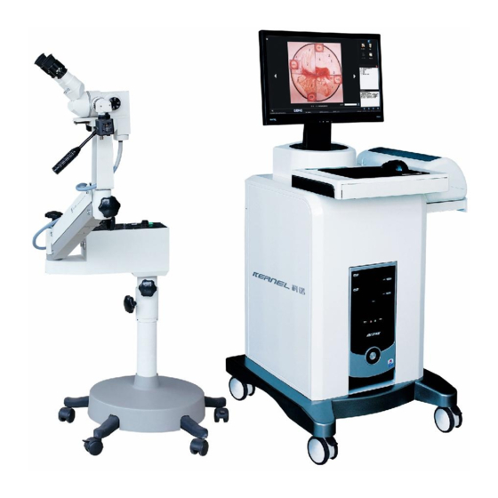
Page 11: Structure And Composition
Colposcope Digital Imaging System 2.2 Structure and composition The system is consists of camera equipment, light source, computers, monitors, printer. Computer mainframe, display screen and printer are optional Display sreen Camera equipment Printer Stand Light source Computer mainframe Pic2-1 2.3 Main technical parameters Display screen:Resolution is not less than 1024 ×... - Page 12 Colposcope Digital Imaging System Ø Light source brightness is adjustable, with the function of filter transfer. Within the working distance, illumination is not less than 1200 Lux. Ø ℃ The irradiated surface temperature does not exceed 41 Ø Guide beam plug diameter:>ф10 ㎜. Ⅱ...
-
Page 13: Chapter Iii Installation And Connection
Colposcope Digital Imaging System Chapter III Installation and Connection 3.1 Check up before installation Take out the instrument from the box, and lay it in a safe, secure and easy to observe position, then lock the universal wheel. of the bottom Check the random parts in accordance with the packing list to ensure the integrity of parts. - Page 14 Colposcope Digital Imaging System Fix the screw A to the position of the bottom which the red arrows indicate, and tighten: Screw A Pic 3-3 Remove the screws B in the bottom of light source: 螺钉 螺钉 螺钉 Screw B Light source Pic 3-4 Fix the light source to the top of strut:...
- Page 15 Colposcope Digital Imaging System Fix the strut and light source with the four removed screws: Screw B Pic 3-6 Remove the screws of the bottom of rocker arm: Screw C Pic 3-7 Fix the rocker arm to the front of light source fixed hole: Rocker arm Cold light source Pic 3-8...
- Page 16 Colposcope Digital Imaging System Fix the screw C to the bottom of rocker arm: Scew C Pic 3-9 10. Remove the screw D from the bottom of eyepiece stand: Screw D Pic 3-10 11. Fix the eyepiece to the front of rocker arm: Eyepiece stand Rocker arm Pic 3-11...
- Page 17 Colposcope Digital Imaging System 12. Install the screw D to the bottom of rocker arm and fix the eyepiece stand: Screw D Pic 3-12 13. Fix the eyepiece to the stand and tighten the screw E : Screw E Eyepiece Pic.3-13 14.
- Page 18 Colposcope Digital Imaging System 15. Insert the fiber port A to the output hole of cold light source: Fiber port A Pic 3-15 16. Lead the fiber into the rocker arm fixed deduction card: fixed deduction card Pic 3-16 17. Insert fiber port B into light input hole in the bottom of eyepiece stand,fixed the screw F:...
-
Page 19: Connection Of Hardware And Software
Colposcope Digital Imaging System 3.2.2 Connection of hardware and software Please refer to installation manual. 3.2.3 AC power connection l Insert the three core power cord into the device's AC power socket, and insert the plug end into the removable porous three-core power socket matching the device. l Insert the removable porous three-core power plug into the AC220V 50Hz three core AC power outlet To ensure reliable grounding. -
Page 20: Chapter Iv The Proposed Clinical Operation
Colposcope Digital Imaging System Chapter IV The proposed clinical operation 4.1 Preparation before the examination 1. Asking about the case history, menstrual history to select the appropriate inspection time. 2. Doing the trichomonas, fungal, Pap smear screening. 3. For the suspected infection patient, should do the culturing of vagina, cervix secretions, and the positive discoverer should be firstly cured according to the disease condition. - Page 21 Colposcope Digital Imaging System When examination, 3% acetic acid solution should be coated on the cervix surface, in the role of acetic acid ,columnar epithelium gets edema, whitish into like grape shape, while the squamous epithelium is slightly pale but not like the grape shapes.
-
Page 22: Chapter V System Operation And Usage
Colposcope Digital Imaging System Chapter V System operation and usage 5.1 Opening and closing of the system The procedure of startuping the system Open the switch of removable porous outlet; Open the switch of monitor power; Press the computer power switch button Insert the smart card "dongle"... -
Page 23: Software Operations
Colposcope Digital Imaging System 5.2 Software Operations Open the software system,enter Figure 5-1. Exit Minimize System time statusline System setup functions Figure 5-1 Introduction of the system interface: System Settings: click this button to enter the system settings interface. Exit the system, Click this button to minimize digital colposcopy system interface. -
Page 24: System Settings
Colposcope Digital Imaging System 5.2.1 System Settings If you use this system for the first time, click the "System Settings" button, open the System Settings form, operate the software settings, video settings and report settings. l Software settings Open the "System Settings", the default is "Software Settings" interface. Or click the "Software Settings"... - Page 25 Colposcope Digital Imaging System Ø foot switch adjustment foot switch's sensitivity, 1-10 may elect. Value sluggishness is higher. select system language, “SPA” is Spanish, “CHS” is Chinese, Ø Language set up “ENG” is English.“VIT” is Vietnamese.. After the revision,restart to work. l Video setup click “video set up”, Figure 5-3 Figure 5-3...
-
Page 26: Images Collection
Colposcope Digital Imaging System Figure 5-4 Ø Hospital name Hospital name which demonstrated in the report. Ø Subtitle Uses for to fill in the subtitle to demonstrate in the report. Ø Report Title Uses for to fill in the report head. Ø... - Page 27 Colposcope Digital Imaging System Real-time Observation function Images colleaction area Video convert Figure 5-5 Note: In real-time image observation area, if does not have the video image, please examine the VIDEO port of the control line connected with capture card whether consistent with the video port in the software set up.
- Page 28 Colposcope Digital Imaging System Figure 5-6 Press this button, freeze the image, this time may carry on the static state to take the images, the image will be quite clear. Click this button to return. Put the pictures together to compare.Click this button, presents the contrast surface, click the arrow to browsing pictures.
- Page 29 Colposcope Digital Imaging System Reduce Clicks on this button, may carry on the reduction to the observation image. Enlarge Clicks on this button, may carry on the enlargement to the observation image. chooses this item, automatic focusing the image or manual control. When manual focusing, carriy out far focusing to the video .
-
Page 30: Atlas Analysis
Colposcope Digital Imaging System mark. After edition completes, click "save". If not, click “back”. Figure 5-9 l Browse click “browse”, as Figure 5-10 "imaging review",operate the enlargement, reduction,resume, save as. Figure 5-10 l Delete click this button to delete the images. l Delete all delete all of the images in the windows area. - Page 31 Colposcope Digital Imaging System Standard atlas Back browsing Patient images Forward browsing Patient atlas diagnosis opinion Standard atlas diagnosis opinion Figure 5-11 The left area of the window is the standard atlas area, right one is the patient atlas area.175 kinds of standard atlases had been provided in the standard atlas area and the corresponding diagnosis opinions to make contras with the patient atlas and convienent to doctor's clinical diagnosis.
-
Page 32: Diagnosis Report
Colposcope Digital Imaging System Figure 5-12 click“+”of each item of the left side in tabulation , launch this tabulation, then choice numerator, assign out the corresponding standard atlas. click" delete” may delete selects the item. Click "close" to shutting down tabulation frame, returns to the normal pattern. NOTE: Only the user's self-definnition altas can be deleted. - Page 33 Colposcope Digital Imaging System Case informations diagnosis opinions area Report type Images display area Figure 5-13 Case report input Ø Case information area: the basic informations of patient in the case area. Ø diagnosis opinions area: Including the cytology inspection, the pathology inspection, the HPV inspection, the colposcope seeing, plan to examine, information,doctor's advice, processing opinion and so on .
- Page 34 Colposcope Digital Imaging System Figure 5-15 Input some common used terminology, click “add to dictionary”, may increase to left side “dictionary”. Laterly use, directly select from the "dictionary",and add into the report. Ø RCI SCORE AREA:User could insert some related infomations about RCI.As Figure 5-16.
- Page 35 Colposcope Digital Imaging System Users can directly mark on it ,or right click to spring out the window of "Biopsy mark",as Figure 5-18. Figure 5-18 Uses can modify the mark type in this window,doing some relatedmark in the Biopsy mark.click "redraw" to clear and click"finish" to save. Ø...
- Page 36 Colposcope Digital Imaging System Figure 5-20 Ø Add printing images: Choose the pictures from the picture display area,click the area that the picture is added,and the picturewill be added into the position. if adding more pictures, and choose picture quantity from report type. Ø...
- Page 37 Colposcope Digital Imaging System Printer button Content of case report Figure 5-21 5.2.5 Case Management Click " " to enter case management,as Figure 5-22 Case data list Function Condition setup Figure 5-22 PDF 文件使用 "pdfFactory Pro" 试用版本创建 ÿ www.fineprint.com.cn...
- Page 38 Colposcope Digital Imaging System Function introduce: click this to dispaly all the case in the case data. Choose one case report from case data,click this button to add informations to disgnosis report,can doing operations of re-examination. Choose one from case list to delete. Choose one case report to view the details,and case report can be renewed and printed.
- Page 39 Colposcope Digital Imaging System l Data output:choose the path,click start "output". l Data input:choose the data you want input,and click"start input" NOTE: ——Do not save case record under system disk and its file —— Do not doing other operations while you back up. ——Data input will cover the existed data,please doing back up of the existed data.
-
Page 40: Observation Systerm Operations
Colposcope Digital Imaging System 5.3 Observation systerm operations Basic parameters 5.3.1 Binocular eyepiece foucus:160mm。 Eyepiece:12.5×,wide-angle vision adjustment±5D。 Work distance:300mm。 Eye-distance adjustment range:50-75mm。 Mico focusing range:0-40mm。 Image collection raster :f4.5-f32。 Eyepiece magnification times and field of view: Input data Magnification times 13.6 21.3 Diameter of filed-of-view... -
Page 41: Light Source Operation
Colposcope Digital Imaging System Five-speed zoom focus knob Binocular eyepiece Digital CCD Before and after the shift knob Optical fiber Handle Pic 5-27 Left of Observation systerm Pic 5-28 Right of Observation systerm Please remove the lence cover and eyepiece cap when use the observation sysyterm.The doctor can directly observe by labeled scale binocular,or can observe through the imaging on the screen got by CCD. - Page 42 Colposcope Digital Imaging System Light switch Indicator light 10A Fuse Light changer 熔断器 2A Fuse Light switch knob Pic 5-30 Open the power switch,the indicator light in on. Adjust light switches, and make it accord with the connecting hole of the fiber optic line.
-
Page 43: Chapter Vi Maintain And Maintenance
Colposcope Digital Imaging System Chapter VI Maintain and Maintenance To ensure the equipment works well, extend equipment life, please pay attention to the maintenance and maintenance. 6.1 Maintenance of equipment and accessories To ensure the normal safety, equipment and accessories should be accepted a preventive check-ups for every 6 months (including the performance of inspection and safety inspection)and maintenance, this in order to prove that the equipment work properly good and in good working condition, for that the medical staff and patients are... -
Page 44: Cleaning Equipment
Colposcope Digital Imaging System comprehensive technical inspection to detect mechanical damage and cable damage every six months. 10. This instrument shall be regularly for maintenance in accordance with the relevant provisions of the hospital 11. If equipment and accessories life expired , in accordance with the relevant provisions dealt with electronics waste. -
Page 45: Storage
Colposcope Digital Imaging System Please clean the lens surface carefully; Please do not use strong or abrasive cleaning agents with the lens surface washing. Please use the lens cleaning paper or cotton swab dipped alcohol. 6.3 Storage If Instruments long time unused, please wipe clean and put into the box, store in a dry and clean place. -
Page 46: Light Source Lamp Change
Colposcope Digital Imaging System Figure 6-1, use a screwdriver unlock the fuse in counterclockwise, remove the fuse as shown in Figure 6-2. Put a new fuse, and then screw into the fuse seat in clockwise as Figure 6-3. 6.6 Light source lamp change WARNING:Power off the light source, and cut off the power line. - Page 47 Colposcope Digital Imaging System Chapter VII Troubles shooting Description Poss ible c ause s and tre atm ent check the cable is in good condition and is in good Can not start computer contact with both the instrument and power socket Check the computer power mangled or not Black screen and alarm Memory bank mangled or not well-inserted...
- Page 48 The schematic,parts,and other technical information,these can be forward to our authorized technical servicer when it's necessary. The lamp bulb do not belong to the warranty area. Contact: Kernel Medical Equipment Co.,Ltd. Add.: Kernel Building, Economic Development District, Xuzhou, Jiangsu, 221004 P.R.C. Tel.:86-516-8773 2209 Fax:86-516-8773 2210 Http://www.kernelmed.com E-mail:kernel@pub.xz.jsinfo.net...
- Page 49 Colposcope Digital Imaging System Appendix Indix of colposcope Index No. 4-1: Diagnoses Opinion: Abnormal Colposcopic Image Pattern 1. Cervical leukoplakia 2. Leukoplakia base 3. Transformation zone(there are Nabothian cyst and gland opening in it) 4. normal cervical mucous membrane 5. Incrassate & rugged cervical leukoplakia 6.
- Page 50 Colposcope Digital Imaging System ------------------------------------------------------------ Index No. 4-13: Diagnoses Opinion: projecting lymph follicle Lymph follicle of cervical surface appears round protuberance (magnify 10X) . Follicle wall is thinner, most distribute dispersedly, there is reticular vascular around it. Most possible is acute inflammation.
- Page 51 Colposcope Digital Imaging System white apophysis like ridge as crater. ------------------------------------------------------------ Index No. 4-18: Diagnoses Opinion: gland openings Magnify picture 4-17 in 16 times, white area of gland opening gradually subsidise. There is a transparent air bubble about cervical os. ------------------------------------------------------------ Index No.
- Page 52 Colposcope Digital Imaging System Index No. 4-22: Diagnoses Opinion: Nabothian cysts There is one Nabothian cysts in posterior lip, content is black-gray, This cyst is black-gray or purple-brown because there is a little bleeding in vesica, then be absorbed, mucus in cyst becomes cloudy.
- Page 53 Colposcope Digital Imaging System Index No. 4-27: Diagnoses Opinion: tree branch-like vascular Chronic cervicitis, vascular hyperplasia on the cervical posterior lip, can see tree branch-like vascular with good ramus, and have many Nabothian cysts which appear round or oval hunching, full of primrose yellow mucus in cyst, distributing thin vascular in surface and allaround.
- Page 54 Colposcope Digital Imaging System ------------------------------------------------------------ Index No. 4-31: Diagnoses Opinion: leukoplakia mosaic It is image of cervical posterior lip, comparted many polygon grids by red vascular on leukoplakia background, appears scalelike, also called tile-like heaping. Can see in tissue beside cancer or chronic cervicitis. ------------------------------------------------------------ Index No.
- Page 55 Colposcope Digital Imaging System Can see red area comparted by white line in central part of image. Opposite to red line of mosaic, it is white line, so called contrary-mosaic. This image can be read usually in acute inflammation or when hyperplasia being active. ------------------------------------------------------------ Index No.
- Page 56 Colposcope Digital Imaging System Cervical surface is covered by white membranoid substance like lard, easy bleeding after erasing. Usually find in advanced cervix cancer, tuberculosis of cervix or when cervical surface being covered one layer of necrotic tissue. ------------------------------------------------------------ Index No. 4-41: Diagnoses Opinion: helical vascular It is one colposcope image of aged woman in post-menopause.
- Page 57 Colposcope Digital Imaging System There are some non-ramus vascular in cervical posterior lip, the change from thick to thin on caliber of vascular is unconspicuous , ramus is so few, nethermost end of vascular increases thick and suddenly intermits. ------------------------------------------------------------ Index No.
- Page 58 Colposcope Digital Imaging System Diagnoses Opinion: ectropium of cervical mucous membrane Top round arc is cervical anterior lip, the surface is pink squamous epithelium, middle and lower part in red area is eversion of cervical mucous membrane. It is covered by one layer of white mucus, it is easy to mis-diagnose with submucous myoma.
- Page 59 Colposcope Digital Imaging System Index No. 5-13: Diagnoses Opinion: cervix in menopausal period Squamous epithelium of cervical surface become thin, squmocoumnar junction shreaks back into cervivical canal, cervix os gets contracture. ------------------------------------------------------------ Index No. 5-14: Diagnoses Opinion: cervix in menopausal period Cervical figure becomes small and slippery, mucous membrane is thin and pale, lacks elasticity, columnar epithelium shreaks back.
- Page 60 Colposcope Digital Imaging System ------------------------------------------------------------ Index No. 5-3: Diagnoses Opinion:young girl’s vulvitis Magnify "5-2" in 16 times,mucous membrane of vestibule congests obviously, can see ramus vascular of hyperplasia, hymen is fleshy and dropsical. ------------------------------------------------------------ Index No. 5-4: Diagnoses Opinion: cervical image of ovulation period in reproductive women Cervical mucous membrance is pink, think with good elasticity, cervix os appears as “transverse-fissure”,being full of transparent mucus.
- Page 61 Colposcope Digital Imaging System varicose vascular. ------------------------------------------------------------ Index No. 5-9: Diagnose Opinion: cervix in premenopausal period Cervical mucous membrane becomes thin and uneven in thickness, cervical surface is not fully covered by pink squamous epithelium, some parts show damask background, appearing the change like "thaw", it is sign of estrogen level beginning turndown.
- Page 62 Colposcope Digital Imaging System much secretion like rice water, focus depth is covered by sage green necrotic membrane substance. ------------------------------------------------------------ Index No. 6-102: Diagnose Opinion: cavernous type of cervical cancer Cervix metamorphoses totally, cancer tissue grows devasculartatingly to tissue depth like digging, as ischemia and edma, cancer tissue apperrs transparent like grape, also called "glassed change", there is old bleeding around cancerous protuberance.
- Page 63 Colposcope Digital Imaging System Urethra orificium is in ringent shape, there is one red polypus-like substance extruding outward from urethra orificium, soft,base part is wider, pathologic result is polypus of urethra orficium. ------------------------------------------------------------ Index No. 6-107: Diagnose Opinion: skin tag of vulva One neoplasm from right labium minus extrudes out, surface is covered by normal squamous epithelium, has thin pedicel connecting with labium minus, softer character, without callosity and pain when pressing, has no any symptom commonly, influnce...
- Page 64 Colposcope Digital Imaging System Index No. 6-110: Diagnose Opinion: vaginal metastatic tuber of choriocarcinoma festered It is metastasis tuber of vaginal anterior wall, extrudes out of vaginal orificium, content of focus extrudes outwards from crevascularse, surface is purple-blue. ------------------------------------------------------------ Index No. 6-111: Diagnose Opinion: gonococcus infection of cervix Cervical mucous membrane evaginates like acute congestion, full of yellow pus in crimson cervical canal, and overflows outwards, covers cervical surface, identified the...
- Page 65 Colposcope Digital Imaging System branch. ------------------------------------------------------------ Index No. 6-13: Diagnose Opinion: condyloma of vulva Multiple small cauliflower-like, surface is offwhite or pink, fundus is wider, top is like burr, this type is most found. Pathologic result is condyloma of vulva. ------------------------------------------------------------ Index No.
- Page 66 Colposcope Digital Imaging System Extrudes like cauliflower, surface is pink, tissue texture is fragile, surface branch is not obvious. Pathologic result is condyloma of urethral orifice. ------------------------------------------------------------ Index No. 6-19: Diagnose Opinion: condyloma of urethral orifice Can see cauliflower-like condyloma in urethral orifice and its right side. Focus in urethral side fossa gets light infection and necrosis, has small parcel of bleeding focus, vaginal muscous membrane also has punctuated and parcel-like bleeding focus.
- Page 67 Colposcope Digital Imaging System -------------------------------------------------------- Index No. 6-23: Diagnose Opinion: angioma of vulva Can see fuchsia round tuber in vulvae, clear boundary with skin. Pathologci result is angioma of vulva. ------------------------------------------------------------ Index No. 6-24: Diagnose Opinion: angioma of vulva Appears fuchsia hunch, some can be seen dilated vascular, tensility is bigger, dsscolos after pressing.
- Page 68 Colposcope Digital Imaging System epidermis keratinization is not obvious, there is clear boundary with surrounding normal skin. --------------------------------------------------------- Index No. 6-29: Diagnose Opinion: Bowen’s disease of vulva Involves anus and its surrounding, there is one sentinel pile at anus, the place corresponding to "at 12 o’lock", focus involves surrounding skin of anus.
- Page 69 Colposcope Digital Imaging System Index No. 6-33: Diagnose Opinion: trichomonal vaginitis Vaginal wall congests and flushy,there are small parcel bleeding spots dispersedly distributing in surface of vaginal wall, and yellow, thin, purulent secretion with air bladder in the vagina. ------------------------------------------------------------ Index No.
- Page 70 Colposcope Digital Imaging System It shows local pathologic change of “6-37” after treatment with acetic acid, mucous membrane of fingerlike tuber appears like white concretion, top is round without branch. Other membrance of small papillary hyperplasia, after treatment acetic acid it appears like white small papilla, top is round and slippery.
- Page 71 Colposcope Digital Imaging System Index No. 6-42: Diagnose Opinion: chronic inflammation hyperplasia of vaginal mucous membrane Top is pointed,fundus is wider, slippery surface of mucous membrance, normal sheen. Attention being differentiate with condyloma. --------------------------------------------------------- Index No. 6-43: Diagnose Opinion: polypi of vaginal wall Mucous membrance of vaginal lateral wall is in flush, extrudes one ligulate polypus, red, has sheen, texture is soft, easy to bleed after touching.
- Page 72 Colposcope Digital Imaging System Index No. 6-47: Diagnose Opinion: condyloma of vaginal wall It is in vaginal side wall, extrudes like cauliflower, fundus is wide, suface is not flat, top is spiny, fragile texture. Pathologic result is vaginal condyloma. ------------------------------------------------------------ Index No.
- Page 73 Colposcope Digital Imaging System Index No. 6-51: Diagnose Opinion: cyst of vaginal wall It is in vaginal posterior wall near vagina orifice, vaginal mucous membrance covering in surface slightly becomes thin, looms offwhite contents in cyst, can see exiguous branch vascular.
- Page 74 Colposcope Digital Imaging System Focus does not penetrate cervical mucous membrance yet, can see many round protuberances in cervical surface like cockscomb. Pathologic result is vaginal adenosis of cervix. ------------------------------------------------------------ Index No. 6-57: Diagnose Opinion: malignant melanoma of the vagina Can see big parcel melanin pigmentation area in vaginal side wall and cervical surface, surrounding distributes punctuated melanin pigmentation like secondary planet.
- Page 75 Colposcope Digital Imaging System Diagnose Opinion: chronic cervicitis Cervix is obese, anterior lip are columnar epithelium, posterior lip are covered by one layer thin squamous epithelium, cervix os appers like "transverse-fissure", there is a nulon thread tail of IUD emerging. ------------------------------------------------------------ Index No.
- Page 76 Colposcope Digital Imaging System Index No. 6-65: Diagnose Opinion: chronic cervicitis during restorations A piece of squamous epithelium on anterior lip appaer white and reticular, but they do not fully syncretized each other, leave punctuated and parcel columnar epithelium area, namely columnar epithelium island.
- Page 77 Colposcope Digital Imaging System ------------------------------------------------------------ Index No. 6-7: Diagnose Opinion: mixed type of vulva dystrophy Skin of labium majus gets incrassation and keratinization, labium minus become white, flat, bad elasticity, epithrlium defects locally. ------------------------------------------------------------ Index No. 6-70: Diagnose Opinion: clustered polypus of cervix Cervix appears erosion,a cluster of clustered small polypi outgrows from cervical canal, pedicel is short and wide, vermeil in color, this type of polypus has soft texture, easy to bleed.
- Page 78 Colposcope Digital Imaging System a cluster of white substance like fibre, it is conglutinated batt) ------------------------------------------------------------ Index No. 6-74: Diagnose Opinion: cervcal white epithelium Can see parcel white protuberance in cervical squamous epithelium field which it is lift side of cervical posterior lip, slightly higher of cervical surface, its surface is flat, perimeter is clear.
- Page 79 Colposcope Digital Imaging System bleeding spot in cervical posterior lip. ------------------------------------------------------------ Index No. 6-79: Diagnose Opinion: submucous myoma of cervix Cervix is obviously obese, one round mass extrudes from cervical posterior lip to cervical canal, fundus is wider, surface is slippery, covered by one layer of columnar epithelium. ------------------------------------------------------------ Index No.
- Page 80 Colposcope Digital Imaging System Index No. 6-83: Diagnose Opinion: blue nevus of cervix Cervix gets erosion in middle degree, there is one blue-brown long and round protuberance in cervical side place, surface is slippery, perimeter is not clear, it is lighter than melanoma in color and lustre.
- Page 81 Colposcope Digital Imaging System It is in cervix os and cervical posterior lip, extrudes like cauliflower, part become white as shaped by snow after treatment with 3% acetic acid, fragile texture,easy to bleed. Pathologic result is condyloma. ------------------------------------------------------------ Index No. 6-89: Diagnose Opinion: cervical malignant melanoma It locates in cervical anterio-lateral lip, atrous, clear perimeter, boundary distributes many branch focus like secondary planet.
- Page 82 Colposcope Digital Imaging System wiping. Pathologic result is cervical cancer in situ. ---------------------------------------------------------- Index No. 6-93: Diagnose Opinion: Cervix Cancer Cervix gets augmentation and transmutation, surface is not flat, primrose yellow, tissue is fragile, easy to bleed. Can see clear and normal squamous epithelium brim around focus. Pathologic result is cervical adenocarcinoma.
- Page 83 Colposcope Digital Imaging System colposcope image of advanced cervix cancer. ------------------------------------------------------------ Index No. 6-98: Diagnose Opinion: Cervix Cancer Cervix appears cauliflower hyperplasia, rugged surface, can see many mastoid processes, saffron yellow backdrop, there are 2 pieces of lardaceous change, and with old and fresh bleeding.
Need help?
Do you have a question about the KN-2200B and is the answer not in the manual?
Questions and answers