Table of Contents
Advertisement
Advertisement
Table of Contents

Related Manuals for Myray ZEN-X


Summary of Contents for Myray ZEN-X
- Page 1 97050937 Rev. 04 2020-04...
-
Page 2: Table Of Contents
Contents 1. INTRODUCTION AND INDICATIONS FOR USE ..................... 3 1.1. DESCRIPTION OF THE MANUAL ........................3 1.2. GENERAL WARNINGS ............................4 1.3. STANDARDS AND REGULATIONS ........................4 1.4. STYLISTIC CONVENTIONS ..........................5 1.5. WARNINGS FOR USE ............................6 1.6. GENERAL SAFETY WARNINGS ......................... 7 1.6.1. -
Page 3: Introduction And Indications For Use
1. INTRODUCTION AND INDICATIONS FOR USE ZEN-X / ZEN-Xi are digital intraoral sensors intended for acquiring intraoral digital images when exposed to X-rays, for diagnostic radiographic examination of the human dentition (teeth, maxillary area and oral structures). The produced digital images are automatically transmitted via digital connection to a PC. -
Page 4: General Warnings
In order to use the ZEN-X / ZEN-Xi sensor, software for capturing and saving the images is needed that is not part of the ZEN-X / ZEN-Xi sensor. Consult the relative manual for information about installation and use of the image management software. -
Page 5: Stylistic Conventions
The sensor has been manufactured in compliance with the IEC standards on safety of electro-medical devices of similar type and particularly with the following technical standards: IEC 60601-1:2005 + A1:2012 - General requirements for basic safety and essential performance. IEC 60601-1-2:2014 (4th Ed.) - General requirements for basic safety and essential performance - Collateral Standard: Electromagnetic disturbances - Requirements and tests. -
Page 6: Warnings For Use
(“drivers” installed in computer and “firmware” in the devices) are compatible with other commercial devices. Therefore use of the ZEN-X / ZEN-Xi sensor and relevant software in conjunction with other commercial devices is not assured or recommended. -
Page 7: General Safety Warnings
necessary to install the sensor with longer cables, a USB repeater must be compulsorily installed every ® 4.5m, and for a maximum of three cable sections (two repeaters). WARNING: The sensor used to capture images is fragile and bothered by electrostatic discharges. -
Page 8: Use Of The Centering Device
Therefore, it is necessary that the PC is ® turned on and the sensor cable (in the case of ZEN-X model) or the electronic interface (in the case of the ZEN-Xi model) is connected to a USB port. If the ZEN-Xi system is installed in a dental chair equipped with integrated ®... -
Page 9: Warranty
1.6.4. WARRANTY The Manufacturer stands behind its products warranting safety, reliability and performance. The warranty is effective from the date of installation of the product. The product is covered for the warranty period indicated in the installation report and, in any case, not less than 12 months. - Page 10 Guidance and Manufacturer's declaration - Electromagnetic emissions The ZEN-X / ZEN-Xi device is designed to operate in the electromagnetic environment specified below. The customer or user of the ZEN-X / ZEN-Xi device should ensure that is used in such environment: Emission test...
- Page 11 Guidance and Manufacturer's declaration - Electromagnetic immunity The ZEN-X / ZEN-Xi device is designed to operate in the specified electromagnetic environment. The customer or the user of the device must ensure its use in an electromagnetic environment with the following features:...
- Page 12 The customer or the user of the ZEN-X / ZEN-Xi device could help in preventing electromagnetic interferences ensuring a minimum distance between the RF mobile and portable communication devices (transmitters) and ZEN-X / ZEN-Xi, as indicated below, in relation to the maximum output power of the radio- communication equipment.
-
Page 13: Protection Against Radiation
1.6.6. PROTECTION AGAINST RADIATION The digital sensor must be used together with an intraoral x-ray system. As such, the system exposes the patient and the operators to the risks deriving from radiation. It must be used in compliance with the safety regulations set out in the radiation protection standards in force in the country of use. - Page 14 Centre for any reason, completely disinfect the outside of the apparatus with a specific product (see paragraph “Cleaning and disinfecting”) and send it back preferably in its original box. No electronic parts of the sensor require maintenance. If the sensor or interface casings are opened to reach the circuits inside, devices may be broken, the protective means for electric safety may be disabled and the warranty will become null and void.
-
Page 15: Cleaning And Disinfection
Scrapping At the end of its lifetime, dispose of the device in accordance with the regulations in force. It is also advisable to disinfect all the external parts of the device before disposal and to separate the materials for differentiated waste collection. - Page 16 All products must be used as directed by the manufacturer. OPERATOR'S MANUAL...
-
Page 17: Cleaning And Disinfection Of The Sensor And Its Power Cable
Cleaning and disinfecting instructions. Clean and disinfect with disposable non-abrasive paper (avoid using recycled paper) or sterile gauze. Do not use sponge cloths or, in any case, any material that can be reused. WARNING: In order to clean equipment connected to the power mains, shut off the devices and disconnect the power supply from the outlet before attempting to clean and disinfect the outside. -
Page 18: Applied Parts
WARNING: Disposable hygienic protections are the main protection means against cross contamination between patients. In order to prevent the transmission of infectious diseases between patients, it is essential to always use disposable protections. Disposable protections are class I medical equipment and cannot be replaced with other protections having lower specifications. -
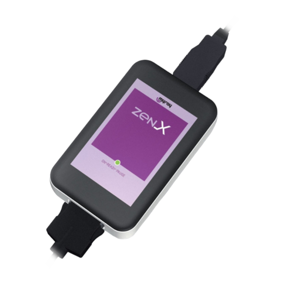
Page 19: Description Of Device And Box Contents
2. DESCRIPTION OF DEVICE AND BOX CONTENTS The X-ray sensor can be purchased in two different sizes (Size 1 and Size 2) in order to adapt to different sizes of oral cavity. The package contains: Sensor package X-ray sensor key with software, driver and electronic user's manual ®... -
Page 20: Description Of Operation
To disengage the sensor from its housing, gently pull the cable from its USB connector to disconnect it from the ® port of the Personal Computer (ZEN-X model) or of the interface (ZEN-Xi model). Do not exert lateral ® movements or stress, and do not pull the cord. -
Page 21: Sensor Integrated In Dental Unit
If the sensor is NOT connected, “SENSOR NOT CONNECTED” will be displayed on a red background in MyRay® Driver TWAIN window. ® If the sensor is connected to a PC where MyRay® Driver TWAIN is NOT active (factory default settings have been ®... - Page 22 WARNING: In order to minimise the temperature on the active surface of the sensor, during periods of non- use it is recommended to disconnect it or take it in stand-by mode, by rotating the sensor support to stand- by position. WARNING: Always use the stand-by position to disconnect the sensor connector.
-
Page 23: Patient Positioning
Closing the door Turn support to position in order to close the door. As ZEN-Xi sensor is physically attached to the dental chair dentist’s board, dirt and sprays may reach the USB connector when the sensor is not connected to ®... -
Page 24: Acquiring An X-Ray Image
Take X-ray picture. The image will appear on the computer screen after a few seconds, and, if enabled, it can be seen in the MyRay® Monitor preview window and right-hand column next to the main window. No other steps are required to capture further images after the first x-ray picture has been taken. - Page 25 A green box indicates that the sensor is enabled and ready to acquire an image. Make an acquisition only when the sensor is in this status. The software also indicates the remaining time before the sensor goes into stand-by mode. WARNING: Always check system status before attempting to take x-rays on a patient.
-
Page 26: Quality Of The X-Ray Images
Sensor in stand-by since sensor support is in “Active” position, but the sensor remained unused for a long time. BLUE FLASHING In this condition it is NOT possible to acquire images. For the sensor to get ready to acquire images, reactivate the sensor by turning sensor support to “Active”... - Page 27 It is important to check and note this limit with your own x-ray system in order to be sure not to exceed it during dental treatment as the images obtained under these conditions will be of poor quality or even unusable. WARNING: Before attempting to take x-ray pictures on patients, it is advisable to take some test pictures on phantoms, comparing the results obtained to the usual ones.
-
Page 28: Specifications
4. SPECIFICATIONS 4.1. TECHNICAL SPECIFICATIONS The device is designed to operate in environmental conditions that are typical of covered working areas and within the parameters under IEC 60601-1. General characteristics Bitmap file 4096 grey levels, compatible with Windows / Mac Image format (PNG, JPG) via USB cable, according to TWAIN®... -
Page 29: Compatibility With X-Ray Generators
Atmospheric pressure from 700 to 1060 hPa Transport and storage conditions Temperature from -20°C to +70°C. RH (humidity) from 0% to 70%. Atmospheric pressure from 700 to 1060 hPa 4.2. COMPATIBILITY WITH X-RAY GENERATORS The features and some of the key functions of the system will largely depend on the characteristics of the X-ray generator and of the software used to display and store the images. -
Page 30: Minimum Prerequisites
If edentulous areas are irradiated, the device may provide images that are too blackened in the missing areas of the irradiated radiographic subject. In these cases, reduce the time indicated in the table by about 1/4. The best results are achieved with a high frequency generator with square collimator and 30cm focus-to-skin distance (refer to the relevant table). -
Page 31: Equipment Identification
5. EQUIPMENT IDENTIFICATION WARNING: Do not remove the identification plates that accompany the product and its accessories. This section includes an example of the identification plates used on the product. For a thorough explanation of the symbols on the identification plates, see 1.4 “STYLISTIC CONVENTIONS”. Position: directly applied on sensor USB cable. -
Page 32: Troubleshooting
6. TROUBLESHOOTING PROBLEM FOUND POSSIBLE CAUSES SOLUTIONS Do not use the sensor on a patient. Conduct tests by capturing an x-ray Doubts concerning sensor image using a phantom. If there are Falling, banging, malfunctioning. efficiency. doubts about proper operation, do not use the sensor and contact the Service Centre. - Page 33 PROBLEM FOUND POSSIBLE CAUSES SOLUTIONS An image is acquired but with poor Use a longer exposure time, make tone range and/or massive Underexposed image. sure the x-ray generator works background noise. correctly. The image is captured but it is very Use a shorter exposure time, check Over-exposed image.
- Page 34 www.cefla.com...
Need help?
Do you have a question about the ZEN-X and is the answer not in the manual?
Questions and answers