Table of Contents
Advertisement
Quick Links
Advertisement
Table of Contents

Related Manuals for St. Jude Medical CardioMEMS CM2000


Summary of Contents for St. Jude Medical CardioMEMS CM2000
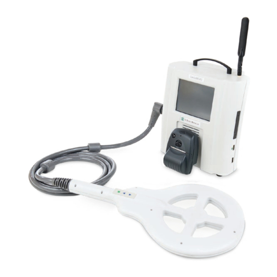
- Page 1 ™ CardioMEMS HF System PA Sensor and Delivery System Model CM2000 User's Manual...
- Page 2 Caution: Federal (U.S.) law restricts this device to sale by or on the order of a physician. Unless otherwise noted, ™ indicates that the name is a trademark of, or licensed to, St. Jude Medical or one of its subsidiaries. ST. JUDE MEDICAL and the nine-squares symbol are trademarks and service marks of St. Jude...
-
Page 3: Table Of Contents
Contents Description Indications Contraindications Clinical Considerations for Patient Selection Warnings Precautions MRI Information Explant and Disposal Potential Adverse Events Instructions for Use Personnel Training Accessories Package Inspection Package Contents Sterilization Pre and Post Procedure Antiplatelet Regimen Implantation Procedure Hospital Electronics System Setup Right Heart Catheterization and Sensor Implant Procedure Patient Identification Card Patient Counseling Information... -
Page 4: Description
Description The CardioMEMS HF System provides pulmonary artery (PA) hemodynamic data used for the monitoring and management of heart failure (HF) patients. The system measures changes in PA pressure which physicians use to initiate or modify heart failure treatment. The system includes the following components: •... -
Page 5: Indications
Table 1. Device model numbers Device Model number PA Sensor and Delivery System CM2000 Patient Electronics System (landline) CM1010 Patient Electronics System (GSM) CM1000 Hospital Electronics System CM3000 Indications The CardioMEMS HF System is indicated for wirelessly measuring and monitoring pulmonary artery (PA) pressure and heart rate in New York Heart Association (NYHA) Class III heart failure patients who have been hospitalized for heart failure in the previous year. -
Page 6: Precautions
of contamination of the device and/or cause patient infection or cross-infection, including, but not limited to, the transmission of infectious disease(s) from one patient to another. Contamination of the device may lead to injury, illness, or death of the patient. •... -
Page 7: Mri Information
• Patients with a reduced ejection fraction should be on stable AHA/ACC guidelines based medical therapy prior to implant. • The PA Sensor is a permanent implant. The sensor does not have any batteries that require replacement or any components that will wear or fail over time. Removal after implantation is not recommended. -
Page 8: Explant And Disposal
Table 3. Artifact Information Pulse sequence T1-SE T1-SE Signal void size 305-mm 34-mm 645-mm 101-mm Plane orientation Parallel Perpendicular Parallel Perpendicular Explant and Disposal The sensor does not require removal before cremation. Do not implant an explanted sensor in another patient as sterility, functionality, and reliability cannot be ensured. Potential Adverse Events Potential adverse events associated with the implantation procedure include, but are not limited to the following:... -
Page 9: Instructions For Use
• Device embolization Instructions for Use Personnel Training Implanting physicians are required to have successfully completed additional training in the use of the PA Sensor and Delivery System prior to implant. Accessories The accessories required to implant the device and set the sensor’s PA pressure baseline are listed in the following table. -
Page 10: Pre And Post Procedure Antiplatelet Regimen
Pre and Post Procedure Antiplatelet Regimen Patients who are currently on anticoagulant therapy, or those in which chronic anticoagulant therapy is indicated for heart failure treatment, will restart treatment after sensor implantation. An INR of <1.5 is recommended prior to sensor implant for patients who were previously on anticoagulant therapy. -
Page 11: Right Heart Catheterization And Sensor Implant Procedure
Right Heart Catheterization and Sensor Implant Procedure 1. Gain percutaneous access to the left or right femoral vein. 2. Introduce 12Fr introducer sheath over a 0.035mm guidewire. 3. Remove the dilator and guidewire. 4. Insert and advance the pulmonary artery (PA) catheter, with balloon inflated, until it reaches a wedge position in the lower lobe region of the left or right pulmonary artery. -
Page 12: Patient Identification Card
14. Under fluoroscopic monitoring, slowly and gently retract and remove the delivery catheter while maintaining guidewire and sensor position. If resistance is encountered, do not forcibly retract the hub or catheter shaft. 15. Insert the PA catheter over the guidewire into the main PA. 16. -
Page 13: Clinical Study Information
Clinical Study Information Introduction Heart failure (HF) is a major public health problem in the United States affecting over 5 million people with over 1 million HF hospitalizations per year. Elevated pulmonary artery (PA) pressures may occur prior to signs and symptoms of HF decompensation and can provide a sound physiologic basis for HF patient management. -
Page 14: Patient Demographics And Disposition
Study Endpoints The primary and secondary endpoints were evaluated after 6 months of patient follow-up. The primary safety endpoints were 1) Freedom from device / system-related complications (DSRC), and 2) Freedom from pressure sensor failures. The primary efficacy endpoint was the rate of HF hospitalizations. All hospitalizations were adjudicated by an independent Clinical Events Committee (CEC) who were blinded to treatment assignment. -
Page 15: Primary And Secondary Endpoint Results
The mean follow-up during Randomized Access was 17.6 months for a total duration of approximately 800 patient years. During the course of Randomized Access, 93 patients in the Treatment group and 110 patients in the Control group exited the study with the primary reason being death. -
Page 16: Primary Efficacy Endpoint
Table 7. Primary Safety Endpoint – Freedom from Pressure Sensor Failures Lower 95.2% Pressure Sensor Objective Performance Confidence p-value Failures (n=550) Criterion (OPC) Limit 0 (0.0%) 550 (100%) 99.3% p<0.0001 Pressure sensor failure counts by group: Treatment (0), Control (0) Exact 95.2% Clopper-Pearson lower confidence limit p-value from exact test of binomial proportions compared to 90% for all patients Primary Efficacy Endpoint... -
Page 17: Medical Management
Medical Management Physicians responded to Treatment patients’ elevated PA pressures by making medication changes to lower PA pressures in an attempt to reduce the risk for HF hospitalization. Physicians documented all medication changes for all patients and indicated whether the change was made in response to PA pressures or standard of care information. -
Page 18: Death Or All Cause Hospitalizations
Death or All Cause Hospitalizations Death or all cause hospitalizations were reduced in the Treatment group (604 in the Treatment group vs. 736 in the Control group, HR 0.84, 95% CI 0.76 – 0.94). The NNT per year to prevent one death or all cause hospitalization was 4. For every 100 patients treated, 29 deaths or all cause hospitalizations would be prevented per year. - Page 19 over the Full Randomized Period (Part 1). They illustrate the apparent difference in treatment effect by gender. As seen in Figure 4, for HF hospitalizations alone, treatment and control women have similar outcomes. However, as seen in Figure 5, control women had an increased early mortality creating a competing risk for HF hospitalizations i.e., fewer control women were alive to have HF hospitalizations.
- Page 20 Also performed were an Anderson-Gill Model with Frailty, Anderson Gill Model with Robust Sandwich Estimates (RSE) and Negative Binomial Regression using an endpoint of time to HF hospitalization or death in Part 1 and Part 1 + Part 2. As seen in the gray rows in Tables 11 and 12 below, all the competing risk analyses taking death into account as a competing risk show that there was no evidence of a treatment-by-gender interaction if a p-value of 0.05 is used.
-
Page 21: Adverse Events
Non-Serious Adverse Device Events There were 17 non-serious adverse device events that occurred over Part 1. There were no additional non-serious adverse device events over Part 2 of the clinical trial. These events were rare and are well known adverse events that occur during right heart catheterization procedures (Table 13). - Page 22 Part 1 Part 2 Treatment (270) Control (280) All Patients (550) All Patients (347) Subjects Events Subjects Events Subjects Events Subjects Events Hepatobiliary disorders 1 (0.4%) 7 (2.5%) 8 (1.5%) 3 (0.9%) Immune system disorders 4 (1.5%) 4 (1.4%) 8 (1.5%) 4 (1.2%) Infections and infestations 76 (28.1%)
- Page 23 Part 1 Part 2 Treatment (270) Control (280) All Patients (550) All Patients (347) Subjects Events Subjects Events Subjects Events Subjects Events Cardiac arrhythmia Cardiomegaly Cardiorenal syndrome Chronic heart failure Congestive cardiac failure aggravated Coronary artery disease progression Coronary atherosclerosis Coronary spasm Decompensated heart failure End stage cardiac failure...
- Page 24 Part 1 Part 2 Treatment (270) Control (280) All Patients (550) All Patients (347) Subjects Events Subjects Events Subjects Events Subjects Events Acute diverticulitis Acute pyelonephritis Bacterial endocarditis Bacterial infection C.difficile colitis Catheter site infection Cellulitis of arm Cellulitis of hand Clostridium difficile infection Community acquired pneumonia Diverticulitis...
- Page 25 Part 1 Part 2 Treatment (270) Control (280) All Patients (550) All Patients (347) Subjects Events Subjects Events Subjects Events Subjects Events Asthma aggravated Bronchitis asthmatic Difficulty breathing Dyspnea exacerbated Exacerbation of asthma Hemoptysis Hypoxia Productive cough Pulmonary mass Respiratory arrest Chronic obstructive pulmonary disease Cough...
- Page 26 Part 1 Part 2 Treatment (270) Control (280) All Patients (550) All Patients (347) Subjects Events Subjects Events Subjects Events Subjects Events Nervous system disorders 29 (10.7%) 28 (10.0%) 57 (10.4%) 27 (7.8%) Syncope Stroke Presyncope Carotid artery stenosis Dizziness Embolic stroke Subarachnoid hemorrhage Anoxic encephalopathy...
- Page 27 Part 1 Part 2 Treatment (270) Control (280) All Patients (550) All Patients (347) Subjects Events Subjects Events Subjects Events Subjects Events Pancreatitis Ascites Dysphagia Emesis Esophagitis Gastroparesis Abdominal bloating Abdominal wall hematoma Chronic epigastric pain Dental caries Esophageal spasm Esophagitis ulcerative Gastric polyps Gastritis erosive...
- Page 28 Part 1 Part 2 Treatment (270) Control (280) All Patients (550) All Patients (347) Subjects Events Subjects Events Subjects Events Subjects Events Cardiac ablation Cardiac resynchronization therapy Cardioversion Cholecystectomy Foot surgery Inguinal hernia repair Abdominal hernia repair Brachytherapy Cardiac pacemaker revision Central line placement Colostomy closure Epicardial lead placement...
- Page 29 Part 1 Part 2 Treatment (270) Control (280) All Patients (550) All Patients (347) Subjects Events Subjects Events Subjects Events Subjects Events Arthritis Arthritis single joint Back pain aggravated Charcot's joint Groin pain Hemarthrosis involving lower leg Lumbar spinal stenosis Lupus erythematosus Muscle necrosis Musculoskeletal chest pain...
- Page 30 Part 1 Part 2 Treatment (270) Control (280) All Patients (550) All Patients (347) Subjects Events Subjects Events Subjects Events Subjects Events Injury to liver Liver disorder Portal hypertension Neoplasms benign, malignant and 5 (1.9%) 4 (1.4%) 9 (1.6%) 5 (1.4%) unspecified (incl cysts and polyps) Lung cancer Large cell lung cancer...
-
Page 31: Fcc Statement
Unanticipated Serious Adverse Device Events There was one event during Part 1 reported as a USADE by the investigator but determined not to be serious or device system related by the CEC which reviewed the event on 27 Jun 2009. There were no additional USADEs over Part 2 of the clinical trial. Serious Adverse Device Events The two SADEs that occurred during Part 1 were hemoptysis during the implant procedure and an in-situ thrombosis during the right heart catheterization procedure. - Page 32 Manufacturer: St. Jude Medical 387 Technology Circle NW Atlanta, Georgia 30313 USA 866-240-3335 sjm.com July 2015 LA-400275-G...
Need help?
Do you have a question about the CardioMEMS CM2000 and is the answer not in the manual?
Questions and answers