Table of Contents
Advertisement
Quick Links
Advertisement
Table of Contents

Related Manuals for St. Jude Medical Optisure LDA220


Summary of Contents for St. Jude Medical Optisure LDA220
- Page 1 Optisure ™ Models LDA220, LDA230, LDA210, LDA220Q, LDA230Q, LDA210Q, LDP220, LDP230, LDP220Q, LDP230Q Quadpole connector/IS-1 and DF-1 connector Active-fixation/Passive-fixation True bipolar Dual-coil/Single-coil Steroid-eluting Endocardial Defibrillation leads User's Manual...
- Page 2 State of California to cause cancer and birth defects or other reproductive harm. For more information, go to www.P65Warnings.ca.gov. Unless otherwise noted, ™ indicates that the name is a trademark of, or licensed to, St. Jude Medical or one of its subsidiaries. ST. JUDE MEDICAL and the nine-squares symbol are trademarks and service marks of St. Jude Medical, LLC and its related companies.
- Page 3 Description ™ Optisure Models LDA220, LDA230, LDA210, LDA220Q, LDA230Q, LDA210Q, LDP220, LDP230, LDP220Q, and LDP230Q transvenous tachyarrhythmia leads are steroid-eluting, active or passive fixation leads, and are designed for long-term attachment to an implantable cardioverter defibrillator (ICD). All leads have two defibrillation electrodes except for models LDA210 and LDA210Q which have one.
- Page 4 (cm) Electrical resistance (Ω): St. Jude Medical IS-1 lead connectors conform to the international connector standard ISO 5841-3. St. Jude Medical DF-1 lead connectors conform to the international connector standard ISO 11318/Amd. 1. MP35N is a trademark of SPS Technologies. 35N LT and 35N LT DFT are registered trademarks of Fort Wayne Metals.
- Page 5 ™ , ETFE, and PTFE St. Jude Medical IS-1 lead connectors conform to the international connector standard ISO 5841-3. St. Jude Medical DF-1 lead connectors conform to the international connector standard ISO 11318/Amd. 1. MP35N is a trademark of SPS Technologies. 35N LT and 35N LT DFT are registered trademarks of Fort Wayne Metals.
- Page 6 Table 2. Passive fixation lead technical specifications Defibrillation electrodes platinum iridium alloy Pacing electrode titanium nitride-coated platinum iridium alloy Steroid-eluting plug dexamethasone sodium phosphate Electrode surface area (mm Pacing tip Pacing ring Distal defibrillation Proximal defibrillation Electrode length: Pacing tip (mm) 0.45 Pacing ring (mm) 2.41...
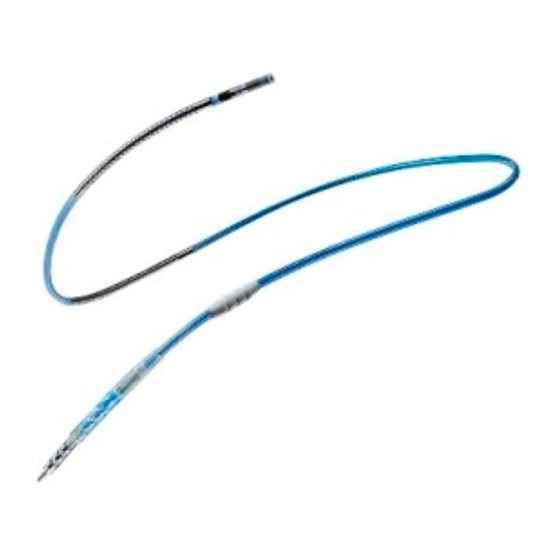
- Page 7 Figure 1. Optisure Models LDA220/LDA230 lead 1. Tip electrode (cathode) 2. Ring electrode (anode) 3. Distal defibrillation electrode 4. Proximal defibrillation electrode 5. Suture sleeve 6. Lead body trifurcation 7. IS-1 connector boot 8. Sense/pace connector (IS-1 bipolar, 3.2 mm) 9.
- Page 8 Figure 2. Optisure Model LDA210 lead 1. Tip electrode (cathode) 2. Ring electrode (anode) 3. Defibrillation electrode 4. Suture sleeve 5. Lead body trifurcation 6. IS-1 connector boot 7. Sense/pace connector (IS-1 bipolar, 3.2 mm) 8. Defibrillation connector (DF-1, 3.2 mm) Figure 3.
- Page 9 Figure 4. Optisure Model LDA210Q lead 1. Tip electrode (cathode) 2. Ring electrode (anode) 3. Defibrillation electrode 4. Suture sleeve 5. DF4 connector Figure 5. Optisure Models LDP220/LDP230 lead 1. Tip electrode (cathode) 2. Ring electrode (anode) 3. Distal defibrillation electrode 4.
- Page 10 Figure 6. Optisure Models LDP220Q/LDP230Q lead 1. Tip electrode (cathode) 2. Ring electrode (anode) 3. Distal defibrillation electrode 4. Proximal defibrillation electrode 5. Suture sleeve 6. DF4 connector Indications The Optisure ™ Models LDA220, LDA230, LDA210, LDA220Q, LDA230Q, LDA210Q, LDP220, LDP230, LDP220Q, and LDP230Q transvenous leads are indicated for use with compatible pulse generators (refer to the applicable defibrillator manual for system indications).
- Page 11 Do not use excessive force or surgical instruments to insert a stylet into a lead as this may damage the lead. Withdraw the stylet after placing the lead to prevent potential malfunction of the lead. Use only St. Jude Medical ™ suture sleeves to immobilize and protect the lead against damage from ligatures.
-
Page 12: Observed Adverse Events
Committee felt that none of these deaths were attributable to the Durata ™ lead. One-hundred sixty additional patients were withdrawn from the study for reasons listed in the following table. The reasons for withdrawal excludes withdrawals for deaths or unsuccessful implants. Table 3. -
Page 13: Potential Adverse Events
Table 4. Observed Adverse Events # Pts with AEs % of Pts with # of AEs AE/pt-years (n (n = 1697) = 3624.92) Impedance >= 100 Ω Elevated pacing thresholds 0.5% 0.002 High DFTs 0.1% 0.000 Lead bent at implant 0.1% 0.000 Migration... -
Page 14: Patients Studied
Table 5. Potential Adverse Events Event Possible Effects skin erosion, tricuspid valve dysfunction, chronic mechanical stimulation of the heart Contamination Infection requiring removal of lead system, pulse generator, or both Post-shock rhythm disturbances Post-shock bradycardia or supraventricular arrhythmias, conduction disturbances Threshold elevation or exit block Loss of efficacy of defibrillation, cardioversion, or pacing therapy... - Page 15 250 to 1100 Ω Values submitted that were greater than 12 mV were excluded from analysis because St. Jude Medical devices truncate R-wave signal amplitudes to 12. The device reports any R-wave amplitude greater than 12 mV as “>12 mV”.
-
Page 16: Personnel Training
Verify that the sterility indicator on the inner package is not purple. Purple indicates that the package has not been sterilized. St. Jude Medical does not recommend use of the product after its expiration date. If the lead package has been breached outside a sterile field or the expiration date has passed, contact St. -
Page 17: Handling The Lead
The package contents have been sterilized with ethylene oxide before shipment. This lead is for single use only and is not intended to be resterilized. If the sterile package has been compromised, contact St. Jude Medical. Handling the Lead Use caution when handling the lead. -
Page 18: Stylet Guide (Funnel, Is-1 Models Only)
® ® To avoid distortion of the Optisure lead tip, do not use a St. Jude Medical Seal-Away™ hemostasis valve. Using the Stylets CAUTION To avoid damaging the lead or body tissue, do not use excessive force or surgical instruments to insert a stylet into a lead. -
Page 19: Selecting And Opening A Vein
Figure 7. Using the clip-on tool While keeping the lead as straight as possible, grasp the connector boot with one hand and with the other hand, rotate the clip-on tool clockwise to extend the helix. (Refer to the Technical Specifications tables for the number of rotations required to extend the helix.) The helix is fully extended when at least 2 turns are visible beyond the lead tip. -
Page 20: Inserting The Lead
vein cutdown or, if a percutaneous subclavian entry is chosen, with a puncture site as lateral as possible (in the area under the lateral two-thirds of the clavicle, lateral to the subclavius muscle). The right subclavian vein and the internal jugular vein can also be used. Inserting the Lead A vein pick is included in the package to aid in inserting the lead into the vein. - Page 21 not placed in the coronary sinus or in a retrograde position, and that the entire distal defibrillation coil is below the tricuspid valve. CAUTION Do not place the lead near another implanted lead. Such close proximity could result in the electrodes making contact with each other and cause electrical interference.
-
Page 22: Positioning The Passive Fixation Lead
affixed lead will come loose easily and must then be repositioned. Allow enough slack in the lead so that it is not under tension as the heart contracts, or the patient breathes deeply or stretches. At the same time for dual-coil leads, ensure that there is not an excess of slack which may allow the proximal electrode to contact the tricuspid valve. -
Page 23: Is4/Df4 Connector Sleeve
CAUTION The stylet must be withdrawn before defibrillation testing to prevent potential malfunction of the lead. However, the sensing and pacing functions of the lead can be evaluated with the stylet still in place. Verify that the tined tip is well fixed in a stable position in the trabeculae of the ventricle by pulling back gently on the lead while observing its position under fluoroscopy. -
Page 24: Testing Defibrillation Efficacy
Figure 11. Attaching the alligator clips NOTE: Remove alligator clips prior to removing the connector sleeve from the lead. Testing Defibrillation Efficacy Once acceptable sensing and pacing performance has been verified, defibrillation lead testing should be performed to determine the voltage/energy requirements for reliable defibrillation, and to ensure that those requirements are well within the output capabilities of the pulse generator. -
Page 25: Connecting The Lead To The Pulse Generator
Figure 12. Suture the lead 1. Vein 2. Fascia 3. Lead body Loop heavy, nonabsorbable suture through the fascia underneath the proximal groove and tie a knot to create a base. Tie sutures firmly around each available groove on the suture sleeve. -
Page 26: Post-Implantation Follow-Up
Post-Implantation Follow-Up St. Jude Medical strongly recommends pre-discharge and chronic follow-up electrophysiology studies, including induction of ventricular fibrillation, in order to verify the long-term performance of the lead system. Follow-up chest x-rays to verify the position of the lead are also recommended. -
Page 27: Technical Support
St. Jude Medical for investigation. It is not recommended that chronically implanted leads be re- used after removal. Cap any abandoned lead and secure the lead caps with sutures to prevent unwanted transmission of electrical signals from the electrode to the heart. Seal the remaining open end of any severed lead with medical adhesive and a lead cap. - Page 30 St. Jude Medical St. Jude Medical Cardiac Rhythm Coordination Center BVBA Management Division The Corporate Village 15900 Valley View Court Da Vincilaan 11 Box F1 Sylmar, CA 91342 USA 1935 Zaventem +1 818 362 6822 Belgium +32 2 774 68 11 sjm.com...
Need help?
Do you have a question about the Optisure LDA220 and is the answer not in the manual?
Questions and answers