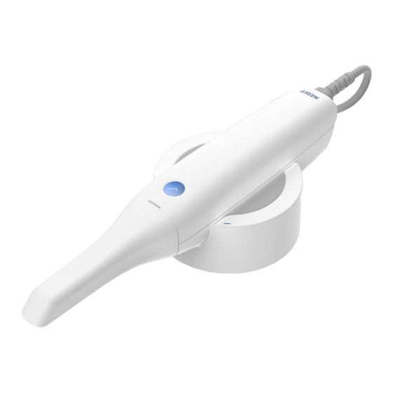
Medit i500 Function Manual
Intraoral scanner
Hide thumbs
Also See for i500:
- User manual (408 pages) ,
- Installation manual (16 pages) ,
- Troubleshooting manual (16 pages)
Table of Contents
Advertisement
Advertisement
Table of Contents

Summary of Contents for Medit i500
- Page 1 Function Manual Intraoral Scanner i500 Revised Date: Feb 2019 Revision No.: 0...
-
Page 2: Table Of Contents
Contents Introduction and overview ............................3 1.1 Intended Use ..................................... 3 1.2 Indication for use ..................................... 3 1.3 Contraindications ....................................3 1.4 Qualifications of the operating user ..............................3 Image Acquisition Software Overview ........................4 2.1 Introduction ...................................... 4 2.2 Installation ......................................4 2.2.1 System Requirement ...................................... -
Page 3: Introduction And Overview
The i500 system must be used in accordance with its accompanying user guide. The user is not allowed to modify the i500 system. Improper use or handling of the i500 system will void its warranty, if any. -
Page 4: Image Acquisition Software Overview
2 Image Acquisition Software Overview 2.1 Introduction The image acquisition software provides a user-friendly working interface to digitally record topographical characteristics of teeth and surrounding tissues using the i500 scanner. 2.2 Installation 2.2.1 System Requirement Laptop Desktop Above Above Intel Core i7-8750H... - Page 5 Select the setup language then click “Next” ➢ Select the installation path ➢...
- Page 6 Read the “License terms and conditions” carefully before checking “I agree to the License ~” then click Install ➢ If the scanner is connected, please disconnect the scanner from the PC by removing the USB cable. ➢ It may take up to several minutes to finish the recommended installation process. Please do not shut down the ➢...
-
Page 7: Model Control In View Screen Using Mouse
After installation is completed, we recommend restarting the PC to ensure optimal program operation. ➢ 2.3 Model Control in View Screen Using Mouse Button Action Usage Starts selection or deletion of entities in view screen when using the polyline Click selection or polyline trimming tool. -
Page 8: User Interface
3 User Interface A. Title Bar B. Main Toolbar C. Guide Message D. Scan Stage E. Scan Information F. Side Toolbar G. Model View Screen H. Command Option... -
Page 9: Title Bar
3.1 Title Bar The Title Bar consists of the Menu, Minimize/Maximize/Restore Window, Exit for the image acquisition software. It also shows patient information. The Menu includes the tools to manage the project, such as New, Open, Save, as well as the Menu tools to change settings and to exit the application. - Page 10 Setting...
- Page 11 Sets whether to start/stop sending anonymous usage statistics to Medit. Statistics About collection of anonymous statistics Medit is striving to constantly improve the product and user experience by collecting certain information such as : Hardware and software configurations such as OS, graphic card, etc.
- Page 12 Adjusts the brightness of the 3D model. The color of the 3D model is optimized in the i500 image acquisition software. Adjust Color Texture When viewing data using other software, the resulting colors may be slightly different from the i500 image acquisition software.
- Page 13 ➢ Turn the dial of the calibration tool to the position ➢ Put the handpiece into the calibration tool. ➢ Click “Next” to start calibration process. ➢ When the handpiece is mounted in the correct position, the system will automatically acquire the data at the position...
- Page 14 ➢ When data acquisition is completed at the position , turn the dial to the next position. ➢ Repeat the steps for positions position. ➢ When data acquisition is completed at the position, the system will automatically calculate and show the calibration result.
-
Page 15: Scan Stage
3.2 Scan Stage The Scan Stage indicates the current working stage. Acquires the 3D image of the Pre-Operation For Maxilla The data in this stage is usually acquired for pre-operation condition of Pre-Operation For maxilla. Maxilla If the Pre-Operation For Maxilla is acquired, this data will be used for the reference of the “Maxilla”. -
Page 16: Scan Pre-Operation For Maxilla
3.2.1 Scan Pre-Operation For Maxilla Acquires the 3D image of the Pre-Operation For Maxilla. The data in this stage is usually acquired for pre-operation condition of maxilla. If the Pre-Operation For Maxilla is acquired, this data will be used for the reference of the “Maxilla”. Command Option Detail Starts the scanning process. -
Page 17: Scan Pre-Operation For Mandible
Controls the level of filtering during scanning. Scanning with low level filtering acquires more amount of data than high level filtering. On the other hand, noisy data such as tongue/cheek/small cluster is acquired also. Filtering Scanning with high level filtering acquires less amount of data than low level filtering. You need more time to complete the scanning of whole model, but the final scan data will be noise-free than the scan data with low level filtering. -
Page 18: Scan Maxilla
Deletes the whole 3D images to start over. Delete If the related data with current stage exists, these data will be deleted also. Undo Undo previous scanning. Redo Redo previous scanning. Controls the level of filtering during scanning. Scanning with low level filtering acquires more amount of data than high level filtering. - Page 19 Stops the scanning process. Stop Scan You can also stop the scanning process using the function button on the device. Aligns 3D images for more accurate scanning. All noise will be deleted after optimization Optimize process. Impression Acquires the data of impression model. Scan Impression data is aligned to intra-oral data in real-time.
-
Page 20: Scan Mandible
3.2.4 Scan Mandible Acquires the 3D image of the mandible. Command Option Detail Starts the scanning process. Start Scan You can also start the scanning process using the function button on the device. Stops the scanning process. Stop Scan You can also stop the scanning process using the function button on the device. Aligns 3D images for more accurate scanning. -
Page 21: Scan Scanbody
Redo Redo previous scanning. Controls the level of filtering during scanning. Scanning with low level filtering acquires more amount of data than high level filtering. On the other hand, noisy data such as tongue/cheek/small cluster is acquired also. Filtering Scanning with high level filtering acquires less amount of data than low level filtering. You need more time to complete the scanning of whole model, but the final scan data will be noise-free than the scan data with low level filtering. -
Page 22: Scan Occlusion
Aligns 3D images for more accurate scanning. Optimize All noise will be deleted after optimization process. High Acquires the data with high resolution for entire or partial area. Resolution When the high resolution and standard resolution area are mixed, result will be merged Scanning smoothly during post-processing. - Page 23 Command Option Detail Starts the scanning process. Start Scan You can also start the scanning process using the function button on the device. Stops the scanning process. Stop Scan You can also stop the scanning process using the function button on the device. Optimize Optimize the align between maxilla and mandible.
-
Page 24: Complete
3.2.7 Complete Completes the scanning process then generates the result data. Provides three method to fill the hole for the result data. 3.3 Main Toolbar The Main Toolbar contains useful commands for editing and analyzing the 3D model, as well as the device status. 3.3.1 Trimming Removes unnecessary data, such as soft tissues and noise. -
Page 25: Brush Trimming
Brush Trimming ✓ Removes all entities on a freehand-drawn path on the screen. The brush comes in 3 different sizes. Select Remove Quick Trimming ✓ Removes island data such as soft tissues by picking. Select Remove... -
Page 26: Tools
3.3.2 Tools Lock Area ✓ Apply by painting the area to lock. Locked (colored) area will not be updated by further scanning. Use this feature to fix retracted gingiva after immediate scanning as it might collapse. You can still trim the locked area. Deleted locked area can be re-scanned. How to lock the area ➢... - Page 27 Clear Clears selection of selected area on a freehand-drawn path on the screen. Selected The brush comes in 3 different sizes. Area Clear All Clears all selected area. Area Manual Alignment ✓ Performs the alignment between maxilla and mandible manually. How to use Manual Alignment ➢...
- Page 28 Command Option Detail Undo selected marker points You can undo the selected maker points using “Remove Marker Point”. Removes marker point. Move Maxilla and Mandible Reset the position of Maxilla and Mandible. Moves maxilla to non-overlapping position from the 3D model for occlusion alignment. Moves mandible to non-overlapping position from the 3D model for occlusion alignment.
-
Page 29: Hd Camera
HD Camera ✓ Takes 2D images with 3D model data and shares the images with a laboratory. Command Option Detail Changes the sharing state. Deletes selected image. Moves to previous/next page. Changes the view style of thumbnail. How to use HD Camera ➢... -
Page 30: Occlusion Analysis
How to change status of sharing ➢ Click and select the image to share (or to stop sharing). ➢ Click the button How to change the name of image ➢ Click and select the image. ➢ Click on the same image. ➢... - Page 31 Command Option Detail Resolution You can change the resolution of the color range by using the slider bar. Low Resolution Medium Resolution High Resolution Acceptable Tolerance You can set the acceptable tolerance range. If the deviation of data is within the tolerance range, it will be displayed in green. Tolerance : 0.2 Tolerance : 0.5...
- Page 32 Switch View Style Changes the viewing style between opened jaw and closed jaw. Opened Jaws Closed Jaws Switch Deviation Style Changes the color deviation style between show all and contact area. Show All Contact Area...
-
Page 33: Undercut Area Analysis
Check the value of deviation When you move the mouse on the colored area of the 3D model, the system will show the value of the deviation. Undercut Area Analysis ✓ Analyzes the undercut area based on insertion direction. You can set the insertion direction via two methods. - Page 34 Command Option Detail Selection Tools Various selection tools are available in the Undercut Analysis command Brush Selects all entities on a freehand-drawn path on the screen. Selection The brush comes in 3 different sizes. Polyline Selects all entities within a polyline shape drawn on the screen. Selection Circle Selects all entities within circular area.
-
Page 35: Swap Maxilla & Mandible
How to calculate the undercut region by the auto direction ➢ Click “Undercut Analysis”. ➢ Set the region of interest to calculate the undercut area. If you do not set the region of interest, the system will calculate the undercut region using all the 3D models in the View Screen. - Page 36 How to use ➢ Click “Result Preview”. ➢ Moves the model into the green rectangle. ➢ Click ➢ System shows the preview result on the pop-up window.
-
Page 37: Scan Replay
Scan Replay ✓ Plays the scanning process of exist model. The scanner tip and scanning area are shown virtually during scanning. User can check the scanning condition such as scanning environment, habit etc. Command Option Detail Show/Hide Shows/Hides scanner tip during play. Scanner Tip Show/Hide Shows/Hides scanning area during play. -
Page 38: Model Display Mode
Model Display Mode ✓ Changes the model display mode. Next three methods are available. Texture On Displays the model with color texture. Texture Off Displays the model without color texture. Allows to see the trend of reliability of scan data. The model shows as green and orange color. -
Page 39: Device Status
Indicates that the i500 needs to be calibrated. Ready Indicates the i500 is ready for use. Scanning Indicates the i500 is currently in the process of scanning. Sleep Indicates the i500 is in sleep mode. Overheating Indicates the i500 is overheated. - Page 40 Zoom Places the model in the middle of the screen. Tool to change the Scan depth Allows to control the scan depth from 12~21mm. The deeper scan depth is useful in general. The shallower scan depth is useful to filter the data which is far away from the tip end.
-
Page 41: Scan Information
3.5 Scan Information Scan Shows scan time during scanning process. Time Scan Shows the number of images taken during scanning process. Images Scan Shows the current scan speed. Speed 3.6 Guide Message Shows information to guide you through the scanning process such as command functions and useful information on the current situation. -
Page 42: Command Option
Analyzes the undercut area based on insertion direction. High level filter produces more accurate initial alignment than low level filter. 3.7 Command Option Shows the available options for the currently running command. For detailed information of the options, please refer to the option description for each command. 3.8 Model View Screen Displays live video and captured images. -
Page 43: Pre-Operation Scan Stage
4.2 Pre-Operation Scan Stage The data in this stage is usually acquired for the pre-operation condition of maxilla or mandible and will be used for the reference data of the maxilla or mandible. How to use ➢ Acquire the data for pre-operation condition. ➢... -
Page 44: High Resolution Scan
➢ Acquire the additional scan data for deleted area. The location between “Pre-Opeartion Model” and “Maxilla(or Mandible)” will be same through whole process. ”Pre-Operation Model” is not used for the occlusion alignment. The occlusion alignment uses maxilla and mandible only. If you move to “Occlusion Stage” after scanning only “Pre-Operation Model”, the “Pre-Operation Model”... -
Page 45: Impression Scan
When the color texture is turned off, high resolution and standard resolution area are shown with different material color. 4.4 Impression Scan “Impression Scan” provides the seamless scanning process to combine the intra-oral and impression scan data. You can easily merge the intra-oral and impression scan data with integrated scanning process. Command option detail Various selection tools are provided to limit the scanning area. - Page 46 How to use impression scan ➢ Acquire the intra-oral scan data ➢ Turn on “Impression Scan” ➢ Mark the area to replace the intra-oral data with the impression data. Marking function is useful to limit the area which should be replaced. You can skip “Marking”...
- Page 47 How to edit impression data When the impression data is taken, unnecessary area needs to be removed before completing the case. For example, if the impression data is acquired like the image below, unnecessary data from the impression scanning should be removed.
- Page 48 ➢ Run the trimming function in impression scan mode. The impression data is shown in the Model View Screen. ➢ Delete unnecessary area in the impression data. ➢ Result will be appeared like below.
-
Page 49: Replace The Margin Of Intra-Oral Data With Impression Data
When “Impression Scan” is useful Replace the margin of intra-oral data with impression data ✓ ➢ Scan base model. ➢ Turn on “Impression Scan” then mark interesting area. In this case, the area for margin is marked. ➢ Acquire the impression data. -
Page 50: Post & Core Case
➢ Result will be generated like below. Post & Core case ✓ In some case of Post & Core, it is very difficult to take the data for post area due to the area is very deep and hard to scan. - Page 51 How to use ➢ Scan base model. ➢ Turn on “Impression Scan” then scan impression model. ➢ Result will be generated like below.
-
Page 52: Occlusion Case
Occlusion case ✓ The case to use impression scan to occlusion alignment. How to use ➢ Scan Maxilla and Mandible. ➢ Turn on “Impression Scan” in the occlusion stage and acquires occlusion data using impression model. In this case, you need to take the impression model 360 degrees for each bite. -
Page 53: Updates To Image Acquisition Software
”High Resolution Scan” is available during impression scan. When the impression data is acquired with high resolution, data will be shown with different color if the model rendering is “Texture off”. 5 Updates to Image Acquisition Software The Image acquisition software automatically checks for updates when the software is running. When a new version of the software is available, the system will automatically download the new version.


Need help?
Do you have a question about the i500 and is the answer not in the manual?
Questions and answers