SonoSite M-Turbo User Manual
Ultrasound system
Hide thumbs
Also See for M-Turbo:
- User manual (280 pages) ,
- Service manual (58 pages) ,
- Quick start manual (14 pages)
Table of Contents
Advertisement
Quick Links
Advertisement
Table of Contents

Related Manuals for SonoSite M-Turbo


Summary of Contents for SonoSite M-Turbo
- Page 1 M-Turbo Ultrasound System User Guide...
- Page 3 M-Turbo Ultrasound System User Guide...
- Page 4 Non-SonoSite product names may be trademarks or registered trademarks of their respective owners. The SonoSite product(s) referenced in this document may be covered by one or more of the following US patents: 5722412, 5817024, 5893363, 6135961, 6203498, 6364839, 6371918, 6383139, 6416475, 6447451, 6471651, 6569101, 6648826, 6575908, 6604630, 6817982, 6835177, 6962566, 7169108, D456509, D461895, D509900, D538432, D544962, D558351, D559390, and by the following counterpart foreign patents: AU727381, AU730822, CA2373065, CN98106133.8, CN98108973.9, DE60021552.0, DE60029777.2, DE60034670.6,...
-
Page 5: Table Of Contents
Contents Introduction Conventions, symbols, and terms ................. vii Customer comments ....................vii Chapter 1: Getting Started About the system ......................1 Preparing the system ....................1 Installing or removing the battery ..............1 Using AC power and charging the battery ..........2 Turning the system on or off ................ - Page 6 OB Calculations setup ....................20 OB Custom Measurements setup ................21 OB Custom Tables setup ................... 22 Presets setup ......................... 22 System Information setup ..................23 USB Devices setup ...................... 23 Limitations of JPEG format ................23 Chapter 3: Imaging Imaging modes ......................
- Page 7 Small Parts calculations ..................64 Transcranial Doppler and Orbital calculations ........65 Vascular calculations ..................67 Patient report ........................68 Vascular and cardiac patient reports ............69 TCD patient report .....................69 OB patient report ....................69 EMED worksheets ....................70 Chapter 5: Troubleshooting and Maintenance Troubleshooting ......................71 Software licensing .......................71 Maintenance .........................72 Cleaning and disinfecting the ultrasound system .........73...
- Page 8 In Situ, derated, and water value intensities ...........105 Tissue models and equipment survey ............106 Acoustic output tables ....................107 Terms used in the acoustic output tables ..........132 Acoustic measurement precision and uncertainty ......133 Labeling symbols ......................134 Chapter 7: References Measurement accuracy ...................139 Sources of measurement errors ................140 Measurement publications and terminology ..........141 Cardiac references ....................141...
-
Page 9: Conventions, Symbols, And Terms
SonoSite at 888‐482‐9449 in the US. Outside the specifications, and safety and acoustic output US, call the nearest SonoSite representative. You information. can also e‐mail SonoSite The user guide is for a reader familiar with at comments@sonosite.com. ultrasound techniques. It does not provide For technical support, please contact SonoSite as training in sonography or clinical practices. follows: Before using the system, you must have ultrasound training. SonoSite Technical Support See the applicable SonoSite accessory user guide for information on using accessories and Phone (US or 877-657-8118 peripherals. See the manufacturer’s instructions Canada): for specific information about peripherals. Phone (Outside 425-951-1330 US and Canada): Or call your local Conventions, symbols, and representative. -
Page 10: Customer Comments
viii Customer comments... -
Page 11: Chapter 1: Getting Started
Figure 2 System Back Connectors: A license key is required to activate the software. (1) DC input connector, (2) I/O connector, (3) Battery, See “Software licensing” on page 71. On and (4) ECG connector occasion, a software upgrade may be required. SonoSite provides a USB device containing the To use the ultrasound system software. One USB device can be used to upgrade 1 Attach a transducer. multiple systems. 2 Turn the system on. (For power switch location, see “System controls” on page 5.) 3 Press the ... -
Page 12: Using Ac Power And Charging The Battery
To install the battery 1 Disconnect the power supply from the ultrasound system. 2 Remove the system from the mini‐dock (if present) and turn it upside down. 3 Place the battery into the battery compartment, at a slight angle. See Figure 4 Slide the battery forward until it locks into place. 5 Push down on the two locking levers to secure the battery. Locking levers To remove the battery 1 Disconnect the power supply from the ultrasound system. 2 Remove the system from the mini‐dock (if present) and turn it upside down. 3 Pull up the two locking levers. 4 Slide the battery back. 5 Lift the battery from the compartment. Using AC power and charging the battery The battery charges when the system is ... -
Page 13: Turning The System On Or Off
Turning the system on or off Caution: Do not use the system if an error message appears on the display. Note the error code and turn off the system. Call SonoSite or your local representative. To turn the system on or off Press the power switch. (See “System controls” on page 5.) To wake up the system To conserve battery life while the system is on, ... -
Page 14: Inserting And Removing Usb Storage Devices
To connect a transducer 1 Remove the system from the mini‐dock (if WARNING: To avoid damaging the USB storage present), and turn it upside down. device and losing patient data from 2 Pull the transducer latch up, and rotate it it, observe the following: clockwise. • Do not remove the USB storage device or turn off the ultrasound 3 Align the transducer connector with the ... -
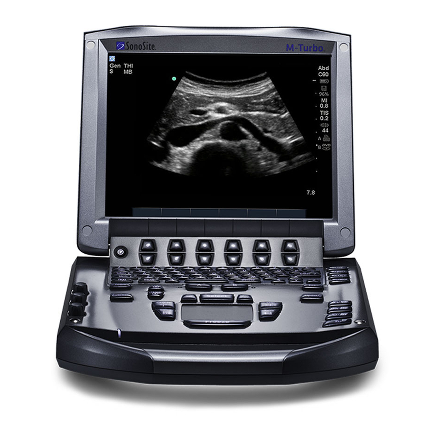
Page 15: System Controls
System controls 12 13 Power switch Turns system on and off. Alphanumeric keys Use to enter text and numbers. Annotation keys “Alphanumeric keyboard” on page ZOOM Magnifies the image 100%. Decreases and increases imaging depth. DEPTH UP, DEPTH DOWN AUTO GAIN Adjusts gain automatically. - Page 16 AC power indicator A steady light indicates that AC power is connected. A flashing light indicates that the system is asleep. CALIPER Displays calipers on-screen for measuring. CALCS Turns the calculations menu on and off. Touchpad Selects, adjusts, and moves items on-screen. FREEZE Stops live imaging and displays a frozen image.
-
Page 17: Screen Layout
Screen layout Figure 1 Screen Layout Mode Data Area Current imaging mode information (for example, Gen, Res, THI, and PW). Orientation Marker Provides indication for image orientation. In dual and duplex images, the orientation marker is green on the active screen. Text Text entered using keyboard. -
Page 18: General Interaction
General interaction Cycle Moves through a list of settings continuously. The upper control key cycles upward. The lower control key cycles downward. Touchpad and cursor Up-Down Moves through a list of settings, Use the touchpad to adjust and move objects stopping at the top or bottom. The upper control on‐screen. The touchpad controls caliper key moves upward. The lower control key moves position, CPD or Color box position and size, the downward. By default, a beep sounds when you cursor, and more. The arrow keys control much reach either end of the range. (See “Audio, Battery of the same functionality as the touchpad. setup” on page 19.) The cursor appears in the setup pages, the patient On-Off Turns a feature on or off. You can press information form, and patient report. You control either control key. In forms, you can instead select the cursor through the touchpad. For example, in the option by using the touchpad and the the patient information form, place the cursor SELECT key. over the last name field and press the SELECT Action Performs an action. You can press either key to activate that field. Additionally, you can control key. Or you can instead select the option use the cursor to select check boxes and items in by using the touchpad and the SELECT key. lists. On-screen options The on‐screen options let you make adjustments ... -
Page 19: Annotation And Text
Annotation and text Alphanumeric keyboard Moves cursor among fields DELETE Removes all text from the in the forms, and tabs screen during text entry between text position in and when not measuring. dual screens. Arrow Keys Move highlighted selection CAPS LOCK Sets the keyboard to in calculations menu, move capital letters. -
Page 20: Preparing Transducers
Caution: To avoid damage to the transducer, Text mode (imaging): Annotation field use only gels recommended by SonoSite. Using gels other than the one recommended by SonoSite can damage the transducer and void the warranty. If you have questions about gel compatibility, contact SonoSite or your local representative. -
Page 21: Training Videos
Key USB device (“Training” appears under are ready to perform the procedure. Type), and then select Select. Note: Image Gallery is an unsupported feature. To apply a transducer sheath To view a video SonoSite recommends the use of market‐cleared, transducer sheaths for intracavitary or surgical 1 Display the list of videos. applications.To lessen the risk of contamination, 2 Select the video. apply the sheath only when you are ready to perform the procedure. 3 Select View on‐screen. 1 Place gel inside the sheath. - Page 22 Abdominal Imaging Applications You can assess multiple pregnancy, fetal hydrops, placental the liver, kidneys, pancreas, spleen, gallbladder, abnormalities, as well as maternal hypertension, bile ducts, transplanted organs, abdominal diabetes, and lupus. vessels, and surrounding anatomical structures WARNING: To prevent injury or misdiagnosis, for the presence or absence of pathology do not use this system for transabdominally. Percutaneous Umbilical Blood Cardiac Imaging Applications You can assess the Sampling (PUBS) or in vitro heart, cardiac valves, great vessels, surrounding Fertilization (IVF) The system has anatomical structures, overall cardiac ...
- Page 23 WARNING: To avoid injury to the patient, use only an Orbital (Orb) or Ophthalmic (Oph) exam type when performing imaging through the eye. The FDA has established lower acoustic energy limits for ophthalmic use. The system will not exceed these limits only if the Orb or Oph exam type is selected.
- Page 24 Intended uses...
-
Page 25: Chapter 2: System Setup
Footswitch (L), Footswitch (R) The function of the system and set preferences. left and right footswitches: Save Clip, Record, Freeze, Save Image, or Print. See also “To connect the footswitch.” Displaying the setup pages To connect the footswitch To display a setup page The SonoSite footswitch allows hands‐free operation with a customizable two‐pedal 1 Press the key. SETUP footswitch. The footswitch is an optional feature. 2 Select the setup page under Setup Pages. WARNING: To avoid contamination, do not use To return to imaging from a setup page, select ... -
Page 26: Security Settings
1 Log in as Administrator. 1 On the Administration setup page, type in the Name box. Administrator 2 Select New. 2 Type the administrator password in the 3 Under User Information, fill in the Name, Password box. Password, and Confirm boxes. (See “Choosing a secure password” on page 18.) If you don’t have the administrator password, contact SonoSite. (See “SonoSite Technical 4 (Optional) In the User box, type the user’s Support” on page vii.) initials to display them in the patient header and the User field in the patient information 3 Select Login. form. To log out as Administrator 5 (Optional) Select the Administration Access Turn off or restart the system. check box to allow access to all administration privileges. -
Page 27: Exporting Or Importing User Accounts
2 Under User List, select the user. 2 Log in as Administrator. 3 Under User Information, make changes as 3 Select Import on‐screen. desired. 4 Select the USB storage device, and select 4 Select Save. Import. Any change to the user name replaces the 5 Restart the system. previous name. All user names and passwords on the system are replaced with the imported data. To delete a user 1 Log in as Administrator. Exporting and clearing the Event log 2 Under User List, select the user. The Event log collects errors and events and can 3 Select Delete. be exported to a USB storage device and read on ... -
Page 28: Logging In As User
Logging in as user 1 In the Exam list on the Annotations setup page, select the exam type whose labels you If user login is required, the User Login screen want to specify. appears when you turn on the system. (See “To require user login” on page 16.) 2 For Group, select A, B, or C for the label group you want associated with that exam. To log in as user The preset labels appear for the selected group. 1 Turn on the system. 3 Do any of the following: 2 In the User Login screen, type your name and password, and select OK. • Add a custom label to the group: Type the label in the Text box, and select Add. To log in as guest •... -
Page 29: Audio, Battery Setup
• In the Serial Port list, select the peripheral. Sleep delay Select Off, or 5 or 10 minutes to Computer (PC) allows patient report data specify the period of inactivity before the system to be sent as ASCII text from the system to goes into sleep mode. a PC. The PC must have third‐party Power delay Select Off, or 15 or 30 minutes to software to acquire, view, or format the specify the period of inactivity before the system data into a report. Check the compatibility automatically turns off. of your software with SonoSite Technical Support. (See also “To send a patient report to a PC” on page 69.) Cardiac Calculations setup Note: Because these peripherals use the same On the Cardiac Calculations setup page, you can RS‐232 connector on the mini‐dock, you can specify measurement names that appear in the connect only one of them at a time. Tissue Doppler Imaging (TDI) calculations menu 2 Restart the system. and on the report page. 3 Attach a serial cable (RS‐232) from the serial See also “Cardiac calculations” on page 50. -
Page 30: Date And Time Setup
IMT Calculations setup To receive storage alerts On the Connectivity setup page, select On the IMT Calculations setup page, you can Internal Storage Capacity Alert. customize the IMT calculations menu. You can The system displays a message if internal specify up to eight measurement names for both storage is near capacity when you end an right side and left side calculations. The exam. The system then deletes archived measurement names also appear in the patient patient exams if specified in DICOM. report. See also “IMT calculations” on page 59. Date and Time setup To customize the IMT calculations menu WARNING: To obtain accurate obstetrics On the IMT Calculations setup page, do the following: calculations, an accurate date and time are critical. -
Page 31: Ob Custom Measurements Setup
To import OB calculation tables Tables that you import are added to those already on the system. 1 Insert the USB storage device that contains the tables. 2 On the OB Calculations setup page, select Import on‐screen. 3 Select the USB storage device, and then select Import. 4 Select OK in the dialog box that appears. The system restarts. Figure 1 OB Calculations Setup Page OB Custom Measurements To specify gestational age and growth setup analysis 1 On the OB Calculations setup page, select the On the OB Custom Measurements setup page, desired OB authors (or select None) in the ... -
Page 32: Ob Custom Tables Setup
3 Select Yes. 4 Select New on‐screen. The exam ends, and any tables and report 5 In the Author box, type a unique name. data associated with the measurement are 6 Enter the data. removed from the system. 7 Select Save on‐screen. To display the measurement for the custom table OB Custom Tables setup in the calculations menu, see “To specify On the OB Custom Tables setup pages, you can gestational age and growth analysis” on page 21. customize growth tables that appear in the To edit or delete an OB custom table calculations menu and patient report. 1 On the OB Calculations or OB Custom Gestational Age Table Measurements The system Measurements setup page, select Tables provides gestational age measurements by ... -
Page 33: System Information Setup
To specify a file format for exported images Assessment of DSA, Ultrasound and CT 1 On the USB Devices setup page, select Export. Digital Images Compressed with the JPEG 2 Under USB Export, select an export type: Protocol,” D Okkalides et al 1994 Phys Med Biol 39 1407‐1421 doi: • SiteLink organizes files in a SiteLink‐style 10.1088/0031‐9155/39/9/008 folder structure. Clips export in H.264 www.iop.org/EJ/abstract/0031‐9155/39/9/008 video saved as MP4 files. To view them, SonoSite recommends QuickTime 7.0 or “Canadian Association of Radiologists, CAR later. Standards for Irreversible Compression in Digital Diagnostic Imaging within • DICOM creates files readable by a DICOM Radiology,” Approved: June 2008. reader. DICOM is an optional feature. www.car.ca/Files/%5CLossy_Compression. 3 Select an image format for your export type. For JPEG image format, also select a JPEG Chapter 2: System Setup... - Page 34 USB Devices setup...
-
Page 35: Chapter 3: Imaging
Chapter 3: Imaging Imaging modes 2D options In 2D imaging, you can select the following The system has a high‐performance display and on‐screen options. advanced image‐optimization technology that significantly simplifies user controls. Imaging Optimize Settings are as follows: modes available depend on the transducer and • Res provides the best possible exam type. See “Imaging modes and exams resolution. available by transducer” on page 31. • Gen provides a balance between resolution and penetration. 2D imaging • Pen provides the best possible 2D is the systemʹs default imaging mode. The ... -
Page 36: M Mode Imaging
Page x/x Indicates which page of options is displayed. Select to display the next This feature depends on transducer page. type. See the SonoSite Biopsy user guide. M Mode imaging Biopsy is not available when the ECG cable is connected. -
Page 37: Cpd And Color Doppler Imaging
3 Set options as desired. 2 Select CPD or Color. Many optimization and depth options The current selection also appears in the available in 2D imaging are also available in upper left‐hand screen. M Mode imaging. See “2D options” on The Color indicator bar on the upper left‐hand page 25. screen displays velocity in cm/s in Color imaging mode only. To display the M Mode trace 3 Using the touchpad, position or resize the ROI 1 Display the M‐line. box as needed. Press the key to toggle SELECT Adjust the depth if necessary. (See “To adjust between position and size. depth” on page 30.) While you position or resize the ROI box, a 3 Press the key. M MODE green outline shows the change. The ROI box indicator on the left‐hand screen shows which The time scale above the trace has small marks ... -
Page 38: Pw And Cw Doppler Imaging
You can use PW/CW Doppler and CPD/Color PRF Scale Select the desired pulse repetition simultaneously. If CPD/Color imaging is on, the frequency (PRF) setting by pressing color ROI box is tied to the D‐line. The key SELECT the control keys. cycles among color ROI box position, color ROI There is a wide range of PRF box size, the D‐line, and (in PW Doppler) angle settings for each Flow Sensitivity correction. The active selection is green. Also, the setting (Low, Med, and High). indicator on the left‐hand screen shows which ... - Page 39 • If using a duplex layout, press the • 0 has an angle correction of 0°. key to toggle between the DOPPLER • +15 has an angle correction of full‐screen D‐line and the duplex layout. +60°. To set a duplex layout, see “Presets setup” You can manually correct the angle on page 22. after selecting a steering angle setting. (See “To display the D-line” PW Doppler options on page 28.) In PW Doppler imaging, you can set the ...
-
Page 40: Adjusting Depth And Gain
box. In PW and CW Doppler imaging, the Live Trace Displays a live trace of the peak or the knob affects Doppler gain. GAIN mean. (See “Presets setup” page 22 to specify peak or mean.) Near and far correspond to the time gain compensation (TGC) controls on other ultrasound systems. Page x/x Indicates which page of options is displayed. Select to display the next page. -
Page 41: Imaging Modes And Exams Available By Transducer
down, left, and right. (You cannot pan in Imaging modes and exams available by Dual.) transducer To exit zoom, press the key again. ZOOM Imaging Mode Imaging modes and exams available by transducer WARNING: To prevent misdiagnosis or harm to the patient, understand your C11x — system’s capabilities prior to use. The diagnostic capability differs for —... -
Page 42: Annotating Images
Imaging Mode Imaging Mode L25x — — — — — TEEx — — 1. Exam type abbreviations are as follows: Abd = Abdomen, Bre = Breast, Crd = Cardiac, Gyn = Gynecology, IMT = — Intima Media Thickness, Msk = Muscle, Neo = Neonatal, Nrv = Nerve, OB = Obstetrical, Oph = Ophthalmic, Orb = —... -
Page 43: Patient Information Form
The default home position depends on the 4 Press the key to set the arrow. ARROW imaging screen layout. You can reset the The arrow changes from green to white. home position. See “To reset the home position” on page 33. To remove the arrow, press the key and ARROW then select Hide. 3 Using the keyboard, type text. To place a pictograph on an image • The arrow keys move the cursor left, right, up, and down. The pictograph set available depends on transducer and exam type. • The key deletes all text. DELETE 1 Press the key. PICTO • The Word option removes a word. 2 Select x/x to display the desired ... - Page 44 To create a new patient information form • ID Patient identification number 1 Press the PATIENT key. • Accession Enter number, if applicable. • Date of birth 2 Select New/End. • Gender Fill in the form fields. See “Patient information form fields” on page 34. • Indications Enter desired text 4 Select Done. • User User initials See also “To append images and clips to a patient • Procedure (button) Available if the DICOM exam” on page 37. Worklist feature is licensed and configured. See the DICOM user guide.
-
Page 45: Images And Clips
Saving the heart rate using a measurement By default, the key saves only the image. As SAVE overwrites this entry. a shortcut during calculations, the key can SAVE save both the image to internal storage and the • Height (Cardiac exam) The patient height in calculation to the patient report. See “Presets feet and inches or meters and centimeters. (To setup” on page 22. change the units, see “Presets setup” on page 22.) To capture and save a clip • Weight (Cardiac exam) The patient weight in Clips, an optional feature, lets you capture, pounds or kilos. (To change the units, see preview, and save clips. “Presets setup” on page 22.) 1 Set Clips options. (See “To set Clips options” • BSA (Cardiac exam) Body Surface Area. on page 35.) Automatically calculated after you enter 2 Press the ... -
Page 46: Reviewing Patient Exams
If the internal storage icon does not Select All selects all patients. appear in the system status area, To deselect patients, select checked boxes or Clear internal storage may be defective. All. Contact SonoSite Technical Support. (See “SonoSite Technical To edit patient information from the patient Support” on page vii.) list You can edit the patient name and ID from the ... -
Page 47: Printing, Exporting, And Deleting Images And Clips
2 Select Edit. Printing, exporting, and deleting images and clips 3 Fill in the form fields, and select OK. WARNING: To avoid damaging the USB To append images and clips to a patient storage device and losing patient exam data from it, observe the following: Although you cannot add images and clips to a • Do not remove the USB storage patient exam that is ended, exported, or archived, ... -
Page 48: Ecg Monitoring
Only available USB devices are selectable. Caution: Use only accessories recommended 5 Select Export. by SonoSite with the system. Your system can be damaged by The files are finished exporting connecting an accessory not approximately five seconds after the USB recommended by SonoSite. animation stops. Removing the USB storage ... - Page 49 ECG Monitoring options Show/Hide Turns on and off ECG trace. Gain Increases or decreases ECG gain. Settings are 0-20. Position Sets the position of the ECG trace. Sweep Speed Settings are Slow, Med, and Fast. Delay Displays Line and Save for clip acquisition delay.
- Page 50 ECG Monitoring...
-
Page 51: Chapter 4: Measurements And Calculations
Chapter 4: Measurements and Calculations You can measure for quick reference, or you can Working with calipers measure within a calculation. You can perform When measuring, you work with calipers, often general calculations as well as calculations in pairs. Results based on the calipers’ position specific to an exam type. appear at the bottom of the screen. The results update as you reposition the calipers by using the Measurements are performed on frozen images. touchpad. In trace measurements, the results For references used, see Chapter 7, “References.” appear after you complete the trace. Outside a calculation, you can add calipers by Measurements pressing the key. You can have multiple CALIPER sets of calipers and can switch from one set to You can perform basic measurements in any another, repositioning them as needed. Each set imaging mode and can save the image with the shows the measurement result. The active measurements displayed. (See “To save an calipers and measurement result are highlighted image” on page 35.) Except for the M Mode HR green. A measurement is complete when you measurement, the results do not automatically finish moving its calipers. save to a calculation and the patient report. If you prefer, you can first begin a calculation and then Within a calculation, calipers appear when you ... -
Page 52: 2D Measurements
To improve precision of caliper placement You can perform a combination of distance, area, circumference, and manual trace measurements Do any of the following: at one time. The total number possible depends • Adjust the display for maximum on their order and type. sharpness. To measure distance (2D) • Use leading edges (closest to the transducer) or borders for starting and You can perform up to eight distance stopping points. measurements on a 2D image. • Maintain a consistent transducer 1 On a frozen 2D image, press the key. CALIPER orientation for each type of measurement. A pair of calipers appears, connected by a • Make sure that the area of interest fills as dotted line. much of the screen as possible. 2 Using the touchpad, position the first caliper, • (2D) Minimize the depth, or zoom. and then press the ... -
Page 53: M Mode Measurements
4 Press the key. 4 Press the key. SELECT SELECT 5 Using the touchpad, complete the trace, and A second vertical caliper appears. press the key. 5 Using the touchpad, position the second See “To save a measurement to a calculation and vertical caliper at the peak of the next patient report” on page 41. heartbeat. See “To save a measurement to a calculation and M Mode measurements patient report” on page 41. Saving the heart rate measurement to the patient report overwrites The basic measurements that you can perform in any heart rate entered on the patient information M Mode imaging are as follows: form. • Distance in cm/Time in seconds See also “To measure fetal heart rate (M Mode)” • Heart Rate (HR) in beats per minute (bpm) on page 63. The time scale above the trace has small marks at 200 ms intervals and large marks at one‐second ... - Page 54 To measure Velocities, Elapsed Time, +/x 4 Using the touchpad, trace the waveform. Ratio, Resistive Index (RI), and Acceleration To make a correction, select Undo on‐screen, (Doppler) backtrack with the touchpad, or press the 1 On a frozen Doppler spectral trace, press the key. BACKSPACE key. CALIPER 5 Press the key. A single caliper appears. The measurement results appear. 2 Using the touchpad, position the caliper to a See “To save a measurement to a calculation and peak systolic waveform. patient report” on page 41. 3 Press the key. SELECT To trace automatically (Doppler) A second caliper appears.
-
Page 55: General Calculations
• Mean Pressure Gradient (PGmean) Menu items followed by ellipses (. . .) have subentries. • Mean Velocity on Peak Trace (Vmean) To select from the calculations menu • Pressure Gradient (PGmax) 1 On a frozen image, press the key. CALCS • Cardiac Output (CO) The calculations menu appears. • Peak Systolic Velocity (PSV) 2 Using the touchpad or arrow keys, highlight • Time Average Mean (TAM)* the desired measurement name. • +/× or Systolic/Diastolic (S/D) To display additional measurement names, • Pulsatility Index (PI) highlight Next, Prev, or a measurement name that has ellipses (. . .). Then press the SELECT • End Diastolic Velocity (EDV) key. •... -
Page 56: Displaying, Repeating, And Deleting Saved Measurements In Calculations
is set to Image/Calcs. (See “Presets setup” Some measurements can be deleted directly from on page 22.) the patient report pages. See “Patient report” on page 68. The calculation saves to the patient report, and the image saves to internal storage EMED calculations with the measurements displayed. The results from EMED calculations automatically appear in the EMED worksheets. Displaying, repeating, and deleting All EMED calculations are available for each saved measurements in calculations exam type. To display a saved measurement To perform an EMED calculation: Do one of the following: 1 Press the key. CALCS • Highlight the measurement name in the 2 Select EMED on‐screen. - Page 57 b Using the touchpad, move the caliper to the trace starting point, and press the Transducer Exam Types key. SELECT c Using the touchpad, trace the desired area. C11x Abdomen To make a correction, select Undo C60x Abdomen on‐screen or press the key. BACKSPACE HFL38x IMT, Small Parts, Vascular d Complete the trace, and press the key. L25x Vascular, Muscle Save the calculation. See “To save a calculation” on page 45. L38x IMT, Small Parts, Vascular The percent area reduction result appears P10x Abdomen on‐screen in the measurement and calculation ...
-
Page 58: Volume Calculations
Volume calculations To calculate volume The volume calculation involves three 2D WARNING: To avoid incorrect calculations, distance measurements: D , D , and D . After all verify that the patient information, measurements are saved, the result appears date, and time settings are on‐screen and in the patient report. accurate. Do the following for each image you need to To avoid misdiagnosis or harming measure: the patient outcome, start a new a On the frozen 2D image, press the ... - Page 59 • Difficulty ensuring uniform insonation of the vessel. Transducer Exam Types The system is limited to the following sample volume sizes: C11x Abdomen • C11x transducer: 1, 2, 3 Gate Size (mm) C60x Abdomen • C60x and P10x transducers: 2, 3, 5, 7, HFL38x Vascular 10, 12 Gate Size (mm) • HFL38x, L25x, L38x, and SLAx L25x Vascular transducers: 1, 3, 5, 7, 10, 12 Gate Size L38x Vascular (mm) P10x Abdomen • P21x transducer: 2, 3, 5, 7, 11.5, 14 Gate Size (mm) P21x Abdomen • Precision in placing the caliper SLAx Vascular • Accuracy in angle correction The following table shows the measurements ...
-
Page 60: Exam-Based Calculations
c Using the touchpad, position the vertical caliper at the beginning of the waveform. Transducer Exam Type If calipers are not positioned correctly, the calculation result is inaccurate. Cardiac d Press the key to display a second SELECT P10x Cardiac vertical caliper. P21x Cardiac e Using the touchpad, position the second vertical caliper at the end of the waveform. TEEx Cardiac Press the key to complete the trace and The following table shows the measurements to display the results. required to complete different cardiac Save the calculation. See “To save a calculations. For definitions of acronyms, see calculation” on page 45. “Glossary” on page 157. To display the volume flow calculation, see Cardiac Calculations “Patient report” on page 68. Cardiac Menu Calculation Measurements... - Page 61 Cardiac Cardiac Menu Calculation Menu Calculation Measurements Measurements Heading Results Heading Results (Imaging Mode) (Imaging Mode) ACS (M Mode) PISA Ann D (2D) PISA Area Radius (Color) LVET (M Mode) LVET MR/VTI (Doppler) MV Rate EF:Slope EF SLOPE MV/VTI (Doppler) Regurgitant (M Mode) Volume...
- Page 62 Cardiac Cardiac Menu Calculation Menu Calculation Measurements Measurements Heading Results Heading Results (Imaging Mode) (Imaging Mode) P. Vein A (Doppler) VMax Vmax (Doppler) Vmax PGmax Adur (Doppler) time VTI (Doppler) S (Doppler) VMax Vmax S/D ratio D (Doppler) PGmax Vmean E (Doppler) PGmean A (Doppler)
- Page 63 To measure LVd and LVs Cardiac Menu Calculation 1 On a frozen 2D image or M Mode trace, press Measurements Heading Results the key. CALCS (Imaging Mode) 2 From the calculations menu, select the TRmax (Doppler) Vmax measurement name. PGmax 3 Position the active (green) caliper at the E (Doppler) starting point. (See “Working with calipers” on page 41.) A (Doppler) E PG 4 Press the key, and position the second SELECT caliper.
- Page 64 c Using the touchpad, trace the left 5 Positioning the calipers, measure the ventricular (LV) cavity. ventricular length. (See “Working with calipers” on page 41.) To make a correction, select Undo on‐screen or press the key. 6 Save the calculation. BACKSPACE d Complete the trace, and press the key. To measure peak velocity Save the calculation. (See “To save a For each cardiac measurement, the system saves calculation” on page 45.) up to five individual measurements and calculates their average. If you take more than To calculate MV or AV area five measurements, the most recent measurement 1 On a frozen 2D image, press the key. replaces the fifth one. If you delete a saved CALCS measurement from the patient report, the next 2 In the calculations menu, locate Area, and ...
- Page 65 4 Using the touchpad, trace the waveform. • In AV, position the caliper at the end diastole. To make a correction, select Undo on‐screen, backtrack with the touchpad, or press the Save the calculation. (See “To save a key. calculation” on page 45.) BACKSPACE 5 Press the key to complete the trace. To calculate Proximal Isovelocity Surface Area (PISA) Save the calculation. (See “To save a calculation” on page 45.) The PISA calculation requires a measurement in 2D, a measurement in Color, and two For information on the automatic trace tool, see measurements in Doppler spectral trace. After all “To trace automatically (Doppler)” on page 44. measurements are saved, the result appears in the patient report. To calculate Right Ventricular Systolic Pressure (RVSP) 1 Measure from Ann D (2D): 1 On a frozen Doppler spectral trace, press the ...
- Page 66 To make a correction, select Undo 4 Press the key. SELECT on‐screen, backtrack with the touchpad, or A second horizontal dotted line with an active press the key. BACKSPACE caliper appears at 300 cm/s. d Press the key to complete the trace. 5 Position the second caliper along the e Save the calculation. waveform at 300 cm/s. For information on the automatic trace tool, see Save the calculation. (See “To save a “To trace automatically (Doppler)” on page 44. calculation” on page 45.) To calculate Isovolumic Relaxation Time To calculate Aortic Valve Area (AVA) (IVRT) The AVA calculation requires a measurement in 2D and two measurements in Doppler. After the 1 On a frozen Doppler spectral trace, press the measurements are saved, the result appears in ...
- Page 67 a From the calculations menu, locate Qp/Qs Position the calipers. (See “Working with and then select LVOT D or RVOT D. calipers” on page 41.) Position the calipers. (See “Working with Save the calculation. (See “To save a calipers” on page 41.) calculation” on page 45.) Save the calculation. (See “To save a Measure from aorta (Doppler). See “To calculation” on page 45.) calculate Velocity Time Integral (VTI)” on page 54. From the calculations menu, select 3 On a frozen Doppler spectral trace, press the AV and then select VTI. key. CALCS For information on the automatic trace tool, see 4 Do the following to measure from LVOT VTI “To trace automatically (Doppler)” on page 44. and again to measure from RVOT VTI: To calculate Heart Rate (HR) a From the calculations menu, select Qp/Qs and then select LVOT VTI or RVOT VTI.
-
Page 68: Gynecology (Gyn) Calculations
1 (CI Only) Fill in the Height and Weight fields on the patient information form. The BSA is WARNING: To avoid incorrect calculations, calculated automatically. (See “To create a verify that the patient information, new patient information form” on page 34.) date, and time settings are Calculate SV. See “To calculate Stroke Volume accurate. (SV) or Stroke Index (SI)” on page 57. To avoid misdiagnosis or harming Calculate HR. See “To calculate Heart Rate the patient outcome, start a new (HR)” on page 57. patient information form before starting a new patient exam and To measure a Tissue Doppler Imaging (TDI) performing calculations. -
Page 69: Imt Calculations
To measure follicles To avoid incorrect calculations, You can save up to six follicular measurements, verify that the patient information, one distance measurement for each of up to six date, and time settings are follicles. accurate. 1 On a frozen 2D image, press the key. To avoid misdiagnosis or harming CALCS the patient outcome, start a new 2 From the calculations menu, select Follicle. patient information form before 3 Do the following for each follicle you want to ... - Page 70 IMT tool options IMT Calculations (2D) When using the IMT tool, you can select the following options on‐screen. Menu Heading Available Measurements Option Description Right-IMT Ant N (Anterior Near Wall) Left-IMT Ant F (Anterior Far Wall) Hide Use to check results. Hides the Lat N (Lateral Near Wall) measurement results and trace line.
-
Page 71: Ob Calculations
To trace IMT manually a Position the caliper at the beginning of the boundary and press the key. SELECT In manually tracing IMT, the user defines the location. b Using the touchpad, mark points by moving the caliper to the next desired 1 On a frozen 2D image, press the key CALCS point and pressing the key. SELECT 2 From the calculations menu, select a To make a correction, select Undo measurement name. on‐screen or press the key to BACKSPACE 3 Select Edit on‐screen, and then select Manual, delete the last segment. and then select Sketch. c Press the key to complete the trace line. A single caliper appears, and Trace appears d If necessary, adjust or edit the next to the measurement. measurement. See “IMT tool options” on 4 Do the following for the desired ... - Page 72 Results from System-Defined OB Measurements To avoid misdiagnosis or harming and Table Authors the patient outcome, start a new patient information form before Calculation Gestational OB Table starting a new patient exam and Result Measurements Authors performing calculations. Starting a new patient information form Gestational —...
- Page 73 page, determines the measurements you must perform to Calculation Gestational OB Table obtain an EFW calculation. (See “OB Calculations setup” Result Measurements Authors page 20.) Individual selections for Hadlock’s EFW equations 1, 2, and 3 are not determined by the user. The selected equation is Estimated Fetal HC, AC, FL Hadlock 1...
-
Page 74: Small Parts Calculations
3 Using the touchpad, position the vertical Cerebral Artery) or UmbA (Umbilical caliper at the peak of the heartbeat. Artery). 4 Press the key. b Position the calipers: SELECT A second vertical caliper appears. • For S/D, RI, position the first caliper at the peak systolic waveform. Press the 5 Using the touchpad, position the second key, and position the second SELECT vertical caliper at the peak of the next caliper at the end diastole on the heartbeat. waveform. Save the calculation. (See “To save a • For S/D, RI, PI, position the caliper at calculation” on page 45.) the beginning of the desired waveform, OB Doppler Calculations and press the key. Use the SELECT touchpad to manually trace the desired Menu area. Press the ... -
Page 75: Transcranial Doppler And Orbital Calculations
5 Position Line A, and save the measurement. Transcranial Doppler and Orbital (See “To save a calculation” on page 45.) calculations Line B (beta line) appears on‐screen, and Line WARNING: To avoid injury to the patient, use B is selected in the calculations menu. only an Orbital (Orb) exam type 6 Position Line B, and save the measurement. when performing imaging through the eye. To calculate d:D ratio Verify that the patient information, 1 On a frozen 2D image, press the ... - Page 76 Transcranial and Orbital Calculations Transcranial and Orbital Calculations TCD and Orb Menu Heading Results Measurements TCD and Orb Menu Heading Results Measurements Dist Prox Prox Dist Bifur* Gate Size Gate Size ECVA ACoA* TICA PCAp1 PCAp2 PCoA Gate Size *Available but not required Siphon WARNING: To avoid injury to the patient, use...
-
Page 77: Vascular Calculations
2 On a frozen Doppler spectral trace, press the Vascular calculations key. CALCS WARNING: To avoid misdiagnosis or harming 3 From the calculations menu, select Left or the patient outcome, start a new Right. patient information form before 4 Do the following for each measurement you starting a new patient exam and want to take: performing calculations. Starting a new patient information form a From the calculations menu, select the ... -
Page 78: Patient Report
3 Do the following for each measurement you want to take: Vascular Calculations a From the calculations menu, select the Menu Vascular Calculation measurement name. Heading Measurement Results b Using the touchpad, position the caliper at the peak systolic waveform. Prox s (systolic), d (diastolic) c Press the key. SELECT s (systolic), A second caliper appears. d (diastolic) d Using the touchpad, position the second Dist s (systolic), caliper at the end diastole on the d (diastolic) waveform. Bulb s (systolic), Save the calculation. (See “To save a ... -
Page 79: Vascular And Cardiac Patient Reports
To send a patient report to a PC touchpad. (The selected measurement is You can send a patient report to a PC as a text file. green.) Ensure correct configuration. See “To 2 Select Delete on‐screen. configure the system for a DVD recorder, PC, Deleted measurements are not included in the or serial bar code scanner” on page 19. summary information. Make sure to use the connection cable supplied by SonoSite. Other connection cables OB patient report may cause audio interference, including an The OB patient report pages have a space for inaudible Doppler signal. signing printed reports. 2 Select Send Rep. on‐screen. To display the OB Twins patient report Vascular and cardiac patient reports On the OB patient report, select one of the following on‐screen: To delete a vascular or cardiac measurement •... -
Page 80: Emed Worksheets
measurement/author or select 1/x on‐screen. For twins, both measurement sets are plotted on the same graph. 3 (Optional) Press the key to save the SAVE current graph page. 4 Select one of the following on‐screen: • Report to return to the previous patient report page • Done to return to live imaging. EMED worksheets Figure 3 OB Anatomy Checklist Page EMED worksheets contain results from EMED To fill out the OB anatomy checklist calculations and checklists that you can complete. You can document reviewed anatomy. To display an EMED worksheet On the Anatomy Checklist page in the OB ... -
Page 81: Chapter 5: Troubleshooting And Maintenance
Chapter 5: Troubleshooting and Maintenance This chapter contains information to help correct Ensure that the DVD recorder is turned on and problems with system operation, to enter a set up properly. See the applicable SonoSite software license, and to take proper care of the accessory user guide and the manufacturers’ system, transducer, and accessories. instructions. External monitor does not work. Troubleshooting Check the monitor connections. If you encounter difficulty with the system, use Check the monitor to ensure that it is turned on the following list to help troubleshoot the and set up properly. See the monitor problem. If the problem persists, contact SonoSite manufacturers’ instructions, if necessary. Technical Support. (See “SonoSite Technical System does not recognize the transducer. Support” on page vii.) Disconnect and reconnect the transducer. System does not turn on.Check all power connections. A maintenance icon appears on the system screen. System maintenance may be required. ... -
Page 82: Maintenance
Support” on page vii.) PCBA serial Transducer bundle version WARNING: Disinfectants and cleaning methods number listed are recommended by SonoSite for compatibility with After you obtain a license key, you must enter it product materials, not for biological into the system. effectiveness. Refer to the To enter a license key disinfectant label instructions for guidance on disinfection efficacy 1 Turn on the system. -
Page 83: Cleaning And Disinfecting The Ultrasound System
WARNING: To prevent contamination, the use Caution: Do not spray cleaners or of sterile transducer sheaths and disinfectant directly on the system sterile coupling gel is surfaces. Doing so may cause recommended for clinical solution to leak into the system, applications of an invasive or damaging the system and voiding surgical nature. -
Page 84: Cleaning And Disinfecting Transducers
instructions for solution strengths and Using a non-recommended disinfectant contact duration. cleaning or disinfection solution, incorrect solution strength, or 5 Wipe surfaces with the disinfectant solution. immersing a transducer deeper or 6 Air dry or towel dry with a clean cloth. for a longer period of time than recommended can damage or Cleaning and disinfecting transducers discolor the transducer and void the transducer warranty. -
Page 85: Cleaning And Disinfecting The Battery
Caution: To avoid damaging the battery, do such as cracks, splitting, or fluid leaks. not allow cleaning solution or If damage is evident, discontinue use of the disinfectant to come in contact transducer, and contact SonoSite or your local with the battery terminals. representative. To clean and disinfect a battery (wipe To clean and disinfect a transducer method) (immersion method) 1 Remove the battery from the system. -
Page 86: Cleaning And Disinfecting Ecg Cables
Cleaning and disinfecting ECG cables Caution: To avoid damaging the ECG cable, do not sterilize. To clean and disinfect the ECG cable (wipe method) 1 Remove the cable from the system. 2 Clean the surface using a soft cloth lightly dampened in a mild soap or detergent cleaning solution. Apply the solution to the cloth rather than the surface. 3 Wipe the surfaces with any of the following products: • Bleach (sodium hypochlorite) • Cidex disinfectants • Green soap 4 Air dry or towel dry with a clean cloth. -
Page 87: Recommended Disinfectants
Before using a disinfectant, confirm that its regulatory status is appropriate for your jurisdiction and use. Verify expiration dates on chemicals. When disposing of chemicals, follow manufacturer recommendations and EPA regulations. See www.sonosite.com for updated cleaning and disinfectant information. Table 1: Disinfectant Compatibility with System and Transducers C60x... - Page 88 Table 1: Disinfectant Compatibility with System and Transducers (continued) C60x ICTx Disinfection and Country L38x C11x/ System Type Active Ingredient HFL38x Cleaning Solutions of Origin P10x L25x Surfaces P21x SLAx Aquatabs (2000) Tablet Sodium Dichloroisocyanurate Aquatabs (5000) Tablet Sodium Dichloroisocyanurate Ascend Liquid Quat Ammonia...
- Page 89 Table 1: Disinfectant Compatibility with System and Transducers (continued) C60x ICTx Disinfection and Country L38x C11x/ System Type Active Ingredient HFL38x Cleaning Solutions of Origin P10x L25x Surfaces P21x SLAx Cidalkan Liquid Alkylamine, isopropanol Cidalkan Lingettes Wipes Ethyl Alcohol Cidex Liquid Gluteraldehyde Cidex OPA...
- Page 90 Table 1: Disinfectant Compatibility with System and Transducers (continued) C60x ICTx Disinfection and Country L38x C11x/ System Type Active Ingredient HFL38x Cleaning Solutions of Origin P10x L25x Surfaces P21x SLAx End-Bac II Liquid Quat. Ammonia Endozime AW Plus Liquid Propanol Envirocide Liquid Isopropyl...
- Page 91 Table 1: Disinfectant Compatibility with System and Transducers (continued) C60x ICTx Disinfection and Country L38x C11x/ System Type Active Ingredient HFL38x Cleaning Solutions of Origin P10x L25x Surfaces P21x SLAx LpHse Liquid O-phenylphenol Lysol Spray Ethanol Lysol IC Liquid O-phenylphenol Madacide 1 Liquid Isopropanol...
- Page 92 Table 1: Disinfectant Compatibility with System and Transducers (continued) C60x ICTx Disinfection and Country L38x C11x/ System Type Active Ingredient HFL38x Cleaning Solutions of Origin P10x L25x Surfaces P21x SLAx Sklar Liquid Isopropanol Sporicidin Liquid Phenol Sporicidin Wipes Wipe Phenol Staphene Spray Ethanol...
- Page 93 Table 1: Disinfectant Compatibility with System and Transducers (continued) C60x ICTx Disinfection and Country L38x C11x/ System Type Active Ingredient HFL38x Cleaning Solutions of Origin P10x L25x Surfaces P21x SLAx Vesphene II Liquid Sodium/ o-Phenylphenate Virex II 256 Liquid Ammonium Chloride Virex TB Liquid Quat.
-
Page 95: Chapter 6: Safety
Chapter 6: Safety This chapter contains information required by regulatory agencies, including information about the ALARA (as low as reasonably achievable) principle, the output display standard, acoustic power and intensity tables, and other safety information. The information applies to the ultrasound system, transducer, accessories, and peripherals. Ergonomic safety These healthy scanning guidelines are intended to assist you in the comfort and effective use of your ultrasound system. WARNING: To prevent musculoskeletal disorders, follow the guidelines in this section. Use of an ultrasound system may be linked to musculoskeletal disorders (MSDs) a,b,c Use of an ultrasound system is defined as the physical interaction between the operator, the ultrasound system, and the transducer. -
Page 96: Position The System
e.Habes, D.J. and S. Baron. “Health Hazard Report 99-0093-2749. ” University of Medicine and Dentistry of New Jersey. (1999). f.Vanderpool, H.E., E.A. Friis, B.S. Smith, and K.L. Harms. “Prevalence of Carpal Tunnel Syndrome and Other Work-related Musculoskeletal Problems in Cardiac Sonographers. ” Journal of Medicine. 35:6 (1993), 605-610. Position the system Promote comfortable shoulder, arm, and hand postures •... -
Page 97: Take Breaks, Exercise, And Vary Activities
Promote comfortable hand, wrist, and finger postures • Hold the transducer lightly in your fingers. • Minimize the pressure applied on the patient. • Keep your wrist in a straight position. Take breaks, exercise, and vary activities • Minimizing scanning time and taking breaks can effectively allow your body to recover from physical activity and help you avoid MSDs. Some ultrasound tasks may require longer or more frequent breaks. However, simply changing tasks can help some muscle groups relax while others remain or become active. • Work efficiently by using the software and hardware features correctly. • Keep moving. Avoid sustaining the same posture by varying your head, neck, body, arm, and leg positions. • Do targeted exercises. Targeted exercises can strengthen muscle groups, which may help you avoid MSDs. Contact a qualified health professional to determine stretches and exercises that are right for you. Electrical safety classification Class I equipment Ultrasound system powered from power supply or part of the Mobile Docking System Internally powered equipment Ultrasound system not connected to the power supply... -
Page 98: Electrical Safety
Electrical safety This system meets EN60601‐1, Class I/internally‐powered equipment requirements and Type BF isolated patient‐applied parts safety requirements. This system complies with the applicable medical equipment requirements published in the Canadian Standards Association (CSA), European Norm Harmonized Standards, and Underwriters Laboratories (UL) safety standards. See Chapter 8, “Specifications.” For maximum safety observe the following warnings and cautions. WARNING: To avoid discomfort or minor risk of patient injury, keep hot surfaces away from the patient. Under certain circumstances, the transducer connector and back of the display enclosure can reach temperatures that exceed EN60601-1 limits for patient contact, therefore only the operator shall handle the system. - Page 99 Do not use the system if an error message appears on the image display: note the error code; call SonoSite or your local representative; turn off the system by pressing and holding the power key until the system powers down.
-
Page 100: Equipment Safety
Equipment safety To protect your ultrasound system, transducer, and accessories, follow these precautions. Caution: Excessive bending or twisting of cables can cause a failure or intermittent operation. Improper cleaning or disinfecting of any part of the system can cause permanent damage. For cleaning and disinfecting instructions, see Chapter 5, “Troubleshooting and Maintenance. - Page 101 If the battery emits an odor or heat, is deformed or discolored, or in any way appears abnormal during use, recharging or storage, immediately remove it and stop using it. If you have any questions about the battery, consult SonoSite or your local representative.
-
Page 102: Clinical Safety
Perform ultrasound procedures prudently. Use the ALARA (as low as reasonably achievable) principle and follow the prudent use information concerning MI and TI. SonoSite does not currently recommend a specific brand of acoustic standoff. If an acoustic standoff is used, it must have a minimum attentuation of .3dB/cm/MHz. -
Page 103: Hazardous Materials
Hazardous materials WARNING: The liquid crystal display (LCD) contains mercury. Dispose of the LCD properly in accordance with local regulations. Electromagnetic compatibility The ultrasound system has been tested and found to comply with the electromagnetic compatibility (EMC) limits for medical devices to IEC 60601‐1‐2:2001. These limits are designed to provide reasonable protection against harmful interference in a typical medical installation. Caution: Medical electrical equipment requires special precautions regarding EMC and must be installed and operated according to these instructions. It is possible that high levels of radiated or conducted radio-frequency electromagnetic interference (EMI) from portable and mobile RF communications equipment or other strong or nearby radio-frequency sources, could result in performance disruption of the ultrasound... -
Page 104: Manufacturer's Declaration
SonoSite could result in malfunctioning of your ultrasound system or other medical electrical devices in the area. Contact SonoSite or your local representative for a list of accessories and peripherals available from or recommended by SonoSite. See the SonoSite accessories user guide. - Page 105 0.5 cycle for 0.5 cycle and voltage environment. If the user of the 40% U 40% U variations on SonoSite ultrasound system power supply requires continued operation (60% dip in U ) for 5 (60% dip in U ) for...
- Page 106 Portable and mobile RF communications equipment IEC 61000-4-6 150 kHz to 80 MHz should be used no closer to any part of the SonoSite ultrasound system including cables, than the recommended separation distance calculated from the equation applicable to the frequency of the transmitter.
-
Page 107: Alara Principle
If the measured field strength in the location in which the SonoSite ultrasound system is used exceeds the applicable RF compliance level above, the SonoSite ultrasound system should be observed to verify normal operation. If abnormal performance is observed, additional measures may be necessary, such as re-orienting or relocating the SonoSite ultrasound system. -
Page 108: Applying Alara
penetration, resolution, and field of view. The default system presets are reset at the start of each new patient. It is the scanning technique of the qualified ultrasound user along with patient variability that determines the system settings throughout the exam. The variables which affect the way the qualified ultrasound user implements the ALARA principle include: patient body size, location of the bone relative to the focal point, attenuation in the body, and ultrasound exposure time. Exposure time is an especially useful variable, because the qualified ultrasound user can control it. The ability to limit the exposure over time supports the ALARA principle. Applying ALARA The system imaging mode selected by the qualified ultrasound user is determined by the diagnostic information required. 2D imaging provides anatomical information; CPD imaging provides information about the energy or amplitude strength of the Doppler signal over time at a given anatomical location and is used for detecting the presence of blood flow; Color imaging provides information about the energy or amplitude strength of the Doppler signal over time at a given anatomical location and is used for detecting the presence, velocity, and direction of blood flow; Tissue Harmonic Imaging uses higher received frequencies to reduce clutter, artifact, and improve resolution on the 2D image. Understanding the nature of the imaging mode used allows the qualified ultrasound user to apply the ALARA principle. Prudent use of ultrasound requires that patient exposure to ultrasound be limited to the lowest ultrasound output for the shortest time necessary to achieve acceptable diagnostic results. Decisions that support prudent use are based on the type of patient, exam type, patient history, ease or difficulty of obtaining diagnostically useful information, and potential localized heating of the patient due to transducer surface temperature. The system has been designed to ensure that temperature at the face of the transducer will not exceed the limits established in Section 42 of EN 60601‐2‐37: Particular requirement for the safety of ultrasound medical diagnostic and monitoring equipment. See “Transducer surface temperature rise” on page 104. In the event of a device malfunction, there are redundant controls that limit transducer power. This is accomplished by an electrical design that limits both power supply current and voltage to the transducer. The sonographer uses the system controls to adjust image quality and limit ultrasound output. The system controls are divided into three categories relative to output: controls that directly affect output, controls that indirectly affect output, and receiver controls. Direct controls The system does not exceed a spatial peak temporal average intensity (ISPTA) of 720 mW/cm for all imaging modes. (For either the Ophthalmic or Orbital exam, the acoustic output is limited to the following values: ISPTA does not exceed 50 mW/cm ; TI does not exceed 1.0, and ... -
Page 109: Indirect Controls
on page 99. Additionally, one means for meeting the ALARA principle is to set the MI or TI values to a low index value and then modifying this level until a satisfactory image or Doppler mode is obtained. For more information on MI and TI, see BS EN 60601‐2‐37:2001: Annex HH. Indirect controls The controls that indirectly affect output are controls affecting imaging mode, freeze, and depth. The imaging mode determines the nature of the ultrasound beam. Tissue attenuation is directly related to transducer frequency. The higher the PRF (pulse repetition frequency), the more output pulses occur over a period of time. Receiver controls The receiver controls are the gain controls. Receiver controls do not affect output. They should be used, if possible, to improve image quality before using controls that directly or indirectly affect output. Acoustic artifacts An acoustic artifact is information, present or absent in an image, that does not properly indicate the structure or flow being imaged. There are helpful artifacts that aid in diagnosis and those that hinder proper interpretation. Examples of artifacts include: • Shadowing • Through transmission • Aliasing • Reverberations • Comet tails For more information on detecting and interpreting acoustic artifacts, see the following reference: Kremkau, Frederick W. Diagnostic Ultrasound: Principles and Instruments. 7th ed., W.B. Saunders Company, (Oct. 17, 2005). Guidelines for reducing MI and TI The following are general guidelines for reducing MI or TI. If multiple parameters are given, ... - Page 110 Table 3: MI Transducer Depth ↑ C11x ↑ C60x ↑ HFL38x ↑ ICTx ↑ L25x ↑ L38x ↑ P10x ↑ P21x ↑ SLAx ↑ TEEx ↓Decrease or lower setting of parameter to reduce MI. ↑Increase or raise setting of parameter to reduce MI. Table 4: TI (TIS, TIC, TIB) Color Power Doppler Settings Transducer...
- Page 111 Table 4: TI (TIS, TIC, TIB) Color Power Doppler Settings Transducer PW Settings Depth Optimize Width Height Depth ↑ ↓ ↑ ↓ (PRF) SLAx — — — ↓ ↓ ↓ (PRF) TEEx — — — — ↓Decrease or lower setting of parameter to reduce MI. ↑Increase or raise setting of parameter to reduce MI.
-
Page 112: Output Display
Output display The system meets the AIUM output display standard for MI and TI (see last reference in “Related guidance documents” below). Table 5 indicates for each transducer and operating mode when either the TI or MI is greater than or equal to a value of 1.0, thus requiring display. Note: The D2x transducer has a static continuous wave (CW) output. This output is fixed. Therefore, TI and MI values cannot be changed by any system controls available to the user. Table 5: TI or MI ≥ 1.0 CPD/ Transducer Model Index M Mode Color Doppler Doppler C11x/8-5 — TIC,TIB, or TIS — C60x/5-2 — TIC, TIB, or TIS — D2x/2 — —... -
Page 113: Mi And Ti Output Display Accuracy
M Mode Color Doppler Doppler TEEx/8-3 TIC, TIB, or TIS Even when MI is less than 1.0, the system provides a continuous real‐time display of MI in all imaging modes, in increments of 0.1. The system meets the output display standard for TI and provides a continuous real‐time display of TI in all imaging modes, in increments of 0.1. The TI consists of three user‐selectable indices, and only one of these is displayed at any one time. In order to display TI properly and meet the ALARA principle, the user selects an appropriate TI based on the specific exam being performed. SonoSite provides a copy of AIUM Medical Ultrasound Safety, which contains guidance on determining which TI is appropriate (See “Related guidance documents” on page 104). MI and TI output display accuracy The accuracy result for the MI is stated statistically. With 95% confidence, 95% of the measured MI values will be within +18% to ‐25% of the displayed MI value, or +0.2 of the displayed value, whichever value is larger. The accuracy result for the TI is stated statistically. With 95% confidence, 95% of the measured TI values will be within +21% to ‐40% of the displayed TI value, or +0.2 of the displayed value, whichever value is larger. The values equate to +1dB to ‐3dB. A displayed value of 0.0 for MI or TI means that the calculated estimate for the index is less than 0.05. Factors that contribute to display uncertainty The net uncertainty of the displayed indices is derived by combining the quantified uncertainty ... -
Page 114: Related Guidance Documents
combination has its own unique characteristic acoustic output, and will not match the nominal output on which the display estimates are based. This variability between systems and transducers introduces an error into displayed value. By doing acoustic output sampling testing during production, the amount of error introduced by the variability is bounded. The sampling testing ensures that the acoustic output of transducers and systems being manufactured stays within a specified range of the nominal acoustic output. Another source of error arises from the assumptions and approximations that are made when deriving the estimates for the display indices. Chief among these assumptions is that the acoustic output, and thus the derived display indices, are linearly correlated with the transmit drive voltage of the transducer. Generally, this assumption is very good, but it is not exact, and thus some error in the display can be attributed to the assumption of voltage linearity. Related guidance documents Information for Manufacturers Seeking Marketing Clearance of Diagnostic Ultrasound Systems and Transducers, FDA, 1997. Medical Ultrasound Safety, American Institute of Ultrasound in Medicine (AIUM), 1994. (A copy is included with each system.) Acoustic Output Measurement Standard for Diagnostic Ultrasound Equipment, NEMA UD2‐2004. Acoustic Output Measurement and Labeling Standard for Diagnostic Ultrasound Equipment, American Institute of Ultrasound in Medicine, 1993. Standard for Real‐Time Display of Thermal and Mechanical Acoustic Output Indices on Diagnostic Ultrasound Equipment, NEMA UD3‐2004. Guidance on the interpretation of TI and MI to be used to inform the operator, Annex HH, BS EN 60601‐2‐37 reprinted at P05699. Transducer surface temperature rise ± Table 6 and Table 7 list the measured surface temperature rise from ambient (23°C 3°C) of transducers used on the ultrasound system. The temperatures were measured in accordance with EN 60601‐2‐37 section 42 with controls and settings positioned to give maximum temperatures Table 6: Transducer Surface Temperature Rise, External Use (°C) Test C11x C60x... -
Page 115: Acoustic Output Measurement
Table 7: Transducer Surface Temperature Rise, Internal Use (°C ) Test ICTx SLAx TEEx Still air Simulated Acoustic output measurement Since the initial use of diagnostic ultrasound, the possible human biological effects (bioeffects) from ultrasound exposure have been studied by various scientific and medical institutions. In October 1987, the American Institute of Ultrasound in Medicine (AIUM) ratified a report from its Bioeffects Committee (Bioeffects Considerations for the Safety of Diagnostic Ultrasound, J Ultrasound Med., Sept. 1988: Vol. 7, No. 9 Supplement). The report, sometimes referred to as the Stowe Report, reviewed available data on possible effects of ultrasound exposure. Another report, “Bioeffects and Safety of Diagnostic Ultrasound,” dated January 28, 1993, provides more current information. The acoustic output for this ultrasound system has been measured and calculated in accordance with “Acoustic Output Measurement Standard for Diagnostic Ultrasound Equipment” (NEMA UD2‐2004), and “Standard for Real‐Time Display of Thermal and Mechanical Acoustic Output Indices on Diagnostic Ultrasound Equipment” (NEMA UDe3‐2004). In Situ, derated, and water value intensities All intensity parameters are measured in water. Since water does not absorb acoustic energy, ... -
Page 116: Tissue Models And Equipment Survey
Attenuation factor (a) for various tissue types are given below: brain = 0.53 heart = 0.66 kidney = 0.79 liver = 0.43 muscle = 0.55 l = skinline to measurement depth in cm f = center frequency of the transducer/system/mode combination in MHz Since the ultrasonic path during the exam is likely to pass through varying lengths and types of tissue, it is difficult to estimate the true In Situ intensity. An attenuation factor of 0.3 is used for general reporting purposes; therefore, the In Situ value commonly reported uses the formula: In Situ (derated) = Water [e ‐(0.069lf) Since this value is not the true In Situ intensity, the term “derated” is used to qualify it. The maximum derated and the maximum water values do not always occur at the same operating conditions; therefore, the reported maximum water and derated values may not be related by the In Situ (derated) formula. For example: a multi‐zone array transducer that has maximum water value intensities in its deepest zone, but also has the smallest derating factor in that zone. The same transducer may have its largest derated intensity in one of its shallowest focal zones. Tissue models and equipment survey Tissue models are necessary to estimate attenuation and acoustic exposure levels In Situ from measurements of acoustic output made in water. Currently, available models may be limited in their accuracy because of varying tissue paths during diagnostic ultrasound exposures and uncertainties in the acoustic properties of soft tissues. No single tissue model is adequate for predicting exposures in all situations from measurements made in water, and continued improvement and verification of these models is necessary for making exposure assessments for specific exam types. A homogeneous tissue model with attenuation coefficient of 0.3 dB/cm MHz throughout the ... -
Page 117: Acoustic Output Tables
Fixed‐path tissue models, in which soft tissue thickness is held constant, sometimes are used to estimate In Situ acoustic exposures when the beam path is longer than 3 cm and consists largely of fluid. When this model is used to estimate maximum exposure to the fetus during transabdominal scans, a value of 1 dB/cm MHz may be used during all trimesters. Existing tissue models that are based on linear propagation may underestimate acoustic exposures when significant saturation due to non‐linear distortion of beams in water is present during the output measurement. The maximum acoustic output levels of diagnostic ultrasound devices extend over a broad range of values: • A survey of 1990‐equipment models yielded MI values between 0.1 and 1.0 at their highest output settings. Maximum MI values of approximately 2.0 are known to occur for currently available equipment. Maximum MI values are similar for real‐time 2D and M Mode imaging. • Computed estimates of upper limits to temperature elevations during transabdominal scans were obtained in a survey of 1988 and 1990 pulsed Doppler equipment. The vast majority of models yielded upper limits less than 1° and 4°C (1.8° and 7.2°F) for exposures of first‐trimester fetal tissue and second‐trimester fetal bone, respectively. The largest values obtained were approximately 1.5°C (2.7°F) for first‐trimester fetal tissue and 7°C (12.6°F) for second‐trimester fetal bone. Estimated maximum temperature elevations given here are for a “fixed path” tissue model and are for devices having I values greater than 500 mW/ SPTA . The temperature elevations for fetal bone and tissue were computed based on calculation procedures given in Sections 4.3.2.1‐4.3.2.6 in “Bioeffects and Safety of Diagnostic Ultrasound” (AIUM, 1993). Acoustic output tables Table 8 through Table 31 indicate the acoustic output for the system and transducer combinations with a TI or MI equal to or greater than one. These tables are organized by transducer model and imaging mode. For a definition of terms used in the tables, see “Terms ... - Page 118 Table 8: Transducer Model: C11x/8-5 Operating Mode: CPD/Color Non-scan Index Label M.I. Scan Non-scan ≤1 >1 aprt aprt Global Maximum Index Value — — — (MPa) (mW) — — 40.50 min of [W (mW) — TA.3 (cm) — (cm) — (cm) —...
- Page 119 Table 9: Transducer Model: C11x/8-5 Operating Mode: PW Doppler Non-scan Index Label M.I. Scan Non-scan ≤1 >1 aprt aprt Global Maximum Index Value — — (MPa) (mW) — 26.29 24.65 min of [W (mW) — TA.3 (cm) — (cm) — (cm) (cm) 0.236...
- Page 120 Table 10: Transducer Model: C60x/5-2 Operating Mode: 2D Non-scan Index Label M.I. Scan Non-scan ≤1 >1 aprt aprt Global Maximum Index Value — — — (MPa) 1.69 (mW) — — min of [W (mW) — TA.3 (cm) — (cm) — (cm) —...
- Page 121 Table 11: Transducer Model: C60x/5-2 Operating Mode: M Mode Non-scan Index Label M.I. Scan Non-scan ≤1 >1 aprt aprt Global Maximum Index Value — — (MPa) 1.62 (mW) — min of [W (mW) — TA.3 (cm) — (cm) — (cm) (cm) (MHz) 2.85 —...
- Page 122 Table 12: Transducer Model: C60x/5-2 Operating Mode: PW Doppler Non-scan Index Label M.I. Scan Non-scan ≤1 >1 aprt aprt Global Maximum Index Value — — (MPa) (mW) — 85.64 min of [W (mW) — TA.3 (cm) — (cm) — (cm) 1.255 (cm) 0.51...
- Page 123 Table 13: Transducer Model: D2x/2 Operating Mode: CW Doppler Non-scan Index Label M.I. Scan Non-scan ≤1 >1 aprt aprt Global Maximum Index Value — — (MPa) (mW) — 90.52 min of [W (mW) — TA.3 (cm) — (cm) — (cm) (cm) 0.66 (MHz)
- Page 124 Table 14: Transducer Model: HFL38x/13-6 Operating Mode: CPD/Color Non-scan Index Label M.I. Non- Scan ≤1 scan >1 aprt aprt Global Maximum Index Value — — — (MPa) 2.556 (mW) 53.49 — — min of [W (mW) — TA.3 (cm) — (cm) —...
- Page 125 Table 15: Transducer Model: HFL38x/13-6 Operating Mode: PW Doppler Non-scan Index Label M.I. Scan Non-scan ≤1 >1 aprt aprt Global Maximum Index Value — — (MPa) 2.37 (mW) — 46.55 46.55 min of [W (mW) — TA.3 (cm) — (cm) —...
- Page 126 Table 16: Transducer Model: ICTx/8-5 Operating Mode: PW Doppler Non-scan Index Label M.I. Scan Non-scan ≤1 >1 aprt aprt Global Maximum Index Value — — (MPa) (mW) — 16.348 min of [W (mW) — TA.3 (cm) — (cm) — (cm) (cm) 0.192 (MHz)
- Page 127 Table 17: Transducer Model L25x/13-6 Operating Mode: PW Doppler Non-scan Index Label M.I. Scan Non-scan ≤1 >1 aprt aprt Global Maximum Index Value — — (MPa) (mW) — 14.02 min of [W (mW) — TA.3 (cm) — (cm) — (cm) (cm) 0.155 (MHz)
- Page 128 Table 18: Transducer Model: L38x/10-5 Operating Mode: CPD/Color Non-scan Index Label M.I. Scan Non-scan ≤1 A >1 aprt aprt Global Maximum Index Value — — — (MPa) 2.89 (mW) 64.88 — — min of [W (mW) — TA.3 (cm) — (cm) —...
- Page 129 Table 19: Transducer Model: L38x/10-5 Operating Mode: PW Doppler Non-scan Index Label M.I. Scan Non-scan ≤1 >1 aprt aprt Global Maximum Index Value 1.04 — — (MPa) 2.345 (mW) — 84.94 84.94 min of [W (mW) — TA.3 (cm) — (cm) —...
- Page 130 Table 20: Transducer Model: P10x/8-4 Operating Mode: 2D Mode Non-scan Index Label M.I. Scan Non-scan ≤1 >1 aprt aprt Global Maximum Index Value — — — (MPa) (mW) — — 35.24 min of [W (mW) — TA.3 (cm) — (cm) —...
- Page 131 Table 21: Transducer Model: P10x/8-4 Operating Mode: Color Non-scan Index Label M.I. Scan Non-scan ≤1 >1 aprt aprt Global Maximum Index Value — — — (MPa) 2.02 (mW) — — 41.38 min of [W (mW) — TA.3 (cm) — (cm) —...
- Page 132 Table 22: Transducer Model: P10x/8-4 Operating Mode: PW Doppler Non-scan Index Label M.I. Scan Non-scan ≤1 >1 aprt aprt Global Maximum Index Value — — (MPa) 2.03 (mW) — 36.25 34.4 31.5 min of [W (mW) — TA.3 (cm) — (cm) —...
- Page 133 Table 23: Transducer Model: P10x/8-4 Operating Mode: CW Doppler Non-scan Index Label M.I. Scan Non-scan ≤1 >1 aprt aprt Global Maximum Index Value — — (MPa) (mW) — 40.72 30.00 min of [W (mW) — TA.3 (cm) — (cm) — (cm) (cm) 0.36...
- Page 134 Table 24: Transducer Model: P21x/5-1 Operating Mode: 2D Non-scan Index Label M.I. Scan Non-scan ≤1 >1 aprt aprt Global Maximum Index Value — — — (MPa) 1.92 (mW) — — 171.53 min of [W (mW) — TA.3 (cm) — (cm) —...
- Page 135 Table 25: Transducer Model: P21x/5-1 Operating Mode: M Mode Non-scan Index Label M.I. Scan Non-scan ≤1 >1 aprt aprt Global Maximum Index Value — — (MPa) 2.10 (mW) — 40.08 29.71 min of [W (mW) — TA.3 (cm) — (cm) —...
- Page 136 Table 26: Transducer Model: P21x/5-1 Operating Mode: CPD/Color Non-scan Index Label M.I. Scan Non-scan ≤1 A >1 aprt aprt Global Maximum Index Value — — — (MPa) 2.19 (mW) 136.91 — — 137.5 min of [W (mW) — TA.3 (cm) —...
- Page 137 Table 27: Transducer Model: P21x/5-1 Operating Mode: PW Doppler Non-scan Index Label M.I. Scan Non-scan ≤1 >1 aprt aprt Global Maximum Index Value — — (MPa) 1.844 (mW) — — 95.55 95.55 min of [W (mW) 120.13 TA.3 (cm) (cm) 2.66 (cm) 3.718 (cm)
- Page 138 Table 28: Transducer Model: P21x/5-1 Operating Mode: CW Doppler Non-scan Index Label M.I. Scan Non-scan ≤1 >1 aprt aprt Global Maximum Index Value — — (MPa) (mW) — — 88.30 101.73 min of [W (mW) 102.54 TA.3 (cm) 1.386 (cm) 1.71 (cm) 1.255...
- Page 139 Table 29: Transducer Model: SLAx/13-6 Operating Mode: PW Doppler Non-scan Index Label M.I. Scan Non-scan ≤1 >1 aprt aprt Global Maximum Index Value — — (MPa) (mW) — 8.75 min of [W (mW) — TA.3 (cm) — (cm) — (cm) 0.65 (cm) 0.13...
- Page 140 Table 30: Transducer Model: TEEx/8-3 Operating Mode: PW Doppler Non-scan Index Label M.I. Scan Non-scan ≤1 >1 aprt aprt Global Maximum Index Value — — (MPa) (mW) — 29.29 min of [W (mW) — TA.3 (cm) — (cm) — (cm) (cm) 0.34 (MHz)
- Page 141 Table 31: Transducer Model: TEEx/8-3 Operating Mode: CW Doppler Non-scan Index Label M.I. Scan Non-scan ≤1 >1 aprt aprt Global Maximum Index Value — — (MPa) (mW) — 27.23 min of [W (mW) — TA.3 (cm) — (cm) — (cm) (cm) 0.39 (MHz)
-
Page 142: Terms Used In The Acoustic Output Tables
Terms used in the acoustic output tables Table 32: Acoustic Output Terms and Definitions Term Definition Derated spatial peak, temporal average intensity in units of milliwatts/cm SPTA TI type Applicable thermal index for the transducer, imaging mode, and exam type. TI value Thermal index value for the transducer, imaging mode, and exam type. -
Page 143: Acoustic Measurement Precision And Uncertainty
Table 32: Acoustic Output Terms and Definitions (Continued) Term Definition Equivalent beam diameter as a function of axial distance z, and is equal to ⁄ π ( ) ) Wo ⁄ z ( ) , where I (z) is the temporal-average intensity as a function of z in centimeters. -
Page 144: Labeling Symbols
Table 33: Acoustic Measurement Precision and Uncertainty Precision Uncertainty Quantity (% of standard deviation) (95% confidence) 1.9% +11.2% 1.9% +12.2% 3.4% +10% 0.1% +4.7% 3.2% +12.5 to -16.8% 3.2% +13.47 to -17.5% Labeling symbols The following symbols are used on the products, packaging, and containers. Table 34: Labeling Symbols Symbol Definition Alternating Current (AC) Class 1 device indicating manufacturer’s declaration of conformance with... - Page 145 Table 34: Labeling Symbols (Continued) Symbol Definition Device complies with relevant Brazilian regulations for electro-medical devices. Canadian Standards Association. The “C” and “US” indicators next to this mark signify that the product has been evaluated to the applicable CSA and ANSI/UL Standards, for use in Canada and the US, respectively.
- Page 146 Table 34: Labeling Symbols (Continued) Symbol Definition Do not stack over 10 high. Electrostatic sensitive devices Device complies with relevant FCC regulations for electronic devices. Fragile Gel sterilized by radiation. STERILE R Indoor use only Device emits a static (DC) magnetic field. Non-ionizing radiation Paper recycle Serial number type of control number...
- Page 147 People’s Republic of China. Contains mercury. (Applies to the LCD and may apply to other components in the ultrasound system.) WARNING: WARNING: Connect Only Connect Only Accessories and Peripherals Accessories and Peripherals Recommended by SonoSite Recommended by SonoSite Chapter 6: Safety...
-
Page 149: Chapter 7: References
Chapter 7: References Measurement accuracy Table 1: 2D Measurement Accuracy and Range The measurements provided by the system do not define a specific physiological or anatomical parameter. Rather, the measurements are of a physical property such as distance for evaluation by the clinician. The accuracy values require that you can place the calipers over one pixel. The Axial < ±2% plus Acquisition Phantom 0-26 cm values do not include acoustic anomalies of the Distance 1% of full scale body. Lateral < ±2% plus Acquisition Phantom 0-35 cm The 2D linear distance measurement results are ... -
Page 150: Sources Of Measurement Errors
1% of plus full scale 1% of full scale a. SonoSite special test equipment was used. b. Full scale for frequency or velocity implies the total frequency Heart Rate < +/- Acquisition Phantom 5-923 or velocity magnitude, displayed on the scrolling graphic image. -
Page 151: Measurement Publications And Terminology
floating‐point versus integer‐type math, which is Body Surface Area (BSA) in m subject to errors introduced by rounding versus Grossman, W. Cardiac Catheterization and truncating results for display of a given level of Angiography. Philadelphia: Lea and Febiger, significant digit in the calculation. (1980), 90. BSA = 0.007184 * Weight 0.425 * Height 0.725 Measurement publications Weight = kilograms and terminology Height = centimeters The following sections list the publications and Cardiac Index (CI) in l/min/m terminology used for each calculation result. Oh, J.K., J.B. Seward, A.J. Tajik. The Echo Manual. Terminology and measurements comply with 2nd Edition, Boston: Little, Brown and Company, AIUM published standards. (1999), 59. Cardiac references CI = CO/BSA where: CO = Cardiac Output Acceleration (ACC) in cm/s BSA = Body Surface Area Zwiebel, W.J. Introduction to Vascular ... - Page 152 Delta Pressure: Delta Time (dP:dT) in Elapsed Time (ET) in msec mmHg/s ET = time between velocity cursors in milliseconds Otto, C.M. Textbook of Clinical Echocardiography. 2nd ed., W.B. Saunders Company, (2000), 117, Heart Rate (HR) in bpm 118. HR = 3 digit value input by user or measured on 32 mmHg/time interval in seconds M Mode and Doppler image in one heart cycle E:A Ratio in cm/sec Interventricular Septum (IVS) Fractional E:A = velocity E/velocity A Thickening, percent Laurenceau, J. L., M.C. Malergue. The Essentials of E/Ea Ratio Echocardiography. Le Hague: Martinus Nijhoff, ...
- Page 153 LVDS = Left Ventricular Dimension Left Ventricular Volume: Single Plane at Systole Method in ml LVEDV = (7.0 * LVDD )/(2.4 + LVDD) Schiller, N.B., P.M. Shah, M. Crawford, et.al. “Recommendations for Quantitation of the Left where: LVEDV = Left Ventricular End Ventricle by Two‐Dimensional Diastolic Volume Echocardiography.” Journal of American Society of LVDD = Left Ventricular Dimension Echocardiography. September‐October 1989, 2:362. at Diastole π ⎛ ⎞ ⎛ ⎞ Left Ventricular Mass in gm ∑ -- - -- - ⎝ ⎠ ⎝ ⎠ Oh, J.K., J.B. Seward, A.J. Tajik. The Echo Manual. ...
- Page 154 Pressure Half Time (PHT) in msec Mitral Valve Area (MVA) in cm Reynolds, Terry. The Echocardiographer’s Pocket Reynolds, Terry. The Echocardiographer’s Pocket Reference. 2nd ed., School of Cardiac Ultrasound, Reference. 2nd ed., School of Cardiac Ultrasound, Arizona Heart Institute, (2000), 391. Arizona Heart Institute, (2000), 391, 452. PHT = DT * 0.29 MVA = 220/PHT where: PHT = pressure half time where: DT = deceleration time Note: 220 is an empirical derived constant and may Proximal Isovelocity Surface Area (PISA) in not accurately predict mitral valve area in mitral prosthetic heart valves. The mitral valve area continuity equation may be utilized in mitral Oh, J.K., J.B. Seward, A.J. Tajik. The Echo Manual. prosthetic heart valves to predict effective orifice area. 2nd ed., Boston: Little, Brown and Company, (1999), 125. MV Flow Rate in cc/sec π...
-
Page 155: Obstetrical References
Right Ventricular Systolic Pressure (RVSP) in Stroke Volume (SV) 2D and M Mode in ml mmHg Oh, J.K., J.B. Seward, A.J. Tajik. The Echo Manual. 2nd ed., Boston: Little, Brown and Company, Reynolds, Terry. The Echocardiographer’s Pocket (1994), 44. Reference. School of Cardiac Ultrasound, Arizona Heart Institute, (1993), 152. SV = (LVEDV – LVESV) RVSP = 4 * (Vmax TR) + RAP where: SV = Stroke Volume where: RAP = Right Atrial Pressure LVEDV = End Diastolic Volume LVEDSV = End Systolic Volume Velocity Time Integral (VTI) in cm Reynolds, Terry. The Echocardiographer’s Pocket Reference. 2nd ed., School of Cardiac Ultrasound, Reynolds, Terry. The Echocardiographer’s Pocket Arizona Heart Institute, (2000), 217. -
Page 156: Gestational Age Tables
23:12 (1996), 885. “Standard Values of Ultrasonographic Fetal Biometry.” Japanese Journal of Medical Ultrasonics, WARNING: The gestational age calculated by 23:12 (1996), 880, Equation 1. your SonoSite system does not match the age in the Gestational Age (GA) by Last Menstrual aforementioned reference at the Period (LMP) 20.0 cm and 30.0 cm abdominal circumference (AC) measurements. - Page 157 Biparietal Diameter (BPD) Hadlock, F., et al. “Estimating Fetal Age: Computer‐Assisted Analysis of Multiple Fetal Chitty, L. S. and D.G. Altman. “New charts for Growth Parameters.” Radiology, 152: (1984), ultrasound dating of pregnancy.” Ultrasound in 497‐501. Obstetrics and Gynecology 10: (1997), 174‐179, Table 3. Hansmann, M., et al. Ultrasound Diagnosis in Obstetrics and Gynecology. New York: Hadlock, F., et al. “Estimating Fetal Age: Springer‐Verlag, (1986), 431. Computer‐Assisted Analysis of Multiple Fetal Growth Parameters.” Radiology, 152: (1984), Osaka University. Ultrasound in Obstetrics and 497‐501. Gynecology. (July 20, 1990), 101‐102. Hansmann, M., et al. Ultrasound Diagnosis in University of Tokyo, Shinozuka, N. FJSUM, et al. Obstetrics and Gynecology. New York: “Standard Values of Ultrasonographic Fetal Springer‐Verlag, (1986), 440. Biometry.” Japanese Journal of Medical Ultrasonics, 23:12 (1996), 886. Osaka University. Ultrasound in Obstetrics and Gynecology. (July 20, 1990), 98. Fetal Trunk Cross-Sectional Area (FTA) University of Tokyo, Shinozuka, N. FJSUM, et al. Osaka University. Ultrasound in Obstetrics and “Standard Values of Ultrasonographic Fetal Gynecology. (July 20, 1990), 99‐100. Biometry.” Japanese Journal of Medical Ultrasonics, ...
-
Page 158: Growth Analysis Tables
Hadlock, F., et al. “Estimating Fetal Age: Biparietal Diameter (BPD) Computer‐Assisted Analysis of Multiple Fetal Chitty, Lyn S. et al. “Charts of Fetal Size: 2. Head Growth Parameters.” Radiology, 152: (1984), Measurements.” British Journal of Obstetrics and 497‐501. Gynaecology 101: (January 1994), 43, Appendix: BPD‐Outer‐Inner. Hansmann, M., et al. Ultrasound Diagnosis in Obstetrics and Gynecology. New York: Hadlock, F., et al. “Estimating Fetal Age: Springer‐Verlag, (1986), 431. Computer‐Assisted Analysis of Multiple Fetal Growth Parameters.” Radiology, 152: (1984), Occipito-Frontal Diameter (OFD) 497‐501. Hansmann, M., et al. Ultrasound Diagnosis in Jeanty P., E. Cousaert, and F. Cantraine. “A Obstetrics and Gynecology. New York: Longitudinal Study of Fetal Limb Growth.” Springer‐Verlag, (1986), 431. American Journal of Perinatology, 1: (January 1984), 136‐144, Table 5. Transverse Trunk Diameter (TTD) (Also published in Hansmann, Hackeloer, Hansmann, M., et al. Ultrasound Diagnosis in Staudach, Wittman. Ultrasound Diagnosis in Obstetrics and Gynecology. New York: Obstetrics and Gynecology. Springer‐Verlag, Springer‐Verlag, (1986), 431. New York, (1986), 176, Table 7.8.) University of Tokyo, Shinozuka, N. FJSUM, et al. “Standard Values of Ultrasonographic Fetal ... -
Page 159: Ratio Calculations
Jeanty P, E. Cousaert, and F. Cantraine. “A Length/Abdominal Circumference Ratio,” Longitudinal Study of Fetal Limb Growth.” American Journal of Roentgenology, 141: (November American Journal of Perinatology, 1: (January 1984), 1983), 979‐984. 136‐144, Table 5. FL/BPD Ratio (Also published in Hansmann, Hackeloer, Hohler, C.W., and T.A. Quetel. “Comparison of Staudach, Wittman. Ultrasound Diagnosis in Ultrasound Femur Length and Biparietal Obstetrics and Gynecology. Springer‐Verlag, Diameter in Late Pregnancy,” American Journal of New York, (1986), 182, Table 7.17.) Obstetrics and Gynecology, 141:7 (Dec. 1 1981), 759‐762. Head Circumference (HC) Chitty, Lyn S., et al. “Charts of Fetal Size: 2. Head FL/HC Ratio Measurements.” British Journal of Obstetrics and Hadlock F.P., R. B. Harrist, Y. Shah, and S. K. Park. Gynaecology 101: (January 1994), 43, Appendix: “The Femur Length/Head Circumference HC‐Derived. Relation in Obstetric Sonography.” Journal of Hadlock, F., et al. “Estimating Fetal Age: Ultrasound in Medicine, 3: (October 1984), 439‐442. Computer‐Assisted Analysis of Multiple Fetal Growth Parameters.” Radiology, 152: (1984), HC/AC Ratio 497‐501. Campbell S., Thoms Alison. “Ultrasound Jeanty P, E. Cousaert, and F. Cantraine. “A Measurements of the Fetal Head to Abdomen longitudinal study of Fetal Head Biometry.” ... - Page 160 Hip Angle/d:D Ratio Percent Diameter Reduction Graf, R. “Fundamentals of Sonographic Handa, Nobuo et al., “Echo‐Doppler Velocimeter Diagnosis of Infant Hip Dysplasia.” Journal of in the Diagnosis of Hypertensive Patients: The Pediatric Orthopedics, Vol. 4, No. 6: 735‐740, 1984. Renal Artery Doppler Technique,” Ultrasound in Medicine and Biology, 12:12 (1986), 945‐952. Morin, C., Harcke, H., MacEwen, G. “The Infant Hip: Real‐Time US Assessment of Acetabular % Diameter Reduction = (1 ‐ D2(cm)/D1(cm)) * Development.” Radiology 177: 673‐677, December 1985. where: D1 = original diameter of the vessel in cm Intima Media Thickness (IMT) D2 = reduced diameter of the vessel Howard G, Sharrett AR, Heiss G, Evans GW, in cm Chambless LE, Riley WA, et al. “Carotid Artery Intima‐Medial Thickness Distribution in General Pressure Gradient (PGr) in mmHG Populations As Evaluated by B‐Mode Oh, J.K., J.B. Seward, A.J. Tajik. The Echo Manual. Ultrasound.” ARIC Investigators. 2nd ed., Lippincott, Williams, and Wilkins, Atherosclerosis Risk in Communities. Stroke. (1999), 64.
- Page 161 RI = abs ((Velocity A – Velocity B)/Velocity A) in measurements where A = velocity cursor + B = velocity cursor x Time Averaged Mean (TAM) in cm/s TAM = mean (mean Trace) Volume (Vol) Beyer, W.H. Standard Mathematical Tables, 28th ed., CRC Press, Boca Raton, FL, (1987), 131. Volume Flow (VF) in l/m Allan, Paul L. et al. Clinical Doppler Ultrasound, 4th ed., Harcourt Publishers Limited. (2000), 36‐38. VF = CSA * TAM * .06 Chapter 7: References...
- Page 162 Measurement publications and terminology...
-
Page 163: Chapter 8: Specifications
Chapter 8: Specifications This chapter contains system and accessory Imaging modes specifications and standards. The specifications • 2D (256 gray shades) for recommended peripherals are in the manufacturers’ instructions. • Color power Doppler (CPD) (256 colors) • Color Doppler (Color) (256 colors) Dimensions • M Mode • Pulsed wave (PW) Doppler System • Continuous wave (CW) Doppler Length: 11.8 in. (29.97 cm) • Tissue Doppler Imaging (TDI) Width: 10.8 in. (27.43 cm) • Tissue Harmonic Imaging (THI) Height: 3.1 in. (7.87 cm) Weight: 8.5 lbs. (3.9 kg) with the C60x transducer Image and clip storage and battery installed Internal storage: The number of images and clips Display you can save depends on imaging mode and file Length: 8.4 in. (21.34 cm) format. -
Page 164: Peripherals
• Power supply Shipping and storage • SiteLink Image Manager System and transducer • SonoCalc IMT ‐35–65°C (‐31–149°F), 15–95% R.H. • System AC power cord (10 ft/3.1 m) 500 to 1060hPa (0.5 to 1.05 ATM) • Triple Transducer Connect Battery Peripherals ‐20–60°C (‐4–140°F), 15–95% R.H. ( For storage longer than 30 days, store at or below room See the manufacturer’s specifications for the temperature.) following peripherals. 500 to 1060hPa (0.5 to 1.05 ATM) Medical grade • Bar code scanner, serial Electrical • Bar code scanner, USB Power Supply Input: 100‐240 VAC, 50/60 Hz, 2.0 • Black‐and‐white printer A Max @ 100 VAC Recommended sources for printer paper: Power Supply Output #1: 15 VDC, 5.0 A Max Contact Sony at 800‐686‐7669 or ... -
Page 165: Emc Standards Classification
EN 60601‐2‐37:2001 + Amendment A1:2005, Airborne equipment European Norm, Particular requirements for the standards safety of ultrasonic medical diagnostic and monitoring equipment. RTCA/DO‐160E:2004, Radio Technical CAN/CSA C22.2, No. 601.1‐M90, Canadian Commission for Aeronautics, Environmental Standards Association, Medical Electrical Conditions and Test Procedures for Airborne Equipment–Part 1. General Requirements for Equipment, Section 21.0 Emission of Radio Safety (including CSA 601.1 Supplement 1:1994 Frequency Energy, Category B. and CSA 601.1 Amendment 2:1998). CEI/IEC 61157:1992, International DICOM standard Electrotechnical Commission, Requirements for the Declaration of the Acoustic Output of NEMA PS 3.15: 2000, Digital Imaging and Medical Diagnostic Ultrasonic Equipment. Communications in Medicine (DICOM)‐Part 15: Security Profiles. UL 60601‐1 (1st Edition), Underwriters Laboratories, Medical Electrical Equipment‐Part 1: General Requirements for HIPAA standard Safety. The Health Insurance and Portability and Accountability Act, Pub.L. No. 104‐191 (1996). EMC standards classification 45 CFR 160, General Administrative ... - Page 166 HIPAA standard...
-
Page 167: Terms
Glossary Terms For ultrasound terms not included in this glossary, refer to Recommended Ultrasound Terminology, Second Edition, published in 1997 by the American Institute of Ultrasound in Medicine (AIUM). as low as reasonably The guiding principle of ultrasound use, which states that you should achievable (ALARA) keep patient exposure to ultrasound energy as low as reasonably achievable for diagnostic results. curved array Identified by the letter C (curved or curvilinear) and a number (60). The transducer number corresponds to the radius of curvature of the array expressed in millimeters. - Page 168 skinline A depth on the display that corresponds to the skin/transducer interface. SonoHD A subset of the 2D imaging mode in which the 2D image is enhanced by reducing speckle noise artifact at tissue margins and improving contrast resolution by reducing artifacts and improving visualization of texture patterns within the image.
-
Page 169: Abbreviations
Abbreviations Abbreviations in User Interface Abbreviation Definition +/× “+” Caliper/”×” Caliper Ratio “A” Wave Peak Velocity A PG “A” Wave Peak Pressure Gradient A2Cd Apical 2 Chamber diastolic A2Cs Apical 2 Chamber systolic A4Cd Apical 4 Chamber diastolic A4Cs Apical 4 Chamber systolic Abdominal Aortic Aneurysm Ascending Aorta Abdomen... - Page 170 Abbreviations in User Interface (Continued) Abbreviation Definition Aorta Aortic Root Diameter Apical Apical View APTD Anteroposterior Trunk Diameter Acceleration (Deceleration) Time Average Ultrasound Age Calculated by averaging the individual ultrasound ages for the fetal biometry measurements performed during the exam. The measurements used to determine the AUA are based on the selected OB calculation authors.
- Page 171 Abbreviations in User Interface (Continued) Abbreviation Definition Cx L Cervix Length Diameter D Apical Distance Apical DCCA Distal Common Carotid Artery DECA Distal External Carotid Artery DICA Distal Internal Carotid Artery Dist Distal dP:dT Delta Pressure: Delta Time “E” Wave Peak Velocity E PG “E”...
- Page 172 Abbreviations in User Interface (Continued) Abbreviation Definition Estimated Fetal Weight Calculated from the measurements performed during the exam. The measurements used to determine EFW are defined by the currently selected EFW calculation author. Endo Endocardial Epicardial EPSS “E” Point Septal Separation Estab.
- Page 173 Abbreviations in User Interface (Continued) Abbreviation Definition Internal Carotid Artery Intima Media Thickness IVRT Iso Volumic Relaxation Time Interventricular Septum IVSd Interventricular Septum Diastolic IVSFT Interventricular Septum Fractional Thickening IVSs Interventricular Septum Systolic Left Atrium LA/Ao Left Atrium/Aorta Ratio LAT F Lateral Far LAT N Lateral Near...
- Page 174 Abbreviations in User Interface (Continued) Abbreviation Definition LVEDV Left Ventricular End Diastolic Volume LVESV Left Ventricular End Systolic Volume LVET Left Ventricular Ejection Time Left Ventricular Opacification LVOT Left Ventricular Outflow Tract LVOT Area Left Ventricular Outflow Tract Area LVOT D Left Ventricular Outflow Tract Diameter LVOT VTI Left Ventricular Outflow Tract Velocity Time Integral...
- Page 175 Abbreviations in User Interface (Continued) Abbreviation Definition MV Area Mitral Valve Area MV Regurgitant Mitral Valve Regurgitant Fraction Fraction MV Regurgitant Volume Mitral Valve Regurgitant Volume MV/VTI Mitral Valve/Velocity Time Integral Mitral Valve Area MV ERO Mitral Valve Effective Regurgitant Orifice MV PISA Area Mitral Valve Proximal Iso Velocity Surface Area MV Rate...
- Page 176 Abbreviations in User Interface (Continued) Abbreviation Definition Pressure Half Time Pulsatility Index PICA Proximal Internal Carotid Artery PISA Proximal Isovelocity Surface Area Plaq Plaque POST F Posterior Far POST N Posterior Near Pulse Repetition Frequency Prox Proximal Peak Systolic Velocity Pulmonic Valve P.
- Page 177 Abbreviations in User Interface (Continued) Abbreviation Definition SonoHD Systolic/Diastolic Ratio Stroke Index Siphon Siphon (internal carotid artery) Submandibular Small Parts Suboccipital Superficial Stroke Volume Time Average Mean Time Average Peak Transcranial Doppler Tissue Doppler Imaging Tissue Harmonic Imaging Thermal Index TICA Terminal Internal Carotid Artery Transorbital...
- Page 178 Abbreviations in User Interface (Continued) Abbreviation Definition Vertebral Artery VArty Vertebral Artery Vascular Venous Volume Flow Vmax Peak Velocity Vmean Mean Velocity Volume Velocity Time Integral Yolk Sac...
-
Page 179: Index
Index Symbols setup 19 specifications 154 +/x measurement 44 beeps 19 biological safety 92 biopsy 26 Numerics bodymarker. See pictographs 2D imaging 25 brightness 26 2D options 25 cables A & B shortcut keys 15 clean and disinfect ECG 76 abbreviations 159 connect power 3 abdominal, intended uses 12... - Page 180 LVd 53 depth LVOT D 53 adjust 30 LVs 53 definition 157 MV/AV area 54 keys 5 overview 50 marker 7 PHT 55 DICOM standard 155 PISA 51 disinfect RVSP 55 battery 75 setup 19 ECG cable 76 SV 57 system 73 TDI 58 transducers 74...
- Page 181 type and transducer 31 export to USB 38 export and import review 37 OB calculation tables 21 imaging modes predefined label groups 18 list of 153 user accounts 17 transducer 31 import. See export and import IMT. See Intima Media Thickness (IMT) in situ, definition 157 infertility, intended uses 12 far 5...
- Page 182 custom tables setup 22 graphs 70 M Mode imaging 26 intended uses 12 maintenance 72 references 145 measurements tables setup 22 +/x Ratio, Doppler 44 on-screen controls 6 See also calculations optimize 25 2D 42 orientation about 41 marker 7 Acceleration, Doppler 44 option 26 accuracy 41, 139...
- Page 183 PW Doppler. See pulsed wave (PW) Doppler imaging EMC classification 155 HIPAA 155 steering CPD 28 Doppler 29 recording problem 71 storage specifications references equipment 154 cardiac 141 images 153 general 149 stroke volume (SV) 57 gestational age tables 146 superficial, intended uses 12 growth analysis tables 148 sweep speed...
- Page 184 ultrasound terminology 157 unfreeze text 18 USB storage device, export to 38 user account 17 user guide, conventions used vii user setup 16 uses, intended 11–13 variance 28 vascular calculations 67 intended uses 13 velocity measurement 44 velocity time integral (VTI) 54 volume calculation 48 Doppler, adjust 29...
- Page 186 P07662-02 *P07662-02*...
Need help?
Do you have a question about the M-Turbo and is the answer not in the manual?
Questions and answers
When we depress the power button, our machine flashes keyboard lights, then clicks and does not turn on.
The cause of the SonoSite M-Turbo not turning on after pressing the power button, with keyboard lights flashing and a clicking sound, could be related to the battery connection. The battery might not be properly seated or connected. It is advised to disconnect the battery by unlocking the two clips, remove and reseat the battery, ensure the connector is properly attached, lock the clips back in place, and then try powering on the system again.
This answer is automatically generated