Table of Contents
Advertisement
Quick Links
Advertisement
Table of Contents

Related Manuals for Meiji Techno ML9000 Series


Summary of Contents for Meiji Techno ML9000 Series
- Page 1 JAPAN...
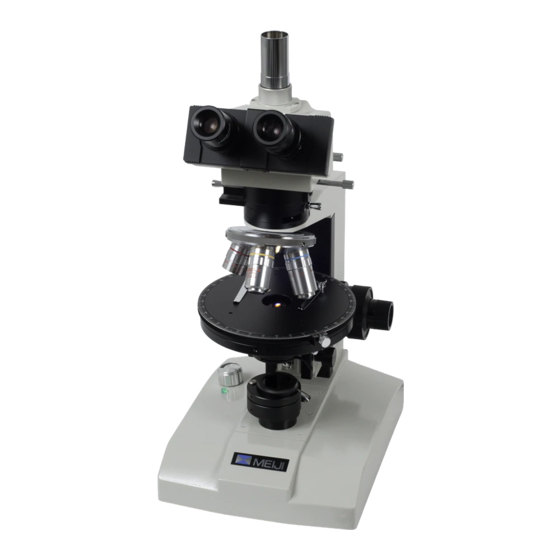
- Page 2 Focusing Cross-line Eyepiece, 10X 10X Eyepiece, Photo Tube High-eyepoint, Widefield MODEL 9300: Trinocular Body MODEL 9200: Binocular Body MODEL 9100: Monocular Body Rotatable 360 /30 Inclination Beam-splitter Lever Analyzer Slider Bertrand Lens/Aperture Ball-bearing Objective Nosepiece Compensator Slot with Objective Mounts Strain Free Microscope Limb Objectives...
-
Page 3: Unpacking, Assembly, Preparation For Use
UNPACKING, ASSEMBLY, PREPARATION FOR USE UNPACKING All MEIJI TECHNO microscopes are usually supplied in an expanded polystyrene, 2-part case and this should be used for storage, possible transport in the future, etc. If your order includes a wooden storage cabinet, release the fixing screws holding the limb and base from the cabinet and withdraw. -
Page 4: Operating Instructions
OPERATING INSTRUCTIONS OPTICAL SET-UP AND ILLUMINATION (1) Turn on the illuminator. Place the specimen slide you wish to examine on the microscope stage and rotate the 10X objective into position for focus. (2) Move the substage condenser up to its top position, using the Rack and pinion focusing control Check to make sure that both the Field iris and the Aperture iris are fully open. - Page 5 After you have focused on the specimen proceed as follows:- (1) Move the sliders on which the two binocular eyepiece tubes are mounted in and out until the distance between them is exactly the same as the distance between the pupils of the observers eyes.
- Page 6 (7) In case the centers of field Iris and the field of view does not coincide, either the two Centering screws on Substage condenser should be loosened and fasten the other to adjust the centering, and vice versa until they coincide. Edge of field iris Perfect centering Centering screws of...
- Page 7 SAFETY AUTO-STOP FOR OBJECTIVES AND STAGE PLATE In order to protect specimen slides and objective lenses from accidental damage, the 20X or bigger power lenses are designed to retract at the tip by built-in spring. Besides, Auto-Stopper is equipped in the microscope limb for further safety.
- Page 8 Please note that the applications and detailed techniques of polarizing microscopy are beyond the scope of this manual. What follows is a description of the special features of the ML9000 Series Polarizing Microscope which are often used in geological, mineralogical, chemical and other optical studies which have long been associated with the polarizing microscope.
-
Page 9: Bertrand Lens
COMPENSATORS A sensitive tint plate (first order red) and mica 1/4 wave plate are supplied with the ML9000 series polarizing microscope as standard. These are fitted in plates with standard DIN dimensions, sliding in a slot cut in the tube just above the objective nosepiece. -
Page 10: Photography And Television
PHOTOGRAPHY AND TELEVISION PHOTOGRAPHY Photographic documentation of microscope visual images is most conveniently achieved by using the trinocular (photo-binocular) bodies for use with 35mm SLR Camera or PMX100 Large Format Camera. In the case of the ML series of biological, metallurgical and polarizing microscopes a trinocular body is equipped with a sliding switch-over beam-splitter component which either (1) allows all of the light to go to the visual eyepieces or (2) directs 80% of the image-forming light upwards to the film plane of a 35mm SLR camera, while still sending 20% of the light to the binocular eyepieces. -
Page 11: Maintenance And Care
MAINTENANCE AND CARE BULB REPLACEMENT When changing light bulbs in the illuminators, always disconnect the plug from the electrical source, and make it sure that the green monitor light is off. Never work on the electrical system without first disconnecting. The bulb is held in a socket block inserted in the rear of the microscope base. (1) Remove the socket block from the microscope base by unscrewing the two screws and pulling the backing plate clear of the instrument. - Page 12 6, Oi-670, Oi-machi, Iruma-gun Saitama 356-0053, Japan Phone : 492-67-0911 Fax : 492-69-0691, 492-69-0692 2186 Bering Drive San Jose, CA., 95131, USA Phone : 408-428-9654 Fax : 408-428-0472 Toll free : 800-832-0060 98.04.1,500 Printed in Japan...
Need help?
Do you have a question about the ML9000 Series and is the answer not in the manual?
Questions and answers