
Related Manuals for Cirs ZERDINE 040GSE


Summary of Contents for Cirs ZERDINE 040GSE
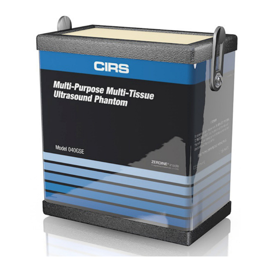
- Page 1 Multi-Purpose, Multi-Tissue Ultrasound Phantom Model 040GSE USER GUIDE 900 Asbury Ave • Norfolk, Virginia 23513 • USA • Tel: 757-855-2765 • WWW.CIRSINC.COM...
-
Page 2: Table Of Contents
TABLE OF CONTENTS 1 OVERVIEW 2 INSTRUCTIONS FOR USE HANDLING AND CARE � � � � � � � � � � � � � � � � � � � � � � � � � � � � � � � � � � � � � � � � � � � � � � � � � � � � � � � � � � � � � � � 2 USE OF REMOVABLE WATER WELL AND COVERS �... -
Page 3: Depth Of Penetration
IEC Technical Standard 62736. CIRS is certified to ISO 13485:2016 standards. We have an in-house test facility to measure acoustic properties of materials. In addition, ultrasound imaging systems are used to inspect each phantom. Every ultrasound phantom that CIRS distributes has passed thorough testing during manufacture and completion to ensure the highest quality product available. - Page 4 If the scanning surface becomes damaged, seal the phantom in an airtight container and IMMEDIATELY contact CIRS for return authorization. Call 800-617-1177, email at rma@cirsinc.com or fax RMA Request form to 757-857-0523.
- Page 5 For curved arrays, the water well may be attached and filled with water to provide better coupling. Side Fire transducers can be particularly chal- lenging to scan with a standard phantom. CIRS has designed a removable endo- cavity cover for these transducers. When this accessory is attached, the phantom should be placed on its back and the cover should be filled with water.
- Page 6 If not, then the conclusions drawn may not be valid. CIRS recommends that you use the most commonly used settings for the type of probe tested- i.e. the liver preset values for an abdominal...
- Page 7 4. Frequency of system assessment: How often each system is evaluated is also up to each facility to determine. CIRS recommends at least annually. Reference the accreditation programs established by the ACR and AIUM at www.acr.org or www.aium.org for further guidance on establishing a QA pro- gram.
- Page 8 3. Adjust the instrument settings (gain, TGC, output, etc.) as for a “normal” technique. Record these settings for use on subsequent testing. 4. Align the probe so that the targets are maximized. 5. Freeze the image and obtain a hard copy. 6.
- Page 9 BEAM PROFILE, FOCAL ZONE AND LATERAL RESPONSE WIDTH The beam profile is the shape of the ultrasound beam. A typical beam profile is shown in Figure 1. The narrowest region within the beam profile is indicative of the focal point. By convention, the region surrounding the focal point with intensity within 3 dB of maximum is the focal zone.
- Page 10 Align the probe so that all the vertical targets are displayed at their maximum intensity level. Freeze the image and obtain a hard copy. Using electronic calipers, measure the distances between two wires at various depths or align the echoes to the display markers for comparison. Record these measurements.
- Page 11 AXIAL AND LATERAL RESOLUTION TESTING (CONTINUED) The Model 040GSE has three combined axial and lateral resolution target groups. The first two groups, at depths of 3 cm and 6.5 cm, are designed for probes of 5 MHz and above. They consist of 13 parallel nylon wires of 80 microns diameter. The third target is located at 10.5 cm depth for evaluation of low frequency probes.
- Page 12 Edge to Edge to adjacent Edge adjacent Edge 3 mm 2 mm Lateral Resolution Figure 3 - Combined Axial/Lateral Resolution Target at 10.5 cm depth (top) and a table listing the distances between them (bottom) Targets C1-D1 C2-D2 C3-D3 C4-D4 C5-D5 Axial Resolution (mm)
- Page 13 In the second method, the anechoic cylinders are used as simulated focal lesions as follows: Apply coupling gel to the scanning surface or fill the water trough with tap water. Adjust the instrument settings (gain, TGC, output, etc.) as for a “normal” tech- nique.
-
Page 14: Grayscale Contrast Sensitivity
Testing for low-contrast target detectability is performed as follows: Apply coupling gel to the scanning surface or fill the water trough with tap water. Position the transducer above the cyst of interest and perpendicular to the wires. You should be imaging the circular cross section of the cylinders. Adjust the instrument settings (gain, TGC, output, etc.) as for a “normal”... -
Page 15: Elasticity Contrast Sensitivity
ELASTICITY CONTRAST SENSITIVITY The Model 040GSE provides elasticity targets for the next generation of ultrasound imagers using elastography. The elasticity value for the background is 20 kPa. The target group at a depth of 1.5 cm has a diameter of 6mm, while the target group at 5 cm has a diameter of 8 mm. - Page 16 DEAD ZONE ASSESSMENT CONTINUED The near field group consists of parallel, 100 micron diameter, nylon, monofila- ment wires horizontally spaced 6 mm apart from center to center (Figure 4, page 13). Vertical distance from the center of each wire to the top edge of the scan- ning surface ranges from 5 mm down to 1 mm in 1 mm increments.
-
Page 17: Specifications
SPECIFICATIONS TARGET LAYOUT PHANTOM Housing ABS Plastic Outer Dimensions 17.8 x 12.7 x 20.3 cm (7 x 5 x 8") Scanning Surface 14 x 9 cm Saran-based laminate Scanning Material Zerdine tissue mimicking gel ® Speed of Sound 1540 m/s Attenuation Low: 0.7 dB/cm/mHz;... - Page 18 WIRE TARGETS Material Nylon monofilament NEAR FIELD GROUP Number of targets Diameter 100 microns Depth range 1 to 5 mm Vertical distance between targets 1 mm VERTICAL DISTANCE GROUP Number of targets Diameter 100 microns Depth range 1 to 16 cm Vertical distance between targets 10 mm HORIZONTAL DISTANCE GROUPS...
- Page 19 ANECHOIC STEPPED CYLINDERS Material Zerdine LEGEND: Diameter (mm) 10.0 X = Apply - = Do Not Apply Table 1 - Cystic Masses Location and Size GRAY SCALE TARGETS Material Zerdine Contrast, dB >15 3 cm, Ø 8mm 11.5 cm, Ø 10mm Table 2 - Gray Scale Targets Location, Contrast and Size ELASTICITY TARGETS...
- Page 20 ® The Model 040GSE is constructed from a patented, solid elastic material devel- oped at CIRS called Zerdine. Phantoms constructed from Zerdine will not melt or leak when punctured and they do not require refrigeration. Zerdine is also more elastic than other materials and allows more pressure to be applied to the scanning surface without subsequent damage to the material.
- Page 21 CIRS will not be responsible for lost or damaged return shipments. Return freight and insurance is to be pre-paid.
- Page 22 APPENDIX 1: QUALITY ASSURANCE RECORD FOR MODEL 040GSE MODEL 040GSE MULTI-PURPOSE MULTI-TISSUE ULTRASOUND PHANTOM QUALITY ASSURANCE RECORD MODEL 040GSE MULTI-PURPOSE MULTI-TISSUE ULTRASOUND PHANTOM QUALITY ASSURANCE RECORD Location: ____________________ Unit: ________________ Probe: ________________ QC Phantom SN: ______________________ Machine Settings: Depth of Field (FOV) ________________________ cm Gain: _______________________ Power: ______________________ Focal Zone(s) ______________ cm ____________ cm ____________ cm __________ cm _________cm Preprocessing__________________ Post Processing ______________________________ Dynamic Range __________________...
- Page 24 COMPUTERIZED IMAGING REFERENCE SYSTEMS, INC. 900 Asbury Ave Norfolk, Virginia 23513 • USA 800.617.1177 TOLL FREE 757.855.2765 TEL: 757.857.0523 FAX: admin@cirsinc.com EMAIL: www.cirsinc.com Technical Assistance 1.800.617.1177 © Computerized Imaging Reference Systems, Inc. has been 2013 Computerized Imaging Reference Systems, Inc. All rights reserved. certified by UL DQS Inc.
Need help?
Do you have a question about the ZERDINE 040GSE and is the answer not in the manual?
Questions and answers