
Related Manuals for Cirs 054GS


Summary of Contents for Cirs 054GS
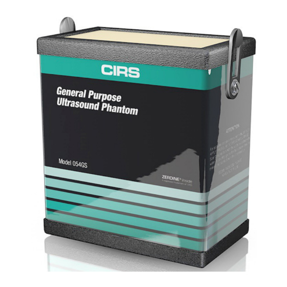
- Page 1 General Purpose Ultrasound Phantom Model 054GS ZERDINE Inside ® A registered trademark of CIRS USER GUIDE 900 Asbury Ave • Norfolk, Virginia 23513 • USA • Tel: 757-855-2765 • WWW.CIRSINC.COM...
-
Page 2: Table Of Contents
DEAD ZONE ASSESSMENT � � � � � � � � � � � � � � � � � � � � � � � � � � � � � � � � � � � � � � � � � � � � � � � � � � � � � � � � � � 1 2 4 SPECIFICATIONS 5 ZERDINE ® 6 WARRANTY 7 APPENDIX: QUALITY ASSURANCE RECORD FOR MODEL 054GS... - Page 3 OVERVIEW The Model 054GS General Purpose Key Tests with Model Ultrasound Phantom is a sturdy, reli- 054GS able phantom for testing the imaging performance of ultrasonic systems. • Uniformity The phantom is made of CIRS' • Depth of Penetration proprietary Zerdine hydrogel poly- ®...
-
Page 4: Instructions For Use
INSTRUCTIONS FOR USE HANDLING AND CARE With proper care, the Model 054GS will withstand years of normal use. Below are some guidelines to follow. The scanning surface is the most important item on the phantom to protect. It can withstand normal scanning pressure but DO NOT press on the scanning surface with your fingernails or any other sharp objects. -
Page 5: Use Of Removable Water Well And Covers
For curved arrays, the water well may be attached and filled with water to provide better coupling. Side Fire transducers can be particularly chal- lenging to scan with a standard phantom. CIRS has designed a removable endo- cavity cover for these transducers. When this accessory is attached, the phantom should be placed on its back and the cover should be filled with water. -
Page 6: General Guidelines For Performing Measurements
If not, then the conclusions drawn may not be valid. CIRS recommends that you use the most commonly used settings for the type of probe tested- i.e. the liver preset values for an abdominal... -
Page 7: Testing Procedures
4. Frequency of system assessment: How often each system is evaluated is also up to each facility to determine. CIRS recommends at least annually. Reference the accreditation programs established by the ACR and AIUM at www.acr.org or www.aium.org for further guidance on establishing a QA program. -
Page 8: Depth Of Penetration
ATTENTION: To register accurate vertical distance measurements, DO NOT APPLY PRESSURE TO THE SCANNING SURFACE! CIRS strongly encourages the user to scan the phantom with the water well filled with water or coupling gel so the transducer does not make direct contact with the scanning surface. As with a patient, even the slightest amount of pressure on the scanning surface will cause incorrect distances to be measured. -
Page 9: Beam Profile/Focal Zone/Lateral Response Width
BEAM PROFILE, FOCAL ZONE AND LATERAL RESPONSE WIDTH The beam profile is the shape of the ultrasound beam. A typical beam profile is shown in Figure 1. The narrowest region within the beam profile is indicative of the focal point. By convention, the region surrounding the focal point with intensity within 3 dB of maximum is the focal zone. -
Page 10: Vertical Distance Measurement
VERTICAL DISTANCE MEASUREMENTS (CONTINUED) 3. Adjust the instrument settings (gain, TGC, output, etc.) as for a “normal” technique. Record these settings for use on subsequent testing. 4. Align the probe so that all the vertical targets are displayed at their maximum intensity level. - Page 11 The Model 054GS has two combined axial and lateral resolution target groups. The first group, at a depth of 3 cm, is designed for probes of 5 MHz and above. It con- sists of 13 parallel nylon wires of 80 microns diameter. The second target is located at 11 cm depth for evaluation of low frequency probes.
-
Page 12: Elevational Resolution
Edge to Edge to adjacent Edge adjacent Edge 2 mm 1 mm Lateral Resolution Figure 3 - Combined Axial/Lateral Resolution Targets at 11 cm depth (top) and a table listing the distances between them (bottom) Targets C1-D1 C2-D2 C3-D3 C4-D4 C5-D5 Axial Resolution (mm) -
Page 13: Low-Contrast Target Detectability
It is desirous for these effects to be minimal. In the Model 054GS, five cylinders having no scatter are provided in the phantom to test a machine's ability to image cyst-like structures over range of depths. The cyl- inders are 8 mm in diameter and located at depths of 4, 7, 10, 13, and 16 cm. -
Page 14: Dead Zone Assessment
GRAYSCALE CONTRAST SENSITIVITY (CONTINUED) 1. Apply coupling gel to the scanning surface or fill the water trough with tap water. 2. Position the transducer above the tumor and perpendicular to the wires. (The tumor should appear as a circular region). 3. -
Page 15: Specifications
Adjust the instrument settings (gain, TGC, output, etc.) to maximize resolution in the near field. Record these settings for use on subsequent testing. Freeze the image while the near field targets are clearly displayed. Count how many wires of the near field target you can see. Subtracting this number from the total number of targets gives you the dead zone measurement. - Page 16 PHANTOM Housing ABS Plastic Outer Dimensions 17.8 x 12.7 x 20.3 cm (7 x 5 x 8") Scanning surface 14 x 9 cm Saran-based laminate Scanning Material Zerdine tissue mimicking gel ® Speed of Sound 1540 m/s Other Compatible with harmonic imaging WIRE TARGETS Material Nylon monofilament...
- Page 17 ZERDINE ® The Model 054GS is constructed from a patented, solid elastic material developed at CIRS called Zerdine. Phantoms constructed from Zerdine will not melt or leak when punctured and they do not require refrigeration. Zerdine is also more elastic than other materials and allows more pressure to be applied to the scanning sur- face without subsequent damage to the material.
- Page 18 CIRS not to comply with documented order specifications. You must return the product to CIRS within 30 calendar days of the issuance of the RMA. All returns should be packed in the original cases and or packaging and must include any accessories, manuals and documentation that shipped with the product.
- Page 19 Anechoic Cylinder Axial and Lateral Resolution 11 cm Gray Scale Duplicate as Needed: CIRS, Inc., 2428 Almeda Avenue, Suite 316, Norfolk, VA 23513 Elevational (800) 617-1177 * (757) 855-2765 or Fax (757) 857-0523 Resolution One Sheet Per System Setup Low-Contrast...
- Page 20 Technical Assistance 1.800.617.1177 Computerized Imaging Reference Systems, Inc. has been © 2013 Computerized Imaging Reference Systems, Inc. All rights reserved. certified by UL DQS Inc. to (ISO) 13485:2016. Certificate Specifications subject to change without notice. Registration No.10000905-MP2016. Publication: 054GS UG 062119...
Need help?
Do you have a question about the 054GS and is the answer not in the manual?
Questions and answers