Subscribe to Our Youtube Channel
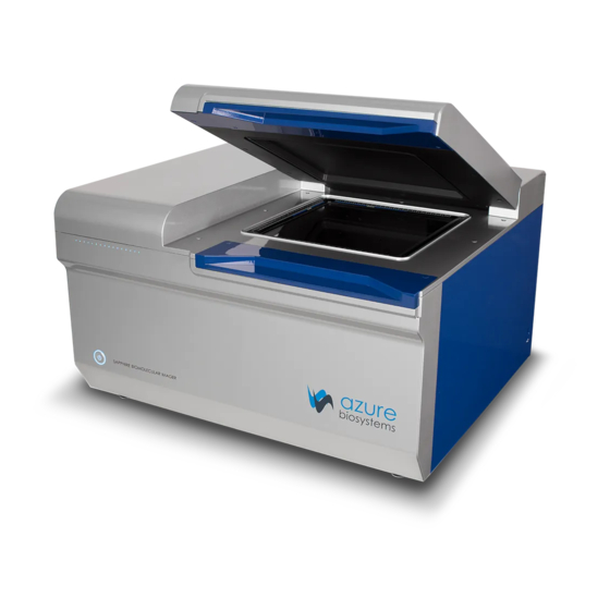
Summary of Contents for Azure Biosystems Sapphire
- Page 1 Sapphire ™ Biomolecular Imager User Manual Rev 20190405 Sapphire Biomolecular Imager User Manual Page 1 ™...
- Page 2 WARNING: Making adjustments or carrying out procedures not specified in this manual, can result in harmful exposure to laser radiation. The Sapphire Biomolecular Imager is a Class I laser instrument that houses four Class IIIB lasers inside the instrument. Under the specified operating procedures, the instrument does not allow operator exposure to laser light. The lasers, with power of 5–25mW, are accessible in the interior of the instrument.
- Page 3 Caution The safety switch in the Sapphire is designed to prevent you from being exposed to the laser radiation. If you open the sample lid while the scanner is in operation, the laser safety switch cuts power to the lasers.
- Page 4 If any defect occurs in the instrument during this warranty period, Azure Biosystems, Inc. will repair or replace the defective parts at its discretion without charge. The following defects, however, are specifically excluded: •...
-
Page 5: Table Of Contents
2.4 Software Installation 2.5 Connecting Other USB Devices to the System Proper Usage – Read Before Use Sapphire Capture Software Overview 4.1 Launch the Sapphire Capture Software 4.2 Fluorescence Imaging 4.2.1 Scanning a Fluorescent Membrane 4.2.2 Scanning a Fluorescent Gel 4.2.3 Scanning a Fluorescent Slide... -
Page 6: Introduction
Q-Module – Add dye flexibility and increase quantitative capability by adding 520 laser channel to Sapphire Biomolecular Imager – NIR • 488 Laser Module – Add dye flexibility by adding 488 laser channel to Sapphire Biomolecular Imager – NIR 1.3 Optional Modifications •... -
Page 7: Table Of Specifications
1.4 Table of Specifications Sapphire NIR Sapphire RGB Sapphire RGBNIR Sapphire PI Part number IS1024 IS1025 IS1026 IS1027 Laser excitation 685, 784 488, 520, 658 488, 520, 658, 784 wavelengths Bit depth 16 bit 16 bit 16 bit 16 bit... -
Page 8: Installation And Setup
2. Turn off master power to the system using the power switch on the back of the instrument. Azure Biosystems recommends leaving the system master power turned on via the switch on the back panel, as this switch may not be easily accessible. -
Page 9: Connecting Other Usb Devices To The System
2.5 Connecting Other USB Devices to the System The external computer is connected to the Sapphire system via a USB 3 connection. You may attach regulatory approved, Windows OS supported USB keyboard, USB mouse or other USB input devices to any of the unused USB ports that remain on the external PC. -
Page 10: Proper Usage - Read Before Use
The heat generated by the PC may damage the unit. Keep the top of the Sapphire Biomolecular Put anything on top of the imager. The Sapphire Imager clear. was not designed to withstand forces acting down on top of the imager. -
Page 11: Sapphire Capture Software Overview
Sapphire Capture Software Overview 4.1 Launch the Sapphire Capture Software Double click the Sapphire desktop icon to launch the scanner software. 4.2 Fluorescence Imaging The Fluorescence module utilizes lasers and PMT or APD detectors to scan fluorescent samples. Sapphire Biomolecular Imager User Manual Page 10 ™... - Page 12 FLUORESCENCE – Select the fluorescence tab at the top of the imaging section. IMAGING AREA – The Imaging Area represents the glass scanning surface of the Sapphire Biomolecular Imager with each square in the Imaging Area grid representing one square centimeter. Use the green corners to select the area of the imaging screen covered by your sample.
- Page 13 Total file size listed in parenthesis. SEQUENTIAL SCAN – By default, the Sapphire Biomolecular Imager scans all channels simultaneously. Turning Sequential Scanning ON scans each channel individually from first channel selected to last instead of scanning simultaneously.
-
Page 14: Scanning A Fluorescent Membrane
Multiplexed scans will appear as a single image, and can be separated into individual signal channels for analysis. If Sequential Scanning was chosen, each individual channel will appear as a grayscale image along with the final multiplexed image. Sapphire Biomolecular Imager User Manual Page 13... -
Page 15: Scanning A Fluorescent Gel
14. During scanning, visualization of the preview image may be adjusted by clicking on the Contrast Settings button. This will open a Channels window that will allow you to select which channels to preview and adjust contrast. Sapphire Biomolecular Imager User Manual Page 14... -
Page 16: Scanning A Fluorescent Slide
11. Clicking PRESCAN will initiate a quick, low resolution scan that can be useful in determining the correct slide positioning, scanning area and intensity setting. Sapphire Biomolecular Imager User Manual Page 15... -
Page 17: Scanning A Fluorescent, Plate-Based Assay
Change the signal Intensity to your preference for the sample you are imaging. Use Intensity 1 (one) to scan strong signals and Intensity 10 (ten) to detect weaker signals. NOTE: Use the PRESCAN function to initiate a quick, low resolution scan to help determine the best Intensity Level to use. Sapphire Biomolecular Imager User Manual Page 16... - Page 18 15. Upon completion of the scan, images will appear according to the color chosen for each channel. Multiplexed scans will appear as a single image, and can be separated into individual signal channels for analysis. Sapphire Biomolecular Imager User Manual Page 17...
-
Page 19: Chemiluminescence Imaging
4.3 Chemiluminescence Imaging The Chemiluminescence Module utilizes a cooled CCD camera to image luminescent samples. CHEMILUMINESCENCE – If your Azure Sapphire configuration includes the optional chemiluminescence module, you will first need to select this option in the “Imaging” section. IMAGING AREA – The Chemi Imaging Area is designated by blue markings on the top left corner of the imaging glass. - Page 20 CAPTURE – Click the capture icon to capture the image according to the set parameters. CCD COOLED INDICATOR – The Sapphire is equipped with a CCD capable of cooling down to 50 degrees C below ambient temperature. The CCD Cooled indicator will turn green when the CCD has reached optimal cooling.
-
Page 21: Taking A Chemi Image
Select Marker to take a separate, white light image of your protein ladder, which can later be overlaid on your chemi blot image. 10. Select CAPTURE. 11. Your images will appear in the gallery tab. Sapphire Biomolecular Imager User Manual Page 20 ™... -
Page 22: Phosphor Imaging
“Imaging” section. IMAGING AREA – The Imaging Area represents the glass scanning surface of the Sapphire Biomolecular Imager with each square in the Imaging Area grid representing one square centimeter. Use the green corners to select the area of the imaging grid covered by your sample. -
Page 23: Scanning A Storage Phosphor Screen
Note: Adjusting the contrast through this window only affects visualization of the captured image. It does not affect captured data. The acquired image will appear in the Gallery Tab once scanning is complete. Sapphire Biomolecular Imager User Manual Page 22... -
Page 24: Visible Imaging
The Visible Imaging module utilizes white light to generate color images of colorimetric samples. VISIBLE – If your Azure Sapphire configuration includes the optional chemiluminescence module, you will also have the ability to take white light images using the Visible tab in the “Imaging” section. -
Page 25: The Image Gallery
Use the Black, White and Gamma sliders to adjust the contrast of your image. Images can also be Inverted, Rotated and Resized. See the Icon Guide below for a complete description of each option. Sapphire Biomolecular Imager User Manual Page 24... - Page 26 Sapphire Biomolecular Imager User Manual Page 25 ™...
- Page 27 SELECT – Generates a box that you can resize to select an area of the captured image to copy or crop. COPY – Copy the whole image, or a selected area of the image, such as a marker or ladder, to the clipboard. Sapphire Biomolecular Imager User Manual Page 26...
- Page 28 Photoshop or Powerpoint, the image appears the same as in the Sapphire software. Other than inverting, this does not change the data in any way.
-
Page 29: Settings
For assistance, please contact support@azurebiosystems.com. Copyright © 2017-2019 Azure Biosystems, Inc. All rights reserved. The Azure Biosystems logo, Azure Biosystems™, and Sapphire™ are trademarks of the Company. Windows is a registered trademark of Microsoft Corporation in the United States and other countries. All other trademarks, service marks and trade names appearing in this brochure are the property of their respective owners.


Need help?
Do you have a question about the Sapphire and is the answer not in the manual?
Questions and answers