Table of Contents
Advertisement
Advertisement
Table of Contents

Summary of Contents for Smith & Nephew Syncera Anthem
- Page 1 Surgical Technique...
- Page 2 In consultation with: Professor T.K. Kim – Seoul, South Korea Professor D. Saris – Utrecht, Netherlands Professor R. Tözün – Istanbul, Turkey Professor Y. Zhou – Beijing, China Professor A. Belooshi – Dubai, U.A.E. Doctor M. Wadhwa – Chandigarh, India Doctor R.
-
Page 3: Table Of Contents
ANTHEM™ PS Total Knee System Surgical Technique Introduction Indications Preoperative planning Femoral preparation Tibial preparation Femoral positioning and sizing A/P and chamfer resection Component trialling Tibial keel preparation Resurfacing patellar preparation Implantation and closure Implant Compatibilities... -
Page 4: Introduction
Introduction The ANTHEM™ PS Total Knee System has been designed to offer orthopaedic surgeons solutions to address a range of intraoperative situations. Proper implant function is dependant on an extensive and accurate surgical technique. The ORTHOMATCH™ Universal Instrumentation system is designed to be used with ANTHEM to provide an easy to use system that facilitates accurate and reproducible surgical outcome. -
Page 5: Preoperative Planning
Preoperative planning On a full lower limb radiological view determine the 6º angle between the anatomical and the mechanical axes. This measurement will be used intraoperatively to select the appropriate valgus angle so that correct 3º 3º limb alignment is restored. (Beware of misleading angles in knees with a flexion contracture or rotated lower extremities.) Note: Many surgeons prefer to simply select... -
Page 6: Femoral Preparation
Femoral preparation Open the femoral canal with the 9.5mm intramedullary drill. The drill has a 12mm step to open the entry point further (Figure 1). Assemble the valgus alignment guide with the valgus bushing (5°,6° or 7°) Right or Left mark facing upwards to match the correct side (hand) of the knee based on preoperative planning and assessment of valgus angle. - Page 7 Once the universal cutting block is secured with pins, remove pins from valgus alignment guide and slide the valgus alignment guide anteriorly to fully remove it. Then remove the IM -handle and valgus bushing from the Intra-medullary canal. Extend the knee fully and insert drop rod guide assembly in to the blade slot of the universal cutting block to check Hip Knee Mechanical Axis (HKA) before resection (Figure 4).
- Page 8 .Once resection level and alignment are determined, insert a third pin into the angled pin hole to secure the universal cutting block in position. Resect the distal femur using an oscillating saw (Figure 7). Figure 7 . Once resection complete then remove pins and the universal cutting block from the femoral bone and remove the cut bones from the distal condyles (Figure 8).
-
Page 9: Tibial Preparation
Tibial preparation Assemble the 3° cut block connector and tibial spiked fixation rod to the extramedullary tibial alignment guide and ankle clamp (Figure 9). Connect the universal cutting block to the 3° cut block connector with the “T” mark facing outwards. This can be attached either centrally or medially depending on preference. -
Page 10: Figure
Align the extramedullary tibial alignment guide parallel to the tibial axis in the coronal and sagittal planes. Rotate the assembly to the medial one-third of the tibial tubercle and impact the anterior spike of the up rod (Figure 10 and 10a). Note: 3°... - Page 11 Check the posterior slope and resection plane with resection check (angel wing) (Figure 11b). Remove the extramedullary tibial alignment guide assembly by loosening the thumbscrews and then using the slaphammer to remove the spike rod superiority while the universal cutting block remains in position with secured pins.
- Page 12 . Remove the universal cutting block from the bone and clean up the proximal tibial surface (Figure 13a). Note: If it has not already been removed, excise completely the entire PCL attachment from the femoral intracondylar notch with either a cautery or scalpel to prevent it from affecting the assessment.
-
Page 13: Femoral Positioning And Sizing
Femoral positioning and sizing The ORTHOMATCH™ femoral sizing guide allows for external rotation to be set from 0-9º based on surgeon Stylus marked to indicate anterior femoral preference and patient anatomy. Rotational alignment flange length/sawblade exit point may be checked by aligning the A/P axis (Whiteside’s Line) with the vertical marks on the sizing guide or by ensuring that the “EPI”... - Page 14 Sizing steps are as follows: Optional: Mark the AP and epicondylar axis on the femur (Figure 15). Place sizing guide onto resected distal femur Pin guide through fixation holes to provide stability Select desired femoral external rotation 0-9deg Note indicated size – if between sizes see sizing step for more detail Select Anterior or Posterior referencing Pin either anterior or posterior referencing holes with...
- Page 15 It is important to use the distal femoral anatomic landmarks (AP axis and Epicondylar Axis) of the knee to guide external rotation and to ensure optimized sizing, ligament balance and patella tracking. This is commonly set at 3°. However, certain patient groups have higher external rotation of the femur, which correlates to a greater degree of tibia vara.
- Page 16 Slide the femoral stylus backward or forward so that the size indicated on the femoral stylus matches the femoral size indicated on the femoral sizer guide. The point at which the femoral stylus contacts the bone marks the peak of the femoral anterior flange (Figure 16d).
-
Page 17: A/P And Chamfer Resection
A/P and chamfer resection Select the appropriate size A/P resection block and place over the pins through the appropriate anterior or posterior referencing holes. Ensure that the cutting block is flush with the resected distal femur (Figure 17). Prior to bone resection, insert the resection check (angel wing) into the anterior blade slot of the A/P resection block to check the plane of the resection and to avoid any chance for notching (Figure 17a). - Page 18 Complete the anterior, posterior and chamfer cuts with an oscillating saw. The block is designed to allow for angling of the sawblade during the cuts. Note: To maintain block stability, the anterior chamfer cut should be completed last. Once all resection cuts are complete, remove the A/P Resection Block and remaining pins from the bone.
-
Page 19: Component Trialling
Component trialling Flex the knee to 90° and insert the appropriate sized femoral trial using the femoral trial impactor. Note: To avoid the trial slipping into flexion, the trial femoral impactor can also be used as a notch Impactor by rotating it 180 degrees and impacting the anterior femoral notch area (Figure 19). - Page 20 Assemble the PS housing reamer by attaching the housing reamer dome and the PS reamer sleeve to the reamer shaft. Ream through the PS housing collet in both anterior and posterior positions until the depth stop of the reamer contacts the PS housing collet (Figure 21a).
- Page 21 Place the appropriate size and desired thickness articular insert trial onto the tibial trial. For Insert thicknesses greater than 9mm select the appropriate shim. Attach the quick connect handle to the tibial trial and insert the assembly into the knee (Figure 22). Note: The best technique is to flex the knee to 120°, push in the insert as far as possible and bring the leg out into full extension.
-
Page 22: Tibial Keel Preparation
Tibial keel preparation Once the trial assessment is complete and final implant sites determined remove the insert trial and, if necessary, the femoral trial (Figure 23). Figure 23 Re-assess the tibial coverage, size and rotation required and pin the tibial trial baseplate in place using two short headed pins (Figure 24). - Page 23 Select the appropriate modular fin punch to prepare the keel and attach to the modular handle. Keel punch through the baseplate. To remove the keel punch, disengage the modular handle and then use the slap hammer to remove from the bone (Figure 24b). Figure 24b Note: An alternative method to setting tibial rotation is to use the tibia trial bullet.
-
Page 24: Resurfacing Patellar Preparation
Resurfacing patellar preparation Rotate the patella to 90°. Trim tissue surrounding the patella using electrocautery. Use a rongeur to remove osteophytes and reduce the patella to its true size. The electrocautery should also be used to release soft tissue attachments to the estimated level of resection. - Page 25 Cut the patella through the dedicated saw guides (Figure 25b). Select the appropriate diameter resurfacing patella drill guide and place it onto the patella. Align patella drill guide to the resected patella. Figure 25b Use the patella peg drill to drill for the three peg holes through the patella drill guide until the drill bottoms out in the guide (Figure 25c).
- Page 26 Remove the patella reamer guide and drill guide from the patella. Place the resurfacing patellar trial onto the resected patella. Use the patella caliper to reassess the patella thickness (Figure 25d and 25e). . Revert back the patella onto the femoral trial to check patella-femoral articulation by flexing and extending the knee several times.
-
Page 27: Implantation And Closure
Implantation and closure Maximally flex the knee and place a thin bent Hohmann retractor laterally and medially and an Aufranc retractor posteriorly to sublux the tibia forward (Figure 26). Apply generous amounts of cement to the dry underside of the baseplate, keel and onto the proximal tibia and keel prep hole (Figure 26). - Page 28 Flex the knee to 90° keeping the thin bent Hohmann laterally and removing the Aufranc retractor. Mix and prepare bone cement for femoral component and distal femur. Apply cement to the femoral component or prepared bone, based on the surgeon’s preference (Figure 26b).
- Page 29 Place the appropriate size Insert trial onto the tibial implant and extend the leg to pressurize the cement. Remove any additional excess cement (Figure 26d). Figure 26d Apply bone cement to the patella. Place the patellar implant onto the patella and clamp into the bone. Remove excess cement (Figure 26e).
- Page 30 .The articular insert is locked into position using the modular tibial impactor. This is best done when the leg is flexed to around 20-30 degrees and impacting on the anterior surface of the insert (Figure 26f). Note: To check insert is fully seated, perform a visual examination on each side of the insert slot.
-
Page 31: Implant Compatibilities
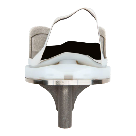
Implant Compatibilities Recommended Combination Femoral Insert Tibia Patella ANTHEM™ ANTHEM ANTHEM GENESIS™ II Standard PS & Narrow PS CoCr PS HF Resurfacing Other Compatible Options ANTHEM™ ANTHEM Standard PS & Narrow PS PS HF Insert Patellas Inserts Tibia Baseplates Femorals GENESIS II Oval Resurfacing GENESIS II PS HF GENESIS II Non porous... - Page 32 Smith & Nephew 1450 Brooks Road Memphis, TN 38116 Telephone: 901-396-2121 Information: 1-800-238-7538 Orders/inquires: 1-800-238-7538 www.smith-nephew.com ©2016 Smith & Nephew. All rights reserved. ™Trademark of Smith & Nephew. 02903 71282019 V2 10/16...


Need help?
Do you have a question about the Syncera Anthem and is the answer not in the manual?
Questions and answers