Table of Contents
Advertisement
Quick Links
Advertisement
Table of Contents

Related Manuals for Smith & Nephew BIRMINGHAM HIP


Summary of Contents for Smith & Nephew BIRMINGHAM HIP
- Page 1 Surgical Technique...
-
Page 3: Table Of Contents
BIRMINGHAM HIP™ Resurfacing System Contents Indications/Contraindications ............4 Warnings and Precautions ............. 5 Surgical Approach ................5 Preoperative Planning ..............6 Intraoperative Templating ............... 7 Acetabular Preparation ..............8 Curved Cup Introducer ..............10 Acetabular Cup Wiring Instruction ..........11 X-Bar .................... -
Page 4: Indications/Contraindications
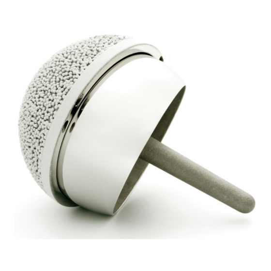
Indications for use The BIRMINGHAM HIP™ Resurfacing (BHR™) System is a single use device intended for hybrid fixation: cemented femoral head component and cementless acetabular component. The BHR System is intended for use in patients requiring primary hip resurfacing arthroplasty due to: Non-inflammatory arthritis (degenerative joint disease) such •... -
Page 5: Warnings And Precautions
The more risk factors the patient has, the greater the risk of procedure failure requiring a revision of the hip. The surgical approach The BIRMINGHAM HIP™ Resurfacing device may be implanted through all normal surgical approaches. The posterior approach is described in this operative technique. -
Page 6: Preoperative Planning
Preoperative planning Templating BHR™ template sets (Figure 1) are used to determine component size and correct implant positioning. The position of the femoral component is a most important pre-operative consideration. Varus positioning must be avoided and slight valgus is recommended (Figure 2). -
Page 7: Intraoperative Templating
Intraoperative templating An assessment is made of the femoral neck diameter using the head/neck template. This provides vital information as to minimum head component size that can be safely used and also the minimum acetabular size that can be utilised. If significant osteophyte formation is present on the femoral neck then this should be removed with rongeurs before definitive assessment of femoral neck diameter is made... -
Page 8: Acetabular Preparation
Acetabular preparation If the anteroinferior capsule is tight an antero- inferior radial capsulotomy is made in line with the psoas tendon. A Hohmann retractor is placed inferior to the radiographic teardrop. The acetabular labrum, transverse ligament and ligamentum teres are excised revealing an unencumbered view of the complete acetabulum and a view of the true floor of the acetabulum. - Page 9 The desired size of acetabular component is mounted on the acetabular introducer and offered up to the acetabular rim. The ace tabular cup is rotated so that its anti-rotation splines are adjacent to the ischium and pubis. The acetabular component is then fully impacted with 15-20˚...
-
Page 10: Curved Cup Introducer
Curved Cup Introducer These instructions provide important information regarding assembly and wiring for use of the BHR™ Curved Cup Introducer. NOTE: This Curved Cup Introducer is for use with BHR Resurfacing cups only. -
Page 11: Acetabular Cup Wiring Instruction
Acetabular Cup wiring instruction The following is the recommended method of attaching the Curved Cup Introducer to the acetabular component. Wire loop 1 Wire loop 3 To ensure correct component fixation, please note that the wire loops are specified as wire Wire loop 2 loops 1, 2 and 3. - Page 12 Acetabular Cup wiring instruction continued Step 3 As in Step 2, now loop wire 3 over the wire grip (Figure 4). Wire loop 3 Figure 4 Step 4 With the two opposing wire loops (2&3) positioned through the wire grip now capture both wires by passing wire loop 1 over the top (Figure 5).
-
Page 13: X-Bar
X-Bar X-Bar (Figure 7) Figure 7 The X-Bar is attached to the curved Cup Introducer. (Figure 8) Figure 8 With the patient positioned correctly align the impactor so that the appropriate bar on the guide, left or right, is parallel to the longitudinal axis of the patient while the vertical bar is perpendicular to the floor. -
Page 14: Femoral Preparation
Femoral preparation The desired position of the femoral alignment pin will be known from the preoperative templating. Identify the tip of the greater trochanter through the tissues with a spinal needle. A ruler is used to measure the desired distance down from the tip of the greater trochanter (Figure 1) and the alignment pin is inserted through the vastus lateralis fibres. -
Page 15: Using The Mcminn Alignment Jig
After the outer cortex is breached the drill is angulated so that the alignment pin is directed towards the femoral head (Figure 3). The alignment pin is left protruding 5mm above the outer fibres of vastus lateralis. NOTE: It is recommended that “Pin in Femur” is placed on the nurse’s swab count board. - Page 16 Femoral preparation continued The adjustable joint in the long arm of the alignment guide is set so that the guide wire will be directed down the mid-lateral axis of the femoral neck (Figure 5a). Bisect the neck with forceps to aid visualisation (Not illustrated). Next the proximal portion of the guide is moved on the femoral head to allow the stylus to be passed around the femoral neck, having first...
-
Page 17: Short Arm Alignment Jig Technique
Short Arm Alignment Jig technique Templating BHR™ template sets are used to determine Short Arm Alignment Jig Ruler component size and correct implant positioning. The position of the femoral component is a most important preoperative consideration. Varus positioning must be avoided and slight valgus is recommended. - Page 18 Short Arm Alignment Jig technique continued The measuring guide is placed on the tip of the lesser trochanter translating the pre operative measurement on to the intertrochanteric crest. The alignment pin insertion point can now be marked (Figure 8). Using the marked insertion point on the intertrochanteric crest, the assembled jig is fixed to the femur by inserting the collared alignment pin through the hole in the distal slot...
- Page 19 A guide wire is inserted when the desired position of the alignment guide has been achieved (Figure 11). The central rod is removed and the guide assembly completely removed. NOTE: Guide wires are intended for single use only The stylus is re-inserted on the guide wire and a final check made to ensure that the stylus passes comfortably around the femoral neck (Figure 12).
- Page 20 Short Arm Alignment Jig technique continued When the desired position of the guide wire has been achieved then the guide wire is overdrilled to the appropriate depth for the implant being inserted (Figure 13). At this stage a hole is drilled and the vent is inserted into the lesser trochanter and connected to the second suction device (not illustrated).
-
Page 21: Using The Sleeve Cutter Stop
Using the Sleeve Cutter Stop The BIRMINGHAM HIP™ Resurfacing (BHR™) Sleeve Cutter Stop was developed to reduce the risk of ‘shoot through’ and therefore femoral neck notching while preparing the femoral head. This is achieved by providing a physical method... - Page 22 Using the Sleeve Cutter Stop continued The sleeve cutter stop stylus is placed on the guide bar. The stylus arm is passed over the femoral head. It is the superior aspect of the femoral neck which is most prone to notching on ‘shoot through’...
- Page 23 When satisfied with the chosen cutting depth an sleeve cutter stop spacer is selected. The correct size of spacer is determined by the space inbetween the base of the instrument and the top of the femoral head. This is achieved using two methods;...
- Page 24 Using the Sleeve Cutter Stop continued Before femoral head preparation, the base of the femoral neck is packed with wet swabs to prevent bone debris entering the periarticular soft tissues. However, it is important to keep these swabs clear of the head so that they do not catch in the femoral cutter instruments.
- Page 25 The peripheral bone and any head/neck osteophytes should be trimmed off taking care not to strip any soft tissue attachments from the femoral neck (Figure 27 & 28). The guide rod is pushed down the femur by hand until it is seated at the bottom of the prepared hole and left in its final position.
- Page 26 Using the Sleeve Cutter Stop continued NOTE: Various methods of templating the desired amount of proximal bone to be removed may be employed. The sleeve cutter is advanced by hand over the previously prepared femoral head until the teeth meet the medial femoral head/neck junction (Figure 29).
- Page 27 The Plan Cutter is then advanced over the guide rod stopping at the marked resection line (Figure 31). Identify the marked resection line with the guide wire to aid visualisation. To ensure correct bone resection, the head/neck template is to be advanced over the guide rod. Meeting the medial head/neck junction, bone has to point to the neutral (0) position of the device (Figure 32).
- Page 28 Using the Sleeve Cutter Stop continued A number of cement keyholes are drilled into the femoral head using the Wroblewski drill (Figure 34). At this stage any cysts are curetted. If the defects are relatively small, they are left and will be filled with cement.
-
Page 29: Using The Stem Drill
Using the Stem Drill The appropriately sized stem drill (tapered reamer) is used to enlarge the parallel hole to suitably fit the tapered stem of the femoral component. There are two sizes of stem drill (tapered reamer) which corresponds to 48-58mm sized groups of femoral components as follows: Size 2 = 46-52mm... - Page 30 Using the Stem Drill continued One minute after the start of cement mixing, the femoral component is impacted into position to the previously made mark (Figure 40). It is important to have a swab positioned anteriorly to collect any extruded cement and to prevent this from flowing into the acetabular component.
-
Page 31: Size Chart
Size Chart The size chart (available as a wall chart) is presented to remind the surgeon of the femoral BHR™ Implant Size Chart head and cup sizes that can be matched HEAD SIZE CUP SIZE (Figure 43). For example, the size 50mm femoral component can be matched with a size 56mm acetabular cup and a size 58mm acetabular cup. -
Page 32: Thrombo-Embolic Prophylaxis
Application of the cemented femoral component of the BIRMINGHAM HIP™ Resurfacing (BHR™) System also raises the femoral intramedullary pressure, but the amount of fat displaced is much less than with a cemented stemmed THR. - Page 33 Notes...
- Page 34 Notes...
- Page 36 Manufacturer Smith & Nephew Orthopaedics Ltd Spa Park Harrison Way Leamington Spa Warwickshire, CV31 3HL ™ Trademark of Smith & Nephew All Trademarks acknowledged. 03189-en (0216-1516) V6 07/19 Not for use in US. 0 1 2 0 0344...
Need help?
Do you have a question about the BIRMINGHAM HIP and is the answer not in the manual?
Questions and answers