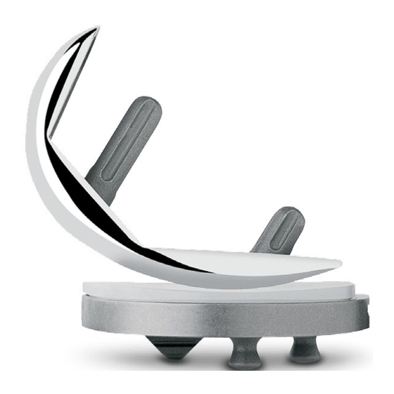
Summary of Contents for smith&nephew ZUK
- Page 1 Minimally Invasive Surgical Techniques for the Medial Compartment Intramedullary, Spacer Block and Extramedullary techniques...
- Page 2 For more information on the ZUK Unicondylar Knee, including its indications for use, contraindications, and product safety information, please refer to the product’s label and the Instructions for Use packaged with the product.
-
Page 3: Table Of Contents
Unicompartmental High Flex Knee Table of Contents Intramedullary (IM) Surgical Technique .......... 2 Introduction ........................2 Rationale ..........................3 Preoperative Planning ......................4 Patient Preparation ......................5 Exposure ..........................5 Step One: Drill Hole in Distal Femur ................... 6 Step Two: Resect the Distal Femoral Condyle ..............7 Step Three: Resect the Proximal Tibia ................ -
Page 4: Intramedullary (Im) Surgical Technique
This guide to the surgical technique The MIS ™ Instruments for the ZUK is a step-by step procedure written Unicompartmental High Flex Knee for a medial compartment UKA. are designed to provide accurate,... -
Page 5: Rationale
Rationale In UKA, the angle of the cuts does The basic goals of unicompartmental The alignment goals for not affect varus/valgus alignment. knee arthroplasty are to improve limb unicompartmental arthroplasty differ Instead, postoperative varus/valgus alignment and function, and to reduce from those that are customary in an alignment is determined by the pain. -
Page 6: Preoperative Planning
Preoperative Planning Take standing weight-bearing A/P Occasionally, in patients who have and lateral radiographs of the affected had total hip arthroplasty with a This technique is written with the knee, and a skyline radiograph of the femoral component that has more distal femoral resection performed first. -
Page 7: Patient Preparation
Patient Preparation Exposure With the patient in the supine position, The incision can be made with the leg A subperiosteal dissection should be test the range of hip and knee flexion. in flexion or extension, according to carried out towards the midline, ending If unable to achieve 120°... -
Page 8: Step One: Drill Hole In Distal Femur
Step One Drill Hole in Distal Femur Without everting the patella, flex the Guides are available for LT MED/RT goal is to be parallel to the cut knee 20°-30° and move the patella LAT or RT MED/LT LAT, with two surface of the tibia after the tibial laterally. -
Page 9: Step Two: Resect The Distal Femoral Condyle
Step Two Resect the Distal Femoral Condyle Make sure that the IM Femoral Note: If a pin is used for fixation of of subsequent guides and for proper Resection Guide is contacting the the IM guide to the distal femur, fit of the implant. -
Page 10: Step Three: Resect The Proximal Tibia
Then slide the dovetail on the Tibial proximal portion will always remain The ZUK Unicompartmental Knee Resector Base onto the proximal end parallel to the distal portion and, System is designed for an anatomic... - Page 11 In the sagittal plane, align the Secure the assembly to the proximal Use the 2mm tip of the Tibial Depth assembly so it is parallel to the anterior tibia by inserting a 48mm Headed Resection Stylus to help achieve the tibial shaft (Fig.15) by using the A/P Screw, or predrilling and inserting a desired depth of cut.
- Page 12 Seat the screw/pin that was inserted Insert a retractor medially to protect into the Tibial Resector Base. Then the medial collateral ligament. Use secure the Tibial Resector to the 1.27mm (0.050-inch) oscillating saw proximal tibia by predrilling and blade through the slot in the cutting inserting Gold Headless Holding guide to make the transverse cut.
-
Page 13: Step Four: Check Flexion/Extension Gaps
Step Four Check Flexion/Extension Gaps To assess the flexion and extension Remove the Flexion/Extension Gap After any adjustment of the flexion gaps, different Flexion/Extension Gap Spacer and flex the knee. Check the and/or extension gap is made, Spacers are available that correspond flexion gap by inserting the thin end use the Flexion/ Extension Gap to the 8mm, 10mm, 12mm and 14mm... -
Page 14: Step Five: Size The Femur
Step Five Size the Femur There are seven sizes of femoral Insert the foot of the guide into the joint Note: Be sure that there is no soft implants and corresponding sizes of and rest the flat surface against the cut tissue or remaining osteophytes Femoral Sizer/Finishing Guides. -
Page 15: Step Six: Finish The Femur
Step Six Finish the Femur The following order is recommended 2. Insert one 33mm Headed Screw Note: For Femoral Sizer/Finishing to maximize the stability and fixation of (gold head) into the angled anterior Guide sizes A and B, the angle of the pin hole is different from the Femoral Sizer/Finishing Guide. - Page 16 3. Insert the Femoral Drill w/Stop into the anterior post hole, and orient it to the proper angle (Fig. 29). Do not attempt to insert or align the drill bit while the drill is in motion. When the proper alignment is achieved, drill the anterior post hole and, if necessary, insert a Femoral Holding Peg for additional stability.
- Page 17 6. Cut the posterior condyle through the cutting slot in the guide (Fig. 32). 7. Remove the screws/pins and the Femoral Sizer/Finishing Guide, and finish any incomplete bone cuts. 8. Ensure that all surfaces are flat. Remove any prominences or uncut bone.
-
Page 18: Step Seven: Finish The Tibia
Step Seven Finish the Tibia Resect the remaining meniscus The Tibial Sizer has a sliding ruler and remove any osteophytes, which facilitates measuring in the A/P especially those interfering with dimension (Fig. 35). Be sure that the the collateral ligament. head of the sizer rests on cortical bone near the edge of the cortex around its Place the head of the Tibial Sizer... - Page 19 Place the corresponding size Tibial There are a number of indicators on Fixation Plate Provisional onto the the Tibial Sizer. If the slider is used cut surface of the tibia. Insert the without the sizer, the etch marks 1 Tibial Plate Impactor into the recess through 6 on the slider indicate the on the provisional and impact it so A/P length of the corresponding...
-
Page 20: Step Eight: Perform Trial Reduction
Step Eight Perform Trial Reduction Remove the IM Patellar Retractor. With Slide the rails on the bottom of the Evaluate soft tissue tension in flexion all bone surfaces prepared, perform Tibial Articular Surface Provisional and extension. Use the 2mm end of a trial reduction with the appropriate into the grooves on the Tibial Fixation the Tension Gauge to help ensure... -
Page 21: Step Nine: Implant Final Components
Step Nine Implant Final Components Obtain the final components and Note: Do not use the Tibial implant the tibial component first. Plate Impactor to impact an all-polyethylene tibial component. Technique tip: With the modest amount of bone removed, particularly Remove the sterile gauze sponge from the tibia, there may be a sclerotic slowly from behind the joint, and cut surface. -
Page 22: Closure
Femoral Component Tibial Articular Surface Closure Apply cement and begin the femoral After the cement has cured, remove Irrigate the knee for the final time component insertion with the leg in any remaining excess cement before and close. Cover the incision with deep flexion. -
Page 23: Spacer Block Surgical Technique
Spacer Block Option After resecting the proximal tibia, bring Attach the Alignment Tower to the the knee to full extension. Insert the Spacer Block (Fig. 3) and insert the 8mm Spacer Block into the joint space Alignment Rod through the Alignment until the anterior stop contacts the Tower. - Page 24 The ZUK Unicompartmental High Flex Knee System has been designed for 5° of posterior tibial slope. The angle on the handle of the Spacer Block is angled 5° relative to the Spacer Block. This ensures that the distal femoral resection is made perpendicular to the long axis of the femur.
-
Page 25: Extramedullary (Em) Surgical Technique
(HTO) where overcorrection is desirable The MIS Instruments for the ZUK the surrounding soft tissue during to displace the weight-bearing forces away from the diseased compartment. -
Page 26: Preoperative Planning
Preoperative Planning Patient Preparation Once the alignment has been set, the This technique is written with the distal With the patient in the supine instrumentation allows reproducible femoral resection performed first. position, test the range of hip and bone resection of the articular However, if preferred, the tibia can be knee flexion. -
Page 27: Exposure
Exposure The incision can be made with the leg A subperiosteal dissection should be in flexion or extension, according to carried out towards the midline, ending preference. Make a medial parapatellar at the patellar tendon insertion. This skin incision extending from the medial will facilitate positioning of the tibial pole of the patella to about 2cm-4cm cutting guide. -
Page 28: Step One: Assemble/Apply The Instrumentation
Tibial Fig. 4 Tibial Resector Resector Base The ZUK Unicompartmental High Flex Knee System is designed for an anatomic position with a 5° posterior slope. It is important that the proximal tibial cut be made accurately. The tibial assembly consists of an Ankle Clamp,... - Page 29 Apply the Instrument Secure the distal portion of the assembly by placing the spring arms of the Ankle Clamp around the ankle proximal to the malleoli (Fig. 6). Loosen the knob at the top of the Distal Telescoping Rod and extend the proximal portion of the assembly to the joint line.
- Page 30 While holding the proximal portion of In the sagittal plane, align the the assembly in place, loosen the knob assembly so it is parallel to the anterior that provides mediolateral adjustment tibial shaft (Fig. 9) by using the A/P of the Distal Telescoping Rod. Adjust slide adjustment at the distal end of the distal end of the rod so it lies the Distal Telescoping Rod.
- Page 31 Optional Technique: If the patient has a slight flexion contracture, cutting less posterior slope may help as it would result in less bone resection posteriorly than anteriorly, thereby opening the extension gap more relative to the flexion gap. This can be accomplished by moving the assembly closer to the leg distally.
-
Page 32: Step Two: Align The Joint
Step Two Align the Joint Spacer Block Note: Avoid aligning the limb in a While maintaining this corrected way that may result in overcorrection. position, use the thumb screw on It is preferable to align the limb in the Tibial Resector Base to move the slight varus for a medial compartment cutting guides superiorly until the arthroplasty, or in slight valgus for a... - Page 33 Secure the Tibial Resector to the proximal tibia by predrilling and inserting Gold Headless Holding Pins, or inserting 48mm Headless Holding Screws, through the two holes (Fig.13). Use electrocautery or the reciprocating saw to score the tibial surface where the sagittal cut will be made.
-
Page 34: Step Three: Resect The Distal Femoral Condyle
Step Three Resect the Distal Femoral Condyle Insert a retractor medially to protect Note: If completing the distal the medial collateral ligament. Using femoral cut after removing the Distal Femoral Resector, the cut must be a narrow, 1.27mm (0.050-inch) thick oscillating saw blade, resect the distal finished with the knee in flexion. - Page 35 Step Four Resect the Proximal Tibia Use a 1.27mm (0.050-inch) oscillating saw blade through the slot in the Tibial Resector to make the transverse cut. The Tibial Resector must remain against the bone during resection. Make the sagittal cut with the knee flexed.
-
Page 36: Step Four: Resect The Proximal Tibia
Step Five Check Flexion/Extension Gaps To assess the flexion and extension Remove the Flexion/Extension Gap gaps, different Flexion/Extension Spacer and flex the knee. Check the Gap Spacers are available that flexion gap by inserting the thin end correspond to the 8mm, 10mm, 12mm, of the selected Flexion/Extension Gap and 14mm tibial articular surface Spacer into the joint (Fig. - Page 37 Step Six Size the Femur There are seven sizes of femoral implants and corresponding sizes of Femoral Sizer/Finishing Guides. The outside contour of the Femoral Sizer/ Finishing Guides matches the contour of the corresponding implant. Insert the prongs on the Insertion Handle into the corresponding holes of the appropriate left or right Femoral Sizer/Finishing Guide (Fig.
-
Page 38: Step Six: Size The Femur
Insert the foot of the guide into the joint and rest the flat surface against the cut distal condyle. Pull the foot of the guide anteriorly until it contacts the cartilage/ bone of the posterior condyle. There should be 2mm-3mm of exposed bone above the anterior edge of the guide (Fig. -
Page 39: Step Seven: Finish The Femur
Step Seven Finish the Femur The following order is recommended 2. Insert one 33mm Headed Screw Note: For Femoral Sizer/Finishing to maximize the stability and fixation of (gold head) into the angled anterior Guide sizes A and B, the angle the Femoral Sizer/Finishing Guide. - Page 40 3. Insert the Femoral Drill w/Stop into the anterior post hole, and orient it to the proper angle (Fig. 27). Do not attempt to insert or align the drill bit while the drill is in motion. When the proper alignment is achieved, drill the anterior post hole and, if necessary, insert a Femoral Holding Peg for additional stability.
- Page 41 6. Cut the posterior condyle through the cutting slot in the guide (Fig. 30). 7. Remove the screws/pins and the Femoral Sizer/Finishing Guide, and finish any incomplete bone cuts. 8. Ensure that all surfaces are flat. Remove any prominences or uncut bone.
-
Page 42: Step Eight: Finish The Tibia
Step Eight Finish the Tibia Resect the remaining meniscus The Tibial Sizer has a sliding ruler and remove any osteophytes, which facilitates measuring in the A/P especially those interfering with dimension (Fig. 33). Be sure that the the collateral ligament. head of the sizer rests on cortical bone near the edge of the cortex around its Place the head of the Tibial Sizer... - Page 43 Place the corresponding size Tibial There are a number of indicators on Fixation Plate Provisional onto the the Tibial Sizer. If the slider is used cut surface of the tibia. Insert the without the sizer, the etch marks 1 Tibial Plate Impactor into the recess through 6 on the slider indicate the on the provisional and impact it so A/P length of the corresponding...
-
Page 44: Step Nine: Perform Trial Reduction
Step Nine Perform Trial Reduction Remove the IM Patellar Retractor. With Slide the rails on the bottom of the Evaluate soft tissue tension in flexion all bone surfaces prepared, perform Tibial Articular Surface Provisional and extension. Use the 2mm end of a trial reduction with the appropriate into the grooves on the Tibial Fixation the Tension Gauge to help ensure... -
Page 45: Step Ten: Implant Final Components
Step Ten Implant Final Components Obtain the final components and Note: Do not use the Tibial implant the tibial component first. Plate Impactor to impact an all-polyethylene tibial component. Technique tip: With the modest amount of bone removed, particularly Remove the sterile gauze sponge from the tibia, there may be a sclerotic slowly from behind the joint, and cut surface. -
Page 46: Closure
Femoral Component Tibial Articular Surface Closure Apply cement and begin the femoral After the cement has cured, remove Irrigate the knee for the final time component insertion with the leg in any remaining excess cement before and close. Cover the incision with deep flexion. -
Page 47: Instrumentation Guide
Finishing Guide 00-5791-041-00 (Headed Screw, 48mm) EM Femoral Resector 7401-3471 (45mm Rimmed SPEED PIN) EM Femoral Resector 00-5791-041-00 (Headed Screw, 48mm) ZUK Additional Disposable Pin IM Femoral Resector 00-5791-054-00 (Collapsing Holding) IM Femoral Resector Driver SPEED PINs 7401-3489 (SPEED PIN Quick Connect) - Page 48 References 1. Cartier P, Seinouiller JL, Grelsamer RP. Unicompartmental knee arthroplasty 10-year minimum follow-up period. J Arthroplasty. 1996;11(7):782-788. Supporting healthcare professionals for over 150 years Smith & Nephew, Inc. www.smith-nephew.com 1450 Brooks Road Memphis, TN 38116 Telephone: 1-901-396-2121 Information: 1-800-821-5700 Orders/inquiries: 1-800-238-7538 The color Pantone 151 Orange for medical instruments is a U.S.


Need help?
Do you have a question about the ZUK and is the answer not in the manual?
Questions and answers