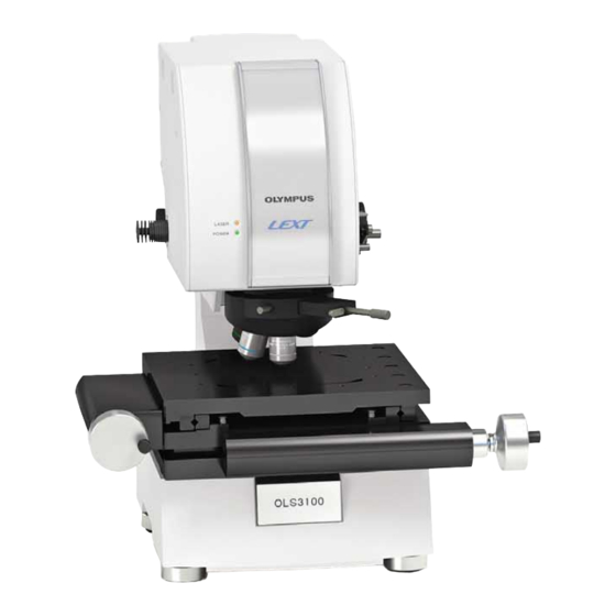
Summary of Contents for Olympus Lext OLS3100
- Page 1 CONFOCAL LASER SCANNING MICROSCOPE OLS3100 The name "LEXT" is formed from the words "Laser" and "Next," and means "next-generation 3D confocal laser microscopes".
- Page 2 Greater simplicity with higher precision: The next step in the evolution of three dimensional laser confocal metrology. LEXT minimizes manual operation, improving ease of use for everyone. Even a first-time user can operate the system like an expert, and obtain fast reliable measurement results. The system features not only improved functionality, but also an even higher level of measurement performance.
- Page 3 W e l c o m e t o t h e w o r l d o f L E X T 3 D...
- Page 4 Automatic operation achieves speedy, high-precision output. Position and magnification settings New, Operation Navigator feature is an online wizard that guides the user in the operation of the LEXT The operation navigator provides animations to guide the user through each step of microscopic observation. You can complete a series of steps by simply operating the mouse in the same way as shown in the animations...
- Page 5 Image capturing Auto Fine View does it all automatically The brightness and contrast of a captured image are automatical- ly adjusted. Image conditioning is typically a manual process requiring an experienced operator. Using the auto fine view function of LEXT, anybody can acquire ideal, high-quality 3D images without special training.
- Page 6 Powerful 3D display facilitates measurement and analysis. Display Adjustable 3D image The angle of a 3D image can be now changed freely with the mouse by grabbing the image. In addition, the 3D image can be scaled up or down in 100 steps using the mouse wheel.
- Page 7 A variety of 3D image presentation patterns A variety of 3D image presentation patterns are provided, including surface texture, real color, wired frame, etc. A 3D image can be rendered to make it more visually effective. Corny layer cells Surface Texture Wired frame Analysis...
- Page 8 Versatile observation methods to handle a wide range of applic Display Brightfield observation Color information can be obtained from brightfield (color) observation. Therefore, brightfield observation can be used effectively to observe a flaw on a color filter or to locate the position of an area of corrosion on metal.
- Page 9 ations. Split screen display An image observed in one observation mode and the same image observed in another observation mode can be displayed simultaneously during live observation. A target point can be located easily by observing a microscopic image with color information and a high-resolution LSM (Laser Scanning Microscope) image simultaneously.
- Page 10 Scan pattern Further advanced, the world’s highest level of repeatability Advanced optical techniques of Olympus accumulated over years have made possible the planar measurement repeatability of 3 = 0.02 µm and the height measurement repeatability of 3 = 0.04 + 0.002L mm (L = measured length in µm).
- Page 11 Scanning Microscope, which enables to improve the optical performance with a 408-nm laser light, was developed. This special objective lens developed with the world-class optical technology of Olympus has made possible the highest level of observational clarity and measurement accuracy at high magnifications.
- Page 12 Full range of measurement/analysis functions to meet virtually The image shown below is a three-dimensional image of a spherocrystal of an injection molded polyamide resin (PA66) product observed using the N-ARC method. Such a spiral-shaped higher-order structure is observed in the spherocrystal growth process after injection molding, although it is of rare occurrence.
- Page 13 W e l c o m e t o t h e w o r l d o f L E X T 3 D any requirement. Solder (after ion etching) Because solder is very soft, years of experience and know-how are required to make specimen preparations before observing the composition of solder under a microscope.
- Page 14 This makes it possible to automate the taking of measurements. Confocal laser scanning microscope for 300-mm wafer observation/OLS3000-300 This product may not be available in your area. Please consult your Olympus dealer. Configuration with a motorized stage Stitched (Tiled) image Measured image...
- Page 15 Specifications Observation method Microscope stand Illumination Laser White light Z stage Vertical movement/Maximum height of specimen Z revolving nosepiece Stroke/Resolution/Repeatability Objective lens Total magnification Field of view Optical zoom Stage * Manual stage/Motorized stage Frame memory Intensity/Height Dimensions Weight *300 mm x 300 mm stage is optional upon special order basis. LEXT unit dimensions Monitor * PC *...
- Page 16 OLYMPUS CORPORATION has obtained ISO9001/ISO14001. Specifications are subject to change without any obligation on the part of the manufacturer. Printed in Japan M1619E-0107B...


Need help?
Do you have a question about the Lext OLS3100 and is the answer not in the manual?
Questions and answers