Table of Contents
Advertisement
Quick Links
Advertisement
Chapters
Table of Contents

Related Manuals for Konica Minolta CS-3


Summary of Contents for Konica Minolta CS-3
- Page 1 CS-3/CS-7 Quality Control Manual Xpress Digital Mammography...
-
Page 3: Table Of Contents
2 .6 .4 Procedure (for CS-3) . . . . . . . . . . . . . 28 Cassettes . . . . . . . . . . . . . . . . . . . . . . 15 2 .6 .5... - Page 4 Contents 2 .10 Compression Force Test . . . . . . . . . . . . . 58 3 .5 Radiation Output Rate . . . . . . . . . . . . . . . 77 2 .10 .1 Test Objective .
- Page 5 Required Equipment . . . . . . . . . . . . 131 3 .18 .4 Procedure (for CS-3) . . . . . . . . . . . . 131 3 .18 .5 Procedure (for CS-7) .
-
Page 7: Introduction
Introduction... -
Page 8: About This Document
Quality Control (QC) is important in any imaging system, but it is especially important in mammography . This document is intended to establish the manufacturers recommended quality control program for Konica Minolta Xpress Digital Mammography System, as part of a full-field digital mammography (FFDM). -
Page 9: Manual Structure
About This Document Manual Structure Following is a description of how this manual is structured: • Introduction This section contains important prefatory material for the intended audiences . • Chapter 1 Overview of Testings (Set-up and Baseline Tests) This section shows the overview of test items in the quality control activities . •... -
Page 10: References
• Konica Minolta REGIUS Model 110HQ Operation Manual • Konica Minolta REGIUS Model 210 Operation Manual • Konica Minolta REGIUS Model 190 Operation Manual • Konica Minolta REGIUS Console CS-3 Operation Manual • Konica Minolta DIRECT DIGITIZER CS-7 Operation Manual... -
Page 11: Acronyms
About This Document Acronyms Following is a list of acronyms used in this document . Automatic Exposure Control Aluminum Contrast to Noise Ratio Computed Radiography Half-Value Layer Magnification Mode Mammography Equipment Evaluation Molybdenum Optical Density PACS Picture Archiving and Communications System PMMA Polymethylmethacrylate Rhodium... -
Page 13: Overview Of Testings (Set-Up And Baseline Tests)
Chapter Overview of Testings (Set-up and Baseline Tests) -
Page 14: Overview Of Testings
1 .1 Xpress Digital Mammography System Overview of Testings Frequency Responsibility Chapter Test No . Test Item Semi- Installation Daily Weekly Monthly Quarterly Annual Annually (Baseline) ü Daily Operational Checks ü (ü) Printer QC (Technologist) View Boxes and Viewing ü (ü) Conditions (Technologist) ü... -
Page 15: Getting Ready For Measurements
2400 divided by the mR of the exposure . S value should be properly calibrated at installation of REGIUS Model 190/210/110HQ prior to any QC activities . For more detailed information about S value, refer to Konica Minolta REGIUS MODEL 210 Technical Commentary/ Image Adjustment Manual . -
Page 16: Baseline Tests
1 .3 Baseline Tests Complete the installation of the mammography X-ray unit, Konica Minolta Xpress Digital Mammography System and the display system (laser printer or diagnostic review workstation) before starting the QC testing . Perform all the tests in Chapter 2 except for "2 .2 Daily Operational Checks" and "2 .9 Repeat Analysis", and all the tests in Chapter 3, as the baseline tests . -
Page 17: Qc Activities For The Technologist
Chapter QC Activities for the Technologist... -
Page 18: Testing Summary
2 .1 Testing Summary This section of the document applies to those procedures required to execute the technologist role in the QC of the Xpress Digital Mammography System . This section provides the following information: • Test objectives . • Recommended frequency to perform the test item . •... -
Page 19: Required Equipment
2 .1 Testing Summary 2 .1 .2 Required Equipment... - Page 20 2 .1 Testing Summary *1 Use the QC pattern specified in the manufacturer's QC manual for your printer or review workstation. *2 Additional equipment may be needed . Follow the manufacturer's QC manual for your printer or review workstation . *3 No special equipment is needed .
-
Page 21: Daily Operational Checks
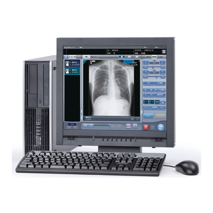
X-ray and CR Control Area Evaluation Ensure the following items: – The monitor of the REGIUS Console CS-3/CS-7 is free of blemishes . – REGIUS login screen is displayed and no errors are reported . – No errors are displayed on the REGIUS Model 190/210/110HQ . -
Page 22: Action
2 .2 Daily Operational Checks 2 .2 .5 Performance Criteria and Corrective Action Performance Criteria: All evaluation items are met . All cassettes are returned . Corrective Action: Restart the Xpress Digital Mammography System and repeat checks . If problems persist reference the appropriate user manuals to determine the solution to the particular error condition . - Page 23 2 .2 Daily Operational Checks TECHNOLOGIST'S MAMMOGRAPHY QC TEST SUMMARY Xpress Digital Mammography System- Konica Minolta Table RT-S1 Daily and Weekly Tests Institution Name: ____________________________________ Room #: _______ Mammography X-ray unit S/N: ________________________ CR Console S/N: ______________ CR Reader S/N: ________________...
-
Page 24: Printer Qc (Technologist)
2 .3 Printer QC (Technologist) 2 .3 .1 Test Objective To ensure that the laser printer used to produce hard copies for final interpretation is performing according to the manufacturer's specifications. FDA recommends the use of devices that have been approved by the FDA Office of Device Evaluation for use in Full Field Digital Mammography . -
Page 25: View Boxes And Viewing Conditions
2 .4 View Boxes and Viewing Conditions (Technologist) 2 .4 .1 Test Objective To follow the guidance given in the "Mammography Quality Control Manual" (ACR, 1999) Radiologic Technologist section procedure 9 . 2 .4 .2 Frequency • Initially before interpreting mammograms •... -
Page 26: Monitor Qc (Technologist)
Corrective Action: Ensure the viewing conditions are appropriate. Remove any objects that are reflecting an unac- ceptable glare . If problems persist, the REGIUS Console CS-3/CS-7 will need to have service to address the issue . The source of the problem shall be identified and corrective action shall be taken before further patient imaging is performed by use of that monitor . -
Page 27: Review Workstation Monitor Qc
2 .5 Monitor QC (Technologist) 2 .5 .2 Review Workstation Monitor QC z Test Objective To ensure that monitors of the review workstation used for final interpretation are performing according to the manu- facturer's specifications. US FDA recommends the use of devices that have been approved by the US FDA Office of Device Evaluation for use in Full Field Digital Mammography . -
Page 28: Contrast To Noise Ratio (Cnr) Check
• Al sheet (Thickness: 0 .2 mm, Size: 60 mm × 60 mm or larger) • Imaging cassette 2 .6 .4 Procedure (for CS-3) Place PMMA (acrylic) phantom of thickness 40 mm on the breast support table . Position the compression paddle as close as possible to the phantom surface . - Page 29 Process the cassette under the Phantom Image Quality exam tag . To identify the analysis tar- get image, take note of the shooting date and time . Start the User Tool . • Click the [Konica Minolta] button . • The System Menu screen is displayed . • Click the [Utility] button .
- Page 30 2 .6 Contrast to Noise Ratio (CNR) Check • Enter the password, and then click the [Go] button . • The User Tool screen is displayed . Start the Analysis Tool . • Click the [CNR/SNR] button . • The Analysis Tool screen is displayed .
- Page 31 • Select [File (F)], and then select the [Open (O)…] from the sub menu . • The file selection screen is displayed. • Select the analysis target image . All images temporarily saved in the REGIUS Console CS-3 are displayed . • Select , and select the [Details] from the sub menu .
- Page 32 2 .6 Contrast to Noise Ratio (CNR) Check Perform the CNR analysis . • Select [QC (Q)], and then select the [CNR (C)] from the sub menu successively . The analysis results are dis- played . • CNR is analyzed and the results are displayed . •...
-
Page 33: Procedure (For Cs-7)
• The CNR value can be calculated by using a measurement tool of a diagnostic review workstation, instead of using the User tool of REGIUS Console CS-3/CS-7 . In that case, process the cassettes under the CNR-WS exam tag and send the image to the diagnostic review workstation . - Page 34 2 .6 Contrast to Noise Ratio (CNR) Check Set the 0 .2 mm Al sheet on the right side of the PMMA and move down the compression pad- dle on the surface of the PMMA . 20mm Chest Side 60mm Al Sheet 20mm 10mm...
- Page 35 2 .6 Contrast to Noise Ratio (CNR) Check • Press [Utility] on the system monitor screen . • The password screen is displayed . • Input the password and press [OK] . • The user tool screen is displayed .
- Page 36 2 .6 Contrast to Noise Ratio (CNR) Check Start the Analysis Tool . • Click the [Analysis] button . • Click the [CNR/SNR] button . • The Analysis Tool screen is displayed .
- Page 37 2 .6 Contrast to Noise Ratio (CNR) Check Load the analysis target image . • Select [File (F)], and then select the [Open (O)…] from the sub menu . • The file selection screen is displayed. • Select the analysis target image . All images temporarily saved in the REGIUS Console CS-7 are displayed . •...
- Page 38 2 .6 Contrast to Noise Ratio (CNR) Check Perform the CNR analysis . • Select [QC (Q)], and then select the [CNR (C)] from the sub menu successively . The analysis results are dis- played . • CNR is analyzed and the results are displayed . •...
-
Page 39: Action
• The CNR value can be calculated by using a measurement tool of a diagnostic review workstation, instead of using the User tool of REGIUS Console CS-3/CS-7 . In that case, process the cassettes under the CNR-WS exam tag and send the image to the diagnostic review workstation . -
Page 40: Documentation
2 .6 Contrast to Noise Ratio (CNR) Check 2 .6 .7 Documentation Record the activity and the results in the Table RT-S1 (TECHNOLOGIST'S MAMMOGRAPHY QC TEST SUMMARY: Daily and Weekly Tests) . HINT • • • • • • • • • • • • • • • • • • • • • • • • • • • • • • • • • • • • • • • • • • • • • • • • • • • • • • • • • • • • • • • • • • • • • • • • • • • • • • • • • • • • • • • • • • • • • • • • • • • • • •... - Page 41 2 .6 Contrast to Noise Ratio (CNR) Check Performance Criteria and Corrective Action • Performance criteria: The new (weekly) image's S value must fall within +/- twenty percent of the baseline S value es- tablished for the same mammography X-ray unit . •...
- Page 42 2 .6 Contrast to Noise Ratio (CNR) Check Table RT-1 (Baseline CNR Value) Institution Name: Date: Room #: Mammography X-ray unit S/N: Cassette Bar Code #: CR Console S/N: CR Reader S/N: Phantom:40 mm 0.2 mm Al 28 kVp CNR1: CNR2: CNR3: CNR4:...
- Page 43 2 .6 Contrast to Noise Ratio (CNR) Check Table RT-1 (Baseline S Value) Institution Name: Date: Room #: Mammography X-ray unit S/N: Cassette Bar Code #: CR Console S/N: CR Reader S/N: Phantom:40mm Exp Count Displayed S value Exp #1 Exp #2 Exp #3 Exp #4...
-
Page 44: Phantom Image Quality (Technologist)
2 .7 Phantom Image Quality (Technologist) 2 .7 .1 Test Objective Acquire a phantom image and evaluate the image quality in an effort to ensure consistent high quality mammography images . 2 .7 .2 Frequency • Initially at installation after appropriate calibration of the equipment to establish a baseline level •... - Page 45 2 .7 Phantom Image Quality (Technologist) For hard copy images as a final interpretation: Measure the following densities. The background density should be measured at the geometric center of the phantom image . The measured OD must not be less than 1 .50 . The DD measurement is obtained by subtracting the density inside the acrylic disc from the density directly adjacent to the disc, in the direction perpendicular to the anode-cathode axis .
-
Page 46: Action
2 .7 Phantom Image Quality (Technologist) IMPORTANT • • • • • • • • • • • • • • • • • • • • • • • • • • • • • • • • • • • • • • • • • • • • • • • • • • • • • • • • • • • • • • • • • • • • • • • • • • • • • • • • • • • • • • • • • • • • • • • • • • • • • •... -
Page 47: Documentation
2 .7 Phantom Image Quality (Technologist) 2 .7 .6 Documentation Record the activity and the results in the Table RT-2 (Phantom Control Chart) and the Table RT-S1 (TECHNOLO- GIST'S MAMMOGRAPHY QC TEST SUMMARY: Daily and Weekly Tests), or use the worksheets and forms refer- enced in the "Mammography Quality Control Manual"... - Page 48 2 .7 Phantom Image Quality (Technologist) Xpress CR Mammography System- Konica Minolta Table RT-2 PHANTOM CONTROL CHART Cassette Bar Code #: ___________________________ kVp: ______ Room #: __________ Mammography X-ray unit S/N: _____________ CR Console S/N: _________ CR Reader S/N: ________...
- Page 49 2 .7 Phantom Image Quality (Technologist) PHANTOM CONTROL CHART (cont'd) Remarks Date Action...
-
Page 50: Visual Inspection
Ensure the following items: – All lights, indicators are function property . – The monitor of the REGIUS Console CS-3/CS-7 is free of smudges and fingerprints. z Imaging cassette and plate Open each REGIUS cassette and inspect the surface of the imaging plate . -
Page 51: Action
2 .8 Visual Inspection 2 .8 .5 Performance Criteria and Corrective Action Performance Criteria: All evaluation items are met . Corrective Action: Restart the Xpress Digital Mammography System and repeat checks . During cassette inspection use the lint free cloth to remove dust blemishes and particulate matter . Anhydrous alcohol should only be used to re- move persistent blemishes or substances . - Page 52 2 .8 Visual Inspection TECHNOLOGIST'S MAMMOGRAPHY QC TEST SUMMARY Xpress CR Mammography System- Konica Minolta Table RT-S2 Monthly, Quarterly, and Semi-Annual Tests Institution Name: _________________________________________ Room #: _________ Mammography X-ray unit S/N: _____________________________________ CR Console S/N: _________________ CR Reader S/N: _________________...
- Page 53 2 .8 Visual Inspection Table RT-3 Monthly Visual Inspection Institution Name: __________________________________________ Room #: __________________ CR Console S/N: _________________ Mammography X-ray unit S/N: _____________ CR Reader S/N: _____________ Date: Initials: Patient Surfaces Mode indicator Angle of Rotation E-Stop is functional Collimators No Wires are exposed...
-
Page 54: Repeat Analysis
2 .9 Repeat Analysis 2 .9 .1 Test Objective Track the repeat and reject activity for the purpose of trend analysis . These analyses are to help maintain delivery of consistent high quality clinical image data . This analysis should be performed on a quarterly basis . 2 .9 .2 Frequency •... -
Page 55: Documentation
2 .9 Repeat Analysis 2 .9 .6 Documentation • Record Repeat/Reject activity on the Table RT-4a (Repeat/Reject Tracking), the RT-4b (Repeat/Reject Results) and the Table RT-S2 (TECHNOLOGIST'S MAMMOGRAPHY QC TEST SUMMARY: Monthly, Quarterly, and Semi- Annual Tests) or use the worksheets and forms found in the "Mammography Quality Control Manual" (ACR, 1999) Radiologic Technologist section procedure 8 . - Page 56 2 .9 Repeat Analysis Table RT-4a Repeat/Reject Tracking Institution Name: __________________________________________ Room #: __________________ Projection Reason for Rejected Date (R-CC, Study ID Cassette ID Technologist Repeat Code (Y/N) L-MLO, etc) Reason Codes: A - Patient Motion D - Incorrect Patient G - Test Image B - Patient Positioning E - Fogged Cassette...
- Page 57 2 .9 Repeat Analysis Table RT-4b Repeat/Reject Results Institution Name: __________________________________________ Department Name: _________________________________________ # of Repeated Percent of Percent Reason Code # of Rejected Images Images Repeat Rejected Repeat Performance Repeated Image Total (X) = (X/T * 100) Reject Performance Rejected Image Total (Y) = (Y/T * 100) Total # of Exposures (T)
-
Page 58: Compression Force Test
2 .10 Compression Force Test 2 .10 .1 Test Objective To ensure the initial power drive compression force is within acceptable operating levels . 2 .10 .2 Frequency • Initially at installation • SEMI-ANNUALLY • As part of an MEE •... -
Page 59: Imaging Plate Fog
2 .11 Imaging Plate Fog 2 .11 .1 Test Objective To verify that plates stored in the exposure room exhibit no evidence of unintentional exposure (fog) . 2 .11 .2 Frequency • Initially before patients are imaged • SEMI-ANUALLY • Whenever the storage space for the cassettes or the radiation shielding is changed 2 .11 .3 Required Equipment •... -
Page 60: Documentation
2 .11 Imaging Plate Fog 2 .11 .6 Documentation Record the results in the Table RT-S2 (TECHNOLOGIST'S MAMMOGRAPHY QC TEST SUMMARY: Monthly, Quar- terly, and Semi-Annual Tests) . -
Page 61: Qc Activities For The Medical
Chapter QC Activities for the Medical Physicist... -
Page 62: Testing Summary
3 .1 Testing Summary This section of the document applies to those procedures required to execute the Medical Physicist role in the QC of the Xpress Digital Mammography System . These tests should only be carried out by a qualified medical physicist in accordance with 21 CFR 900 .12(a)(3) which defines these qualifications. -
Page 63: Test Items
3 .1 Testing Summary 3 .1 .1 Test Items z Xpress Digital Mammography System Medical Physicist QC activities Test No . Test Item Action Criteria Physical Inspection Tube Voltage Measurement and Reproducibility Beam Quality Radiation Output Rate Average Glandular Dose View Boxes and Viewing Conditions Printer QC Monitor QC... -
Page 64: Testing Frequency
3 .1 Testing Summary 3 .1 .2 Testing Frequency All tests contained in this section are to be carried out a) initially upon installation prior to patient examination, b) an- nually thereafter, c) after any major repair, upgrade, or modification on a component of the imaging chain which may affect the performance to be tested, and d) when problems are suspected. -
Page 65: Requied Equipment
3 .1 Testing Summary 3 .1 .3 Requied Equipment... - Page 66 3 .1 Testing Summary *1 Use the QC pattern specified in the manufacturer's QC manual for your printer or review workstation. *2 Additional equipment may be needed . Follow the manufacturer's QC manual for your printer or review workstation . *3 The purity of the Al ≥...
-
Page 67: Physical Inspection
3 .2 Physical Inspection 3 .2 .1 Test Objective To ensure the overall safety and mechanical stability of the Xpress Digital Mammography System . 3 .2 .2 Frequency • Initially at installation • ANNUALLY • As part of an MEE 3 .2 .3 Required Equipment See "Mammography Quality Control Manual"... -
Page 68: Tube Voltage Measurement And
Survey Date: Mammography X-ray unit S/N: Model: Date of Installation: Room #: Inspector's Name: Signature: Konica Minolta QC manual version(s) at facility (check all that apply): CR Reader S/N: CR Console S/N: Printer S/N: Monitor S/N: PASS/FAIL Physical Inspection MP-1... -
Page 69: Physical Inspection
3 .2 Physical Inspection 3.2 Physical Inspection Table MP-1 Institution Name: Date: Inspectors Name: Equipment Room #: Mammography X-ray unit S/N: CR Reader S/N: CR Console S/N: Cassette Bar Code #: Evaluation Item: Pass/Fail - Assemblies move freely and cassette is secure in any position of the C-arm - Support stands and compression paddles change normally - Compression release and emergency stop function properly - Compression thickness display is accurate within 5 mm of true value... -
Page 70: Test Objective
3 .3 Tube Voltage Measurement and Reproducibility 3 .3 .1 Test Objective To ensure the accuracy and reproducibility of the displayed tube voltage of the generator . 3 .3 .2 Frequency • Initially at installation of the mammography X-ray unit •... -
Page 71: Action
3 .3 Tube Voltage Measurement and Reproducibility Make an exposure . (On some units, a cassette must be placed in the cassette holder to enable exposure .) Record the results for the measured and displayed values in the correct fields on the Table MP-2 (Tube Voltage Measurement and Reproducibility) . - Page 72 3 .3 Tube Voltage Measurement and Reproducibility 3.3 Tube Voltage Measurement and Reproducibility Table MP-2 Date: Inspectors Name: Mammography X-ray unit S/N: Rep #1 Measured kVp value at the lowest measureable level Rep #1 Displayed kVp value at the lowest measureable level Pass/Fail % error %( / -1) Criteria...
-
Page 73: Beam Quality
3 .4 Beam Quality 3 .4 .1 Test Objective To measure half value layer of the device . 3 .4 .2 Frequency • Initially at installation of the mammography X-ray unit • ANNUALLY • As part of an MEE • After any service or maintenance on the mammography X-ray unit which may affect beam quality 3 .4 .3 Required Equipment •... - Page 74 3 .4 Beam Quality Position the paddle as close to the tube as possible, all the way up . Make the first exposure and record the mGy. Make the second exposure and record the mGy . Calculate the average of the two mGy values and record it . Add 0.3 mm of Al filtration between the tube and the detector of the dose meter and repeat the process, from Step 5 through Step 7 .
-
Page 75: Action
3 .4 Beam Quality 3 .4 .5 Performance Criteria and Corrective Action Performance Criteria: After entering the data on the Table MP-3 (Beam Quality), the table will calculate Pass/Fail for this test . For 28kVp Mo/Mo the HVL must be over 0 .30mm Al equivalent, and is typically <0 .40 mm Al . Typical HVL values for Mo/Mo and other target/filter combinations, as published by EUREF, are provided below . - Page 76 3 .4 Beam Quality 3.4 Beam Quality Table MP-3 Mammography X-ray unit S/N: Target/Filter: Mo/Mo Measured tube voltage: 28kVp Pre-set tube load: t l i o i t Rep #1 Rep #2 Average exposure: The direct exposure reading is denoted as Y and Y are the exposure readings with added Al thickness of X and X...
-
Page 77: Radiation Output Rate
3 .5 Radiation Output Rate 3 .5 .1 Test Objective To measure the radiation output rate of the equipment . 3 .5 .2 Frequency • Initially at installation of the mammography X-ray unit • ANNUALLY • As part of an MEE 3 .5 .3 Required Equipment •... -
Page 78: Action
3 .5 Radiation Output Rate Make three exposures and record the mGy and the exposure time in the appropriate fields on Table MP-4 (Radiation Output Rate) . The Table MP-4 will denote Pass/Fail . 3 .5 .5 Performance Criteria and Corrective Action Performance Criteria: The system shall be able to produce a minimum output of 7 .0mGy air kerma per second (800mR per second) when operating at 28 kVp in the standard mammography mode (Mo/Mo) . - Page 79 3 .5 Radiation Output Rate 3.5 Radiation Output Rate Table MP-4 Date: Inspectors Name: Mammography X-ray unit S/N: * Measurements should be taken using a minimum 3 sec. exposure Measured tube voltage: 28kVp Total Measured Radiation Radiation exposure time Output Rate Output mGy mGy/s Repeat #...
-
Page 80: Average Glandular Dose
3 .6 Average Glandular Dose 3 .6 .1 Test Objective To determine the average glandular dose delivered for a particular object . 3 .6 .2 Frequency • Initially at installation of the mammography X-ray unit • ANNUALLY • As part of an MEE 3 .6 .3 Required Equipment •... -
Page 81: Action
3 .6 Average Glandular Dose The detector should be the same height as the top of the RMI phantom . Expose the phantom and the detector of the dose meter with the kVp, target material and fil- tration at which the system is most commonly used clinically (Using AEC) . Make four (4) exposures and record the mR and mAs from the dose meter and console of the mammography X-ray unit . - Page 82 3 .6 Average Glandular Dose 3.6 Average Glandular Dose Table MP-5 Date: Inspectors Name: Mammography X-ray unit S/N: AEC Mode All Cells Active Target/Filter Measured tube voltage Repeat # Rep #1 Rep #2 Rep #3 Rep #4 Mean Pass/Fail Calculated Average Glandular Dose: Criteria: ≤3 mGy P Pass...
-
Page 83: View Boxes And Viewing Conditions
3 .7 View Boxes and Viewing Conditions (Medical Physicist) 3 .7 .1 Test Objective To follow the guidance given in the "Mammography Quality Control Manual" (ACR, 1999) Medical Physicist section procedure 11 . 3 .7 .2 Frequency • Initially before interpreting mammograms •... -
Page 84: Printer Qc (Medical Physicist)
3 .8 Printer QC (Medical Physicist) 3 .8 .1 Test Objective To ensure that the laser printer used to produce hard copies for final interpretation is performing according to the manufacturer's specifications. US FDA recommends the use of devices that have been approved by the US FDA Office of Device Evaluation for use in Full Field Digital Mammography . -
Page 85: Monitor Qc (Medical Physicist)
Corrective Action: Ensure the viewing conditions are appropriate. Remove any objects that are reflecting an unac- ceptable glare . If problems persist the REGIUS Console CS-3/CS-7 will need to have service to address the issue . The source of the problem shall be identified and corrective action shall be taken before further patient imaging is performed by use of that monitor . -
Page 86: Review Workstation Monitor Qc
3 .9 Monitor QC (Medical Physicist) 3 .9 .2 Review Workstation Monitor QC z Test Objective To ensure that monitors of the review workstation used for final interpretation are performing according to the manu- facturer's specifications. US FDA recommends the use of devices that have been approved by the US FDA Office of Device Evaluation for use in Full Field Digital Mammography . -
Page 87: (Cr Console Monitor)
3 .9 Monitor QC (Medical Physicist) 3.9 CR Console Monitor QC Table MP-6 Date: Inspectors Name: Monitor S/N: SMPTE Assessment Pass/Fail 5 % patch Visible 95 % patch Visible All Gradation steps distinguishable P Pass F Fail Comments:... -
Page 88: Dark Noise
The REGIUS cassettes should be erased prior to use in this test . Register a test patient on the REGIUS Console CS-3/CS-7 . Select the COMP QA/QC exam tag . This will populate the screen with all the QA/QC test tags required for the complete test . -
Page 89: Performance Criteria And Corrective
3 .10 Dark Noise 3 .10 .5 Performance Criteria and Corrective Action Performance Criteria: The acquired image on Dark Noise 1 and Dark Noise 2 should display an S value of 5000 . Corrective Action: Ensure the ROI has not been inadvertently moved out of the image area . The ROI should be cov- ering the entire plate and can be moved by the end user to correct . - Page 90 3 .10 Dark Noise 3.10 Dark Noise Table MP-7 Date: Inspectors Name: Cassette Bar Code #: CR Reader S/N: S value Pass/Fail Dark Noise 1 Dark Noise 2 Criteria S = 5000 P Pass F Fail Comments:...
-
Page 91: Ghost Image Evaluation
3 .11 Ghost Image Evaluation 3 .11 .1 Test Objective To ensure that images being produced from the reader are free of any latent image and are fully erased following an erasure performed during normal reading . 3 .11 .2 Frequency •... -
Page 92: Performance Criteria And Corrective
3 .11 Ghost Image Evaluation Remove the RMI-156 phantom and replace it with the 2cm of PMMA (acrylic) phantom . Register the cassette under the Erase 2 exam tag . Expose the cassette using 28kVp 40mAs . Process the cassette and view the acquired image . 3 .11 .5 Performance Criteria and Corrective Action Performance Criteria: The Erase 2 image should be free of any visible indication of the RMI phantom from the previ-... - Page 93 3 .11 Ghost Image Evaluation 3.11 Ghost Image Evaluation Table MP-8 Date: Inspectors Name: Cassette Bar Code #: CR Reader S/N: l i a Criteria; No Latent image is Detected P Pass F Fail Comments:...
-
Page 94: S Value Response Indicator
3 .12 S value Response Indicator 3 .12 .1 Test Objective To ensure the displayed S value of the CR Image is within specification. 3 .12 .2 Frequency • Initially before patients are imaged • ANNUALLY • After major repair, upgrade, or modification of the REGIUS Model 190/210/110HQ or the mammography X-ray unit, and whenever its calibration problem is suspected 3 .12 .3 Required Equipment... -
Page 95: Performance Criteria And Corrective
3 .12 S value Response Indicator Wait 2 minutes and then process the cassette . Record the mR and the displayed S value on table MP-9 (S value Response Indicator) . Repeat steps 1 through 5 three more times . IMPORTANT •... - Page 96 3 .12 S value Response Indicator 3.12 S value Response Indicator Table MP-9 Date: Inspectors Name: Cassette Bar Code #: CR Reader S/N: Stability and Reproducibility S=2400/ Displayed Measured X(mR) S value dose (mR) Exp Count S= 2400/ X(mR) Exp #1 X(mR) is the value of the average exposure value stated in mR.
-
Page 97: Aec Performance Checks
• Calibrated dose meter z Procedure Register the patient on the REGIUS Console CS-3/CS-7 . Set the REGIUS Console CS-3/CS-7 for a minimum of four exposures under the AEC Checks exam tag . HINT • • • • • • • • • • • • • • • • • • • • • • • • • • • • • • • • • • • • • • • • • • • • • • • • • • • • • • • • • • • • • • • • • • • • • • • • • • • • • • • • • • • • • • • • • • • • • • • • • • • • •... -
Page 98: Aec Performance Checks
3 .13 AEC Performance Checks The detector should be the same height as the top of the RMI phantom . Make the exposure with 28 kVp in AEC mode . Process the cassettes in the REGIUS Model 190/210/110HQ . Note the mAs and mR of the image . Repeat the process, from Step 1 through Step 8, three more times . -
Page 99: Thickness Tracking
• Al sheet (Thickness: 0 .2 mm, Size: 60 mm × 60 mm or larger) • Imaging cassette z Procedure (for CS-3) Place PMMA (acrylic) phantom of thickness 2 cm on the breast support table . Position the compression paddle as close as possible to the phantom surface . - Page 100 Process the cassette under the Phantom Image Quality exam tag . To identify the analysis tar- get image, take note of the shooting date and time . Start the User Tool . • Click the [Konica Minolta] button . • The System Menu screen is displayed . • Click the [Utility] button .
- Page 101 3 .13 AEC Performance Checks • Enter the password, and then click the [Go] button . • The User Tool screen is displayed . Start the Analysis Tool . • Click the [CNR/SNR] button . • The Analysis Tool screen is displayed . Load the analysis target image .
- Page 102 3 .13 AEC Performance Checks • Select the analysis target image . All images temporarily saved in the REGIUS Console CS-3 are displayed . • Select , and select the [Details] from the sub menu . • The list is displayed in the detail view .
- Page 103 • The CNR value can be calculated by using a measurement tool of a diagnostic review workstation, instead of using the User tool of REGIUS Console CS-3/CS-7 . In that case, process the cassettes under the CNR-WS exam tag and send the image to the diagnostic review workstation .
- Page 104 3 .13 AEC Performance Checks z Procedure (for CS-7) Place PMMA (acrylic) phantom of thickness 2 cm on the breast support table . Position the compression paddle as close as possible to the phantom surface . Move the AEC sensor to its most forward position (closest to the chest side) . Set the 0 .2 mm Al sheet on the right side of the PMMA and move down the compression pad- dle on the surface of the PMMA .
- Page 105 3 .13 AEC Performance Checks Start the User Tool . • Click [System] on the patient registration screen or the list screen . Patient registration screen List screen • The system monitor screen is displayed .
- Page 106 3 .13 AEC Performance Checks • Press [Utility] on the system monitor screen . • The password screen is displayed . • Input the password and press [OK] . • The user tool screen is displayed .
- Page 107 3 .13 AEC Performance Checks Start the Analysis Tool . • Click the [Analysis] button . • Click the [CNR/SNR] button . • The Analysis Tool screen is displayed .
- Page 108 3 .13 AEC Performance Checks Load the analysis target image . • Select [File (F)], and then select the [Open (O)…] from the sub menu . • The file selection screen is displayed. • Select the analysis target image . All images temporarily saved in the REGIUS Console CS-7 are displayed . •...
- Page 109 3 .13 AEC Performance Checks Perform the CNR analysis . • Select [QC (Q)], and then select the [CNR (C)] from the sub menu successively . The analysis results are dis- played . • CNR is analyzed and the results are displayed . •...
- Page 110 • The CNR value can be calculated by using a measurement tool of a diagnostic review workstation, instead of using the User tool of REGIUS Console CS-3/CS-7 . In that case, process the cassettes under the CNR-WS exam tag and send the image to the diagnostic review workstation .
- Page 111 • Imaging cassette • Calibrated dose meter Procedure 1 Register the test patient on the REGIUS Console CS-3/CS-7 . 2 Set the REGIUS Console CS-3/CS-7 for three exposures . HINT • • • • • • • • • • • • • • • • • • • • • • • • • • • • • • • • • • • • • • • • • • • • • • • • • • • • • • • • • • • • • • • • • • • • • • • • • • • • • • • • • • • • • • • • • • • • • • • • •...
- Page 112 3 .13 AEC Performance Checks 3.13 AEC Performance Checks Table MP-10 (CNR) Date: Inspectors Name: Mammography X-ray unit S/N: Stability and Reproducibility 28kVp, 4 cm acrylic Exp Count Exp #1 Exp #2 Exp #3 Exp #4 Mean Standard Deviation (SD) Pass/Fail Coeff of variation (%) ≤...
- Page 113 3 .13 AEC Performance Checks 3.13 AEC Performance Checks Table MP-10 (S value) Date: Inspectors Name: Mammography X-ray unit S/N: Stability and Reproducibility 28 kVp Exp Count Exp #1 Exp #2 Exp #3 Exp #4 Mean Standard Deviation(SD) Pass/Fail Coeff of variation(%) Criteria ≤5% ≤5%...
-
Page 114: Collimator Assessment
3 .14 Collimator Assessment 3 .14 .1 Test Objective To ensure that the collimator assembly performs in such a manner that the X-ray field coincides with the light field and is aligned with the image receptor . 3 .14 .2 Frequency •... - Page 115 3 .14 Collimator Assessment Place the (4) pennies on the edge of the light field as shown in the picture below. Make sure not to cover the cells used for AEC . Replace the compression paddle and ensure that it is 4 .2 cm above the breast support table . Use the digital readout to ensure 4 .2 cm .
-
Page 116: Performance Criteria And Corrective
3 .14 Collimator Assessment Repeat steps 1 through 10 for all routinely used collimator/bucky/compression paddle com- binations and target materials . When testing the large image receptor, the top anode may be positioned diagonally to capture all four edges of the X-ray field, or, two large cassettes may be used on top of the image receptor . - Page 117 3 .14 Collimator Assessment Performance Criteria (X-ray Field/Image Area Alignment): Chest Side Chest Side Cassette B Cassette B X-ray field Cassette A X-ray field / Image area alignment • Measure the deviation (a2-d2) of the coins position in relation to the exposed field. •...
-
Page 118: Documentation
3 .14 Collimator Assessment 3 .14 .6 Documentation • Record this activity in the Table MP-11 (Collimator Assessment) and the Table MP-S (MEDICAL PHYSICIST'S MAMMOGRAPHY QC TEST SUMMARY) . - Page 119 3 .14 Collimator Assessment 3.14 Collimator Assessment Table MP-11 Date: Inspectors Name: Mammography X-ray unit S/N: Quarter Coin Chest Side Chest Side Cassette B Cassette A Cassette A Light X-ray Field field Cassette A Alignment of chest wall edges of X-ray field / Image area alignment X-ray field/Light field alignment compression paddles and image...
-
Page 120: Cr Reader Scanner Performance
3 .15 CR Reader Scanner Performance 3 .15 .1 Test Objective To ensure there are no geometric distortions being introduced into the processed image . To ensure there are no laser jitter being introduced into the processed image . 3 .15 .2 Frequency •... -
Page 121: Procedure
3 .15 CR Reader Scanner Performance 3 .15 .4 Procedure z Geometric Distortion Set the Al plate under the X-ray tube . Place a cassette into the cassette holder . Place the steel scale on the breast support table lengthwise with the table . Place one marker at zero mark and the other one at 150 mm mark of the steel scale . - Page 122 3 .15 CR Reader Scanner Performance z Jitter Place a REGIUS cassette on the breast support table . Place another REGIUS cassette into the cassette holder to activate the exposure . Place the steel scale approximately in the center at right angles to the chest wall side of cas- sette .
-
Page 123: Performance Criteria And Corrective
3 .15 CR Reader Scanner Performance 3 .15 .5 Performance Criteria and Corrective Action Performance Criteria (Geometric Distortion): The error in length should be within +/- 5% of the true value. Performance Criteria (Jitter): The borders of the scale should appear straight and smooth . No jitter along the border should be observed. - Page 124 3 .15 CR Reader Scanner Performance 3.15 CR Reader Scanner Performance Table MP-12 Date: Inspectors Name: Cassette Bar Code #: CR Reader S/N: Geometric Distortion True Value Measured Value Measurement Objects actual value of the distance between the paper clips Pass/Fail measured distance between the paper clips % Difference...
-
Page 125: Spatial Resolution
3 .16 Spatial Resolution 3 .16 .1 Test Objective To observe the limiting resolution of the system using a resolution test tool . 3 .16 .2 Frequency • Initially before patients are imaged • ANNUALLY • Whenever the system spatial resolution is suspected 3 .16 .3 Required Equipment •... -
Page 126: Performance Criteria And Corrective
3 .16 Spatial Resolution 3 .16 .5 Performance Criteria and Corrective Action Performance Criteria: Evaluate the processed image looking for 7 LP/mm or greater . Corrective Action: If this test yields results outside the action limits as specified, the source of the problem shall be identified and corrective action shall be taken on the component(s) that caused the failure before further patient im- aging is performed . - Page 127 3 .16 Spatial Resolution 3.16 Spatial Resolution Table MP-13 Date: Inspectors Name: AEC cells position All three cells recommended Cassette Bar Code # Acquisition Mode 18×24 cm, 24×30 cm Density setting Target- Filter kVp setting Limiting resolution in line-pair per mm ( ≥7LP/mm): Pass/Fail Bars parallel to A-C axis LP/mm...
-
Page 128: Artifact Evaluation
3 .17 Artifact Evaluation 3 .17 .1 Test Objective To ensure the images being produced are free of any artifacts that can compromise the interpretation of the clinical image. At least one image should be obtained for each imaging mode and target filter combination in use. This test should be performed for all imaging cassettes in use . -
Page 129: Performance Criteria And Corrective
3 .17 Artifact Evaluation 3 .17 .5 Performance Criteria and Corrective Action Performance Criteria: No significant artifacts should be observed. Corrective Action: If artifact is present on the image, rotate acrylic 180 degrees, if artifact moves, then it is on the acrylic . - Page 130 3 .17 Artifact Evaluation 3.17 Artifact Evaluation Table MP-14 Date: Inspectors Name: Unit Evaluation Cassette Evaluation Artifact present Acceptable Bar Code # Acquisition mode: 18 × 24 cm (Yes/No) (Yes/No) Cassette Bar Code# Selected Filter: AEC or Manual Artifacts Detected: Acquisition mode: 24 ×...
-
Page 131: Inter-Plate Consistency
• Calibrated dose meter • All REGIUS cassettes in use 3 .18 .4 Procedure (for CS-3) Select a REGIUS cassette used for mammography . Erase the plate using ERASE MODE 2 on the REGIUS Model 190/210/110HQ . Set the Al plate under X-Ray tube . - Page 132 3 .18 Inter-Plate Consistency Start the User Tool . • Click the [Konica Minolta] button . • The System Menu screen is displayed . • Click the [Utility] button . • The Password screen is displayed . • Enter the password, and then click the [Go] button .
- Page 133 • Select [File (F)], and then select the [Open (O)…] from the sub menu . • The file selection screen is displayed. • Select the analysis target image . All images temporarily saved in the REGIUS Console CS-3 are displayed . • Select , and select the [Details] from the sub menu .
- Page 134 3 .18 Inter-Plate Consistency • Select the file indicating the shooting date and time that you have took note of, and click the [Open (O)] button. • The analysis target image is displayed . Perform the SNR analysis . • Select [QC (Q)], and then select the [SNR (S)] from the sub menu successively . The analysis results are dis- played .
-
Page 135: Procedure (For Cs-7)
• The SNR value can be calculated by using a measurement tool of a diagnostic review workstation, instead of using the User tool of REGIUS Console CS-3/CS-7 . In that case, process the cassettes under the SNR-WS exam tag and send the image to the diagnostic review workstation . - Page 136 3 .18 Inter-Plate Consistency Start the User Tool . • Click [System] on the patient registration screen or the list screen . Patient registration screen List screen • The system monitor screen is displayed . • Press [Utility] on the system monitor screen . •...
- Page 137 3 .18 Inter-Plate Consistency • Input the password and press [OK] . • The user tool screen is displayed . Start the Analysis Tool . • Click the [Analysis] button . • Click the [CNR/SNR] button . • The Analysis Tool screen is displayed .
- Page 138 3 .18 Inter-Plate Consistency Load the analysis target image . • Select [File (F)], and then select the [Open (O)…] from the sub menu . • The file selection screen is displayed. • Select the analysis target image . All images temporarily saved in the REGIUS Console CS-7 are displayed . •...
- Page 139 3 .18 Inter-Plate Consistency Perform the SNR analysis . • Select [QC (Q)], and then select the [SNR (S)] from the sub menu successively . The analysis results are dis- played . • SNR is analyzed and the results are displayed . •...
-
Page 140: Performance Criteria And Corrective
• The SNR value can be calculated by using a measurement tool of a diagnostic review workstation, instead of using the User tool of REGIUS Console CS-3/CS-7 . In that case, process the cassettes under the SNR-WS exam tag and send the image to the diagnostic review workstation . -
Page 141: Documentation
3 .18 Inter-Plate Consistency 3 .18 .7 Documentation Record the results in the Table MP-15 (SNR) (Inter-Plate Consistency) and the Table MP-S (MEDICAL PHYSICIST'S MAMMOGRAPHY QC TEST SUMMARY) . • • • • • • • • • • • • • • • • • • • • • • • • • • • • • • • • • • • • • • • • • • • • • • • • • • • • • • • • • • • • • • • • • • • • • • • • • • • • • • • • • • • • • • • • • • • • • • • • • • • • • HINT •... - Page 142 3 .18 Inter-Plate Consistency 3.18 Inter-Plate Consistency Table MP-15 (SNR) Date: Inspectors Name: kVp: AEC Mode: mean pixel value/standard deviation in pixel value Mean Standard Deviation(SD) Coefficient SD/Mean Pass/Fail P Pass Criteria; +/- 15% F Fail Comments:...
- Page 143 3 .18 Inter-Plate Consistency 3.18 Inter-Plate Consistency Table MP-15 (S value) Date: kVp: AEC Mode: Difference Displayed from IP Bar Code # S value Mean Pass Fail Mean P Pass Criteria +/- 15% F Fail Comments:...
-
Page 144: Phantom Image Quality
3 .19 Phantom Image Quality (Medical Physicist) 3 .19 .1 Test Objective Acquire a phantom image and evaluate the image quality in an effort to ensure temporal changes in images quality. 3 .19 .2 Frequency • Initially at installation, after appropriate calibration of the equipments to acquire a baseline image •... - Page 145 3 .19 Phantom Image Quality (Medical Physicist) Depending on the facility, make a choice between hard copy or soft copy . For hard copy images as a final interpretation: Measure the following densities. The background density should be measured in the geometric center of the phantom image . The DD measurement is obtained by subtracting the den- sity inside the acrylic disc from the density directly adjacent to the disc, in the direction perpendicular to the anode- cathode axis .
-
Page 146: Performance Criteria And Corrective
3 .19 Phantom Image Quality (Medical Physicist) IMPORTANT • • • • • • • • • • • • • • • • • • • • • • • • • • • • • • • • • • • • • • • • • • • • • • • • • • • • • • • • • • • • • • • • • • • • • • • • • • • • • • • • • • • • • • • • • • • • • • • • • • • • • •... -
Page 147: Documentation
3 .19 Phantom Image Quality (Medical Physicist) 3 .19 .6 Documentation Record the activity and the results in the Table MP-16 (Phantom Image Quality) and Table MP-S (MEDICAL PHYSI- CIST'S MAMMOGRAPHY QC TEST SUMMARY) or use the worksheets and forms referenced in the "Mammography Quality Control Manual"... -
Page 148: Phantom Image Quality
3 .19 Phantom Image Quality (Medical Physicist) 3.19 Phantom Image Quality Table MP-16 Phantom Type & S/N: Cassette Bar Code #: If AEC used: AEC cells position: All three cells recommended Density setting: Mode: 18×24 cm, 24×30 cm Criteria Previous Current Pass/Fail Comments... -
Page 149: Appendices
Chapter Appendices... -
Page 150: Rt-Tables
4 .1 RT-Tables TECHNOLOGIST'S MAMMOGRAPHY QC TEST SUMMARY Xpress Digital Mammography System- Konica Minolta Table RT-S1 Daily and Weekly Tests Institution Name: ____________________________________ Room #: _______ Mammography X-ray unit S/N: ________________________ CR Console S/N: ______________ CR Reader S/N: ________________ Year... - Page 151 4 .1 RT-Tables Table RT-1 (Baseline CNR Value) Institution Name: Date: Room #: Mammography X-ray unit S/N: Cassette Bar Code #: CR Console S/N: CR Reader S/N: Phantom:40 mm 0.2 mm Al 28 kVp CNR1: CNR2: CNR3: CNR4: CNR5: Mean CNR Value (Baseline) +/- 20% 20mm Chest Side...
- Page 152 4 .1 RT-Tables Table RT-1 (Baseline S Value) Institution Name: Date: Room #: Mammography X-ray unit S/N: Cassette Bar Code #: CR Console S/N: CR Reader S/N: Phantom:40mm Exp Count Displayed S value Exp #1 Exp #2 Exp #3 Exp #4 Exp #5 Mean S value (Baseline) +/- 20%...
- Page 153 4 .1 RT-Tables Xpress CR Mammography System- Konica Minolta Table RT-2 PHANTOM CONTROL CHART Cassette Bar Code #: ___________________________ kVp: ______ Room #: __________ Mammography X-ray unit S/N: _____________ CR Console S/N: _________ CR Reader S/N: ________ Month: Date: Initials: S value +/-20% +0.05...
- Page 154 4 .1 RT-Tables PHANTOM CONTROL CHART (cont'd) Remarks Date Action...
- Page 155 4 .1 RT-Tables TECHNOLOGIST'S MAMMOGRAPHY QC TEST SUMMARY Xpress CR Mammography System- Konica Minolta Table RT-S2 Monthly, Quarterly, and Semi-Annual Tests Institution Name: _________________________________________ Room #: _________ Mammography X-ray unit S/N: _____________________________________ CR Console S/N: _________________ CR Reader S/N: _________________...
- Page 156 4 .1 RT-Tables Table RT-3 Monthly Visual Inspection Institution Name: __________________________________________ Room #: __________________ CR Console S/N: _________________ Mammography X-ray unit S/N: _____________ CR Reader S/N: _________________ Date: Initials: Patient Surfaces Mode indicator Angle of Rotation E-Stop is functional Collimators No Wires are exposed Motor...
- Page 157 4 .1 RT-Tables Table RT-4a Repeat/Reject Tracking Institution Name: __________________________________________ Room #: __________________ Projection Reason for Rejected Date (R-CC, Study ID Cassette ID Technologist Repeat Code (Y/N) L-MLO, etc) Reason Codes: A - Patient Motion D - Incorrect Patient G - Test Image B - Patient Positioning E - Fogged Cassette H - Other...
- Page 158 4 .1 RT-Tables Table RT-4b Repeat/Reject Results Institution Name: __________________________________________ Department Name: _________________________________________ # of Repeated Percent of Percent Reason Code # of Rejected Images Images Repeat Rejected Repeat Performance Repeated Image Total (X) = (X/T * 100) Reject Performance Rejected Image Total (Y) = (Y/T * 100) Total # of Exposures (T)
-
Page 159: Mp-Tables
Survey Date: Mammography X-ray unit S/N: Model: Date of Installation: Room #: Inspector's Name: Signature: Konica Minolta QC manual version(s) at facility (check all that apply): CR Reader S/N: CR Console S/N: Printer S/N: Monitor S/N: REQUIRED TESTS PASS/FAIL Physical Inspection... -
Page 160: Physical Inspection
4 .2 MP-Tables 3.2 Physical Inspection Table MP-1 Institution Name: Date: Inspectors Name: Equipment Room #: Mammography X-ray unit S/N: CR Reader S/N: CR Console S/N: Cassette Bar Code #: Evaluation Item: Pass/Fail - Assemblies move freely and cassette is secure in any position of the C-arm - Support stands and compression paddles change normally - Compression release and emergency stop function properly - Compression thickness display is accurate within 5 mm of true value... -
Page 161: Tube Voltage Measurement And Reproducibility
4 .2 MP-Tables 3.3 Tube Voltage Measurement and Reproducibility Table MP-2 Date: Inspectors Name: Mammography X-ray unit S/N: Rep #1 Measured kVp value at the lowest measureable level Rep #1 Displayed kVp value at the lowest measureable level Pass/Fail % error %( / -1) Criteria +/-5% Measured kVp at the most commonly used kvp setting... -
Page 162: Beam Quality
4 .2 MP-Tables 3.4 Beam Quality Table MP-3 Mammography X-ray unit S/N: Target/Filter: Mo/Mo Measured tube voltage: 28kVp Pre-set tube load: t l i o i t Rep #1 Rep #2 Average exposure: The direct exposure reading is denoted as Y and Y are the exposure readings with added Al thickness of X and X... -
Page 163: Radiation Output Rate
4 .2 MP-Tables 3.5 Radiation Output Rate Table MP-4 Date: Inspectors Name: Mammography X-ray unit S/N: * Measurements should be taken using a minimum 3 sec. exposure Measured tube voltage: 28kVp Total Measured Radiation Radiation exposure time Output Rate Output mGy mGy/s Repeat # Rep #1... -
Page 164: Average Glandular Dose
4 .2 MP-Tables 3.6 Average Glandular Dose Table MP-5 Date: Inspectors Name: Mammography X-ray unit S/N: AEC Mode All Cells Active Target/Filter Measured tube voltage Repeat # Rep #1 Rep #2 Rep #3 Rep #4 Mean Pass/Fail Calculated Average Glandular Dose: Criteria: ≤... - Page 165 4 .2 MP-Tables 3.9 CR Console Monitor QC Table MP-6 Date: Inspectors Name: Monitor S/N: SMPTE Assessment Pass/Fail 5 % patch Visible 95 % patch Visible All Gradation steps distinguishable P Pass F Fail Comments:...
-
Page 166: Dark Noise 1
4 .2 MP-Tables 3.10 Dark Noise Table MP-7 Date: Inspectors Name: Cassette Bar Code #: CR Reader S/N: S value Pass/Fail Dark Noise 1 Dark Noise 2 Criteria S = 5000 P Pass F Fail Comments:... -
Page 167: Ghost Image Evaluation
4 .2 MP-Tables 3.11 Ghost Image Evaluation Table MP-8 Date: Inspectors Name: Cassette Bar Code #: CR Reader S/N: l i a Criteria; No Latent image is Detected P Pass F Fail Comments:... -
Page 168: S Value Response Indicator
4 .2 MP-Tables 3.12 S value Response Indicator Table MP-9 Date: Inspectors Name: Cassette Bar Code #: CR Reader S/N: Stability and Reproducibility S=2400/ Displayed Measured X(mR) S value dose (mR) Exp Count S= 2400/ X(mR) Exp #1 X(mR) is the value of the average exposure value stated in mR. Exp #2 Exp #3 Exp #4... -
Page 169: Aec Performance Checks (Stability And Reproducibility
4 .2 MP-Tables 3.13 AEC Performance Checks Table MP-10 (CNR) Date: Inspectors Name: Mammography X-ray unit S/N: Stability and Reproducibility 28kVp, 4 cm acrylic Exp Count Exp #1 Exp #2 Exp #3 Exp #4 Mean Standard Deviation (SD) Pass/Fail Coeff of variation (%) ≤... -
Page 170: Aec Performance Checks
4 .2 MP-Tables 3.13 AEC Performance Checks Table MP-10 (S value) Date: Inspectors Name: Mammography X-ray unit S/N: Stability and Reproducibility 28 kVp Exp Count Exp #1 Exp #2 Exp #3 Exp #4 Mean Standard Deviation(SD) Pass/Fail Coeff of variation(%) Criteria ≤5% ≤5%... -
Page 171: Collimator Assessment
4 .2 MP-Tables 3.14 Collimator Assessment Table MP-11 Date: Inspectors Name: Mammography X-ray unit S/N: Quarter Coin Chest Side Chest Side Cassette B Cassette A Cassette A Light X-ray Field field Cassette A Alignment of chest wall edges of X-ray field / Image area alignment X-ray field/Light field alignment compression paddles and image receptors... -
Page 172: Cr Reader Scanner Performance
4 .2 MP-Tables 3.15 CR Reader Scanner Performance Table MP-12 Date: Inspectors Name: Cassette Bar Code #: CR Reader S/N: Geometric Distortion True Value Measured Value Measurement Objects actual value of the distance between the paper clips Pass/Fail measured distance between the paper clips % Difference da/dm Criteria... -
Page 173: Spatial Resolution
4 .2 MP-Tables 3.16 Spatial Resolution Table MP-13 Date: Inspectors Name: AEC cells position All three cells recommended Cassette Bar Code # Acquisition Mode 18×24 cm, 24×30 cm Density setting Target- Filter kVp setting Limiting resolution in line-pair per mm ( ≥7LP/mm): Pass/Fail Bars parallel to A-C axis LP/mm... -
Page 174: Artifact Evaluation
4 .2 MP-Tables 3.17 Artifact Evaluation Table MP-14 Date: Inspectors Name: Unit Evaluation Cassette Evaluation Artifact present Acceptable Bar Code # Acquisition mode: 18 × 24 cm (Yes/No) (Yes/No) Cassette Bar Code# Selected Filter: AEC or Manual Artifacts Detected: Acquisition mode: 24 ×... -
Page 175: Inter-Plate Consistency
4 .2 MP-Tables 3.18 Inter-Plate Consistency Table MP-15 (SNR) Date: Inspectors Name: kVp: AEC Mode: mean pixel value/standard deviation in pixel value Mean Standard Deviation(SD) Coefficient SD/Mean Pass/Fail P Pass Criteria; +/- 15% F Fail Comments:... - Page 176 4 .2 MP-Tables 3.18 Inter-Plate Consistency Table MP-15 (S value) Date: kVp: AEC Mode: Difference Displayed from IP Bar Code # S value Mean Pass Fail Mean P Pass Criteria +/- 15% F Fail Comments:...
-
Page 177: Phantom Image Quality
4 .2 MP-Tables 3.19 Phantom Image Quality Table MP-16 Phantom Type & S/N: Cassette Bar Code #: If AEC used: AEC cells position: All three cells recommended Density setting: Mode: 18×24 cm, 24×30 cm Criteria Previous Current Pass/Fail Comments Date kVp setting S value S value Change... - Page 180 A47FNA02EN01 2016-10-04 (JD)