Optika Italy B-380 Series Instruction Manual
Hide thumbs
Also See for B-380 Series:
- Instruction manual (240 pages) ,
- Instruction manual (80 pages) ,
- Instruction manual (84 pages)

Related Manuals for Optika Italy B-380 Series


Summary of Contents for Optika Italy B-380 Series
- Page 1 B-380 Series inStruction Manual Model B-382PL-ALC B-383PL B-382PLI-ALC B-383PLI B-382PH-ALC B-383PH B-382PHI-ALC B-383PHI B-383FL B-383LD1 B-383LD2 Ver. 4.0 2019...
-
Page 2: Table Of Contents
Summary Warning Symbols and conventions Safety information intended use overview B-382Pl-alc / B-382Pli-alc B-383Pl / B-383Pli B-382PH-alc / B-382PHi-alc B-383PH / B-383PHi B-383Fl B-383lD1 / B-383lD2 unpacking assembling B-382Pl-alc / B-382Pli-alc B-383Pl / B-383Pli B-382PH-alc / B-382PHi-alc B-383PH / B-383PHi B-383lD1 / B-383lD2 B-383Fl assembling the microscope... -
Page 3: Symbols And Conventions
Warning This microscope is a scientific precision instrument designed to last for many years with a minimum of maintenance. It is built to high optical and mechanical standards and to withstand daily use. We remind you that this manual contains important information on safety and maintenance, and that it must therefore be made accessible to the instrument users. -
Page 4: Overview
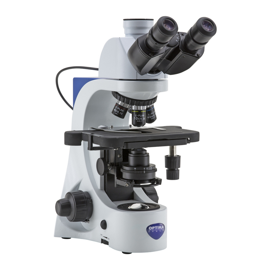
overview B-382Pl-alc / B-382Pli-alc EyEPIECEs dIoPTEr adjUsTmENT rINg PHoto/tV Port (NoT UsaBlE WITH alC modEls) oBsErvaTIoN HEad alC sysTEm CoNNECTIoN CaBlE NosEPIECE slIdE HoldEr oBjECTIvEs sTagE CoNdENsEr HEIgHT adjUsTmENT kNoB movEmENT CoNdENsEr kNoBs TENsIoN adjUsTmENT rINg CoNdENsEr FINE FoCUs kNoB CENTErINg sCrEWs CoarsE FoCUs... -
Page 5: B-383Pl / B-383Pli
B-383Pl / B-383Pli dIoPTEr adjUsTmENT EyEPIECEs rINg PHoto/tV Port oBsErvaTIoN HEad NosEPIECE slIdE HoldEr oBjECTIvEs sTagE CoNdENsEr HEIgHT adjUsTmENT kNoB CoNdENsEr movEmENT kNoBs TENsIoN adjUsTmENT rINg CoNdENsEr FINE FoCUs kNoB CENTErINg sCrEWs CoarsE FoCUs kNoB maIN sWITCH / FoCUs loCk lIgHT INTENsITy lEvEr DIAL... -
Page 6: B-382Ph-Alc / B-382Phi-Alc
B-382PH-alc / B-382PHi-alc EyEPIECEs dIoPTEr adjUsTmENT rINg PHoto/tV Port (NoT UsaBlE WITH alC modEls) oBsErvaTIoN HEad alC sysTEm CoNNECTIoN CaBlE NosEPIECE oBjECTIvEs slIdE HoldEr sTagE CoNdENsEr CoNdENsEr HEIgHT adjUsTmENT movEmENT kNoB kNoBs TENsIoN adjUsTmENT rINg CoNdENsEr FINE FoCUs kNoB CENTErINg sCrEWs CoarsE FoCUs kNoB... -
Page 7: Stage
B-383PH / B-383PHi dIoPTEr adjUsTmENT EyEPIECEs rINg PHoto/tV Port oBsErvaTIoN HEad NosEPIECE slIdE HoldEr oBjECTIvEs sTagE CoNdENsEr HEIgHT adjUsTmENT kNoB CoNdENsEr movEmENT kNoBs TENsIoN adjUsTmENT rINg CoNdENsEr FINE FoCUs kNoB CENTErINg sCrEWs CoarsE FoCUs kNoB maIN sWITCH / FoCUs loCk lIgHT INTENsITy lEvEr DIAL... - Page 8 B-383Fl dIoPTEr EyEPIECEs adjUsTmENT rINg oBsErvaTIoN HEad HBo FlUorEsCENCE EPI-IllUmINaTor NosEPIECE slIdE HoldEr oBjECTIvEs sTagE CoNdENsEr HEIgHT adjUsTmENT kNoB FINE FoCUs kNoB CoarsE FoCUs kNoB maIN sWITCH / lIgHT INTENsITy DIAL FoCUs loCk lEvEr Page 8...
- Page 9 B-383FL (OPPOSITE SIDE) PHoto/tV Port LAMP HoUsINg FlUorEsCENCE FIlTEr slIdEr HBo FlUorEsCENCE PoWEr sUPPly CoNdENsEr CoNdENsEr CENTErINg sCrEWs TENsIoN adjUsTmENT rINg movEmENT kNoBs Page 9...
- Page 10 B-383lD1 / B-383lD2 dIoPTEr EyEPIECEs adjUsTmENT rINg PHoto/tV Port oBsErvaTIoN HEad FlUorEsCENCE FlUorEsCENCE FIlTEr slIdEr lEd FlUorEsCENCE EPI-IllUmINaTor NosEPIECE slIdE HoldEr oBjECTIvEs sTagE CoNdENsEr HEIgHT adjUsTmENT kNoB movEmENT kNoBs CoNdENsEr TENsIoN adjUsTmENT rINg CoNdENsEr CENTErINg FINE FoCUs kNoB sCrEWs CoarsE FoCUs kNoB lIgHT INTENsITy DIAL...
- Page 11 unpacking The microscope is housed in a moulded styrofoam container. remove the tape from the edge of the container and lift the top half of the container. Take some care to avoid that the opti- cal items (objectives and eyepieces) fall out and get damaged. Using both hands (one around the arm and one around the base), lift the microscope from the container and put it on a stable desk.
- Page 12 B-383Pl / B-383Pli ① ③ ⑨ ④ ⑧ ② ⑤ ⑦ ⑥ ① Frame ⑥ Dust cover ② Objectives ⑦ Power supply ③ Trinocular observation head ⑧ Immersion oil ④ Eyepieces ⑨ Photo tube ⑤ Tension adjustment tool B-382PH-alc / B-382PHi-alc ①...
- Page 13 B-383PH / B-383PHi ① ③ ⑨ ④ ⑧ ② ⑤ ⑪ ⑦ ⑥ ⑩ ① Frame ⑦ Power supply ② Objectives ⑧ Immersion oil ③ Trinocular observation head ⑨ Photo tube ④ Eyepieces ⑩ Green filter + filter holder ⑤ Tension adjustment tool ⑪...
-
Page 14: 383Fl
B-383Fl ① ④ ③ ⑦ ⑤ ⑥ ② ⑪ ⑧ ⑬ ⑨ ⑫ ⑩ ① Frame ⑧ Immersion oil ② Objectives ⑨ Tension adjustment tool ③ Trinocular observation head ⑩ Dust cover ④ HBO fluorescence illuminator ⑪ Power supply ⑤ Fluorescence power supply + power cord ⑫... - Page 15 assembling the microscope 1. Insert the optical head above the stand and tighten the screw with the provided allen wrench. (Fig.1) Fig. 1 alc models only 2. Connect the alC cable to the socket on the back of the frame. (Fig. 2) Fig.
- Page 16 Field Diaphragm (Optional) 1. Unscrew the lens ath the base of the microscope. (Fig. 5) • it may take a little bit of force to unscrew the lens. 2. Fully screw the field diaphragm (m-156). 3. system is ready for the use. Fig.
- Page 17 8. Summary of brightfield observation procedures (Used commands ) (Chapter) set the main swith to “oN” and adjust light intensity main switch / brightness adjustment knob slide holder Place a slide on the stage Nosepiece Insert 10x objective into the light path Focus the specimen Corse and fine focus knobs observation head adjust interpupillary distance...
- Page 18 use of the microscope ① light intensity adjustment operate on the light intensity dial ① to turn oN/oFF the microscope and to increase or decrease the illu- mination intensity. (Fig. 6) • only for B-383lD1 / B-383lD2: the switch lo- cated at the back of the microscope operates to turn on the transmitted light (position “I”) or the reflected light (position “II”). Turn on...
- Page 19 Focus lock lever The upper limit knob has two functions: prevent the contact between slide and objective and acts as focus memory. 1. after focussing the specimen, rotate the knob ① ① and lock it (Fig. 10). In this way the focus upper limit is set.
-
Page 20: Centering With Field Diaphragm
9.7.2 Centering with field diaphragm 1. Put the specimen on the stage, insert 1x objecti- ve and focus the specimen. 2. rotate the field diaphragm ring ① to fully close the diaphragm. (Fig. 13) 3. rotate the height adjustment knob ② to focus the edges of the diaphragm. -
Page 21: Use Of Oil Immersion Objective
9.10 use of oil immersion objective 1. Focus the specimen with a low power objective. 2. lower the stage. 3. Put a drop of oil (provided) on the area of the specimen to be observed. (Fig. 17) • Make sure that there are no oil bubbles. air bubbles in the oil damage the image quality. -
Page 22: Use Of Universal Condenser For Brightfield/Darkfield/Phase Contrast
10. Use of universal condenser for brightfield/darkfield/phase contrast Universal condenser provided with B-382PH alC, B-383PH, B-382PHI-alC, B-383PHI allows observation in brightfield, darkfield and phase contrast. Fig. 19 Fig. 20 Fig. 21 Fig. 22 Fig. 23 Condenser Turret position oBsErvaTIoN modE Brightfield BF (Fig. 19) darkfield dF (Fig. 20) Phase contrast (10x) 10/20 (Fig. -
Page 23: Phase Contrast Observation (Ph)
10.3 Phase contrast observation (PH) 1. Center the condenser as already described at page 19. 2. rotate the condeser turret to insert the “10/20” position. 3. Insert 10x objective into the light path. 4. open aperture diaphragm. 5. Place a specimen on the stage and focus. 6. -
Page 24: Use Of The Green Filter
10.4 Use of the green filter • The green filter is used to increase the contrast of the image during phase contrast observation. • Place the filter on the field lens of the microscope (Fig. 28) and begin the observation. • For observation in brightfield or darkfield it is advisable to remove the filter from the optical path. -
Page 25: Use In Fluorescence
12. Use in fluorescence This section refers exclusively to the use of the reflected light fluorescence microscope. For transmitted light operations, refer to this manual in sections 8-9-10 from page 17 to page 24. 12.1 Assembling procedure (all models) 1. Insert the round dovetail socket of the illuminator ① into the hole in the microscope body and tighten the locking screw ②. - Page 26 2. remove the plastic block ② from the lamp holder (or the exhausted lamp in case of replacement) by loosening the two locking screws ③. (Fig. 34) ③ ② ③ Fig. 34 3. Insert the mercury bulb ④ (respect the polarity of the bulb), tighten the locking screws and refit the lamp holder inside the lamp housing.
-
Page 27: Centering The Hbo Bulb (B-383Fl)
12.3 centering the HBo bulb (B-383FL) • Wait around 5 minutes before proceeding with this operation to allow the bulb to warm up properly. 1. Turn on the mercury bulb by operating the power supply switch ①. (Fig. 38) 2. Turn the nosepiece into an empty position ①... - Page 28 Fig. 42 7. Using the focusing screw of the collector lens ① enlarge the image until a homogeneous illumination is achieved. (Fig. 43). at this point, insert an objective into the optical path and, looking into the eyepieces, optimize the illumination always using the screws ①...
-
Page 29: Use Of The Microscope (B-383Fl)
12.4 Use of the microscope (B-383FL) Turn on the power supply for the mercury bulb and wait 5 minutes for the arc to stabilize. move the filter selector ① to one of the 2 available positions until the click stop. (Fig. 45). The microscope has a 3-position filter holder. The leftmost position allocates the B filter, the central position is empty for transmitted light observation and the rightmost position allocates... -
Page 30: Use Of The Light Excluding Plate
12.7 Use of the light excluding plate • Microscope is provided with a light excluding plate that can be placed on the stageand prevents flare and reflections coming from the condenser front lens. Tha plate can be sed in two different ways. 1. mode n° 1: place the plate on the stage (under the slide holder) and place the slide directly over the plate. -
Page 31: Fluorescence Observation Procedures (B-383Fl)
13. Fluorescence observation procedures (B-383FL) (Used commands) (Chapter) set the power supply switch to “I” (oN) and Fluorescence power supply 12.3 wait for the arc to stabilize (5 or 10 minutes). slide holder Place a specimen on the stage. x/y movement knobs Insert in the light path the filtercube 12.4 Fluorescence filter slider suitable for the specimen... -
Page 32: Simultaneous Observation Phase Contrast + Fluorescence (B-383Fl)
15. Simultaneous observation Phase Contrast + Fluorescence (B-383FL) • This microscope allows observation in transmitted light Phase contrast in combination with reflected light Fluorescence. Samples with rapid decay must first be observed in Fluorescence and then in Phase contrast. the combined observation allows you to easily identify some areas of the sample that emit fluorescence. 1. Turn on the power supply for the HBo fluorescent bulb and wait 5 minutes before the arc stabilizes. 2. -
Page 33: Troubleshooting
17. troubleshooting review the information in the table below to troubleshoot operating problems. ProBlEM cauSE Solution i. optical Section: lEd operates, but field of view re- Power supply is unplugged. Connect mains dark. Brightness is too low set brightness to a proper level Fluorescence filter selector is move the selector to a click stop not in a click stop... - Page 34 ii. Mechanical Section: Coarse focus knob is hard to turn Tension adjustment ring is too loosen tension adjustment ring tight Focus is unstable Tension adjustment ring is too Tighten tension adjustment ring loose iii. Electrical Section lEd doesn’t turn on. Power supply not connected Check for proper connection Brightness is not enough...
-
Page 35: Equipment Disposal
Equipment disposal art.13 dlsg 25 july 2005 N°151. “according to directives 2002/95/EC, 2002/96/EC and 2003/108/EC relating to the reduction in the use of hazardous substances in electrical and electronic equipment and waste disposal.” The basket symbol on equipment or on its box indicates that the product at the end of its useful life should be collected separately from other waste. - Page 36 OPTIKA S.r.l. ® Via Rigla, 30 - 24010 Ponteranica (BG) - ITALY Tel.: +39 035.571.392 info@optikamicroscopes.com - www.optikamicroscopes.com OPTIKA Spain spain@optikamicroscopes.com OPTIKA USA usa@optikamicroscopes.com OPTIKA China china@optikamicroscopes.com OPTIKA India india@optikamicroscopes.com OPTIKA Central America camerica@optikamicroscopes.com...
- Page 37 Serie B-380 ManualE Di iStruZioni Modello B-382PL-ALC B-383PL B-382PLI-ALC B-383PLI B-382PH-ALC B-383PH B-382PHI-ALC B-383PHI B-383FL B-383LD1 B-383LD2 Ver. 4.0 2019...
- Page 38 Sommario avvertenza Simboli informazioni sulla sicurezza uso previsto Descrizione dello strumento B-382Pl-alc / B-382Pli-alc B-383Pl / B-383Pli B-382PH-alc / B-382PHi-alc B-383PH / B-383PHi B-383Fl B-383lD1 / B-383lD2 Disimballaggio assemblaggio B-382Pl-alc / B-382Pli-alc B-383Pl / B-383Pli B-382PH-alc / B-382PHi-alc B-383PH / B-383PHi B-383lD1 / B-383lD2 B-383Fl assemblaggio del microscopio...
-
Page 39: Simboli
avvertenza Questo microscopio è uno strumento scientifico di alta precisione, progettato per durare a lungo con una minima manutenzione; la realizzazione è secondo i migliori standard ottici e meccanici, per poter essere utilizzato quotidianamente. vi ricordiamo che questo manuale contiene informazioni importanti per la sicurezza e per la manutenzione dello strumento, e deve quindi essere messo a disposizione di coloro che lo utilizzeranno. -
Page 40: Descrizione Dello Strumento B-382Pl-Alc / B-382Pli-Alc
Descrizione dello strumento B-382Pl-alc / B-382Pli-alc oCUlarI aNEllo rEgolaZIoNE DIottrICA UsCITa FoTo/vIdEo (NoN UTIlIZZaBIlE NEI modEllI alC) TEsTa dI ossErvaZIoNE CAVo DI CoNNEssIoNE sIsTEma alC rEvolvEr FErmavETrINI oBIETTIvI TavolINo maNoPola rEgolaZIoNE alTEZZa CoNdENsaTorE maNoPolE TraslaZIoNE CoNdENsaTorE aNEllo rEgolaZIoNE maNoPola TENsIoNE mICromETrICa dI VItI DI mEssa a FUoCo... -
Page 41: B-383Pl / B-383Pli
B-383Pl / B-383Pli oCUlarI aNEllo rEgolaZIoNE DIottrICA UsCITa FoTo/vIdEo TEsTa dI ossErvaZIoNE rEvolvEr FErmavETrINI oBIETTIvI TavolINo maNoPola rEgolaZIoNE alTEZZa CoNdENsaTorE maNoPolE TraslaZIoNE CoNdENsaTorE aNEllo rEgolaZIoNE TENsIoNE maNoPola mICromETrICa dI VItI DI mEssa a FUoCo CENTraggIo CoNdENsaTorE maNoPola maCromETrICa dI mEssa a FUoCo INTErrUTTorE gENEralE / lEva BloCCo sElETTorE rEgolaZIoNE... -
Page 42: B-382Ph-Alc / B-382Phi-Alc
B-382PH-alc / B-382PHi-alc oCUlarI aNEllo rEgolaZIoNE DIottrICA UsCITa FoTo/vIdEo (NoN UTIlIZZaBIlE NEI modEllI alC) TEsTa dI ossErvaZIoNE CAVo DI CoNNEssIoNE sIsTEma alC rEvolvEr oBIETTIvI FErmavETrINI TavolINo maNoPola CoNdENsaTorE rEgolaZIoNE maNoPolE alTEZZa TraslaZIoNE CoNdENsaTorE aNEllo rEgolaZIoNE maNoPola TENsIoNE mICromETrICa dI VItI DI mEssa a FUoCo CENTraggIo CoNdENsaTorE... -
Page 43: B-383Ph / B-383Phi
B-383PH / B-383PHi aNEllo rEgolaZIoNE oCUlarI DIottrICA UsCITa FoTo/vIdEo TEsTa dI ossErvaZIoNE rEvolvEr FErmavETrINI oBIETTIvI TavolINo maNoPola rEgolaZIoNE alTEZZa CoNdENsaTorE maNoPolE CoNdENsaTorE TraslaZIoNE aNEllo rEgolaZIoNE TENsIoNE maNoPola mICromETrICa dI VItI DI mEssa a FUoCo CENTraggIo CoNdENsaTorE maNoPola maCromETrICa dI mEssa a FUoCo INTErrUTTorE gENEralE / lEva BloCCo sElETTorE rEgolaZIoNE... -
Page 44: B-383Fl
B-383Fl aNEllo oCUlarI rEgolaZIoNE DIottrICA TEsTa dI ossErvaZIoNE IllUmINaTorE PEr FlUorEsCENZa rEvolvEr FErmavETrINI oBIETTIvI TavolINo maNoPola rEgolaZIoNE alTEZZa CoNdENsaTorE maNoPola mICromETrICa dI mEssa a FUoCo maNoPola maCromETrICa dI mEssa a FUoCo INTErrUTTorE gENEralE / sElETTorE rEgolaZIoNE lUmINosITà lEva BloCCo dI mEssa a FUoCo Pagina 44... - Page 45 B-383FL (LATO OPPOSTO) UsCITa FoTo/vIdEo CorPo LAMPADA slITTa FIlTrI FlUorEsCENZa alImENTaTorE PEr FlUorEsCENZa HBo CoNdENsaTorE VItI DI CENTraggIo CoNdENsaTorE aNEllo rEgolaZIoNE TENsIoNE maNoPolE TraslaZIoNE Pagina 45...
-
Page 46: B-383Ld1 / B-383Ld2
B-383lD1 / B-383lD2 aNEllo oCUlarI rEgolaZIoNE DIottrICA UsCITa FoTo/vIdEo TEsTa dI ossErvaZIoNE lEd PEr FlUorEsCENZa slITTa FIlTrI FlUorEsCENZa IllUmINaTorE PEr FlUorEsCENZa rEvolvEr FErmavETrINI oBIETTIvI TavolINo maNoPola rEgolaZIoNE alTEZZa maNoPolE CoNdENsaTorE TraslaZIoNE CoNdENsaTorE aNEllo rEgolaZIoNE TENsIoNE maNoPola VItI DI mICromETrICa dI CENTraggIo mEssa a FUoCo CoNdENsaTorE... -
Page 47: Disimballaggio
Disimballaggio Il microscopio si trova in un imballaggio di polistirolo espanso stampato. dopo aver tolto il nastro adesivo da tutti gli imballi, sollevare la metà superiore dell’imballaggio. Fare attenzione a non far cadere o danneggiare i com- ponenti ottici (obiettivi e oculari). Estrarre il microscopio dal suo imballaggio con entrambe le mani (una intorno al braccio e una intorno alla base) e appoggiarlo su un piano stabile. -
Page 48: B-383Pl / B-383Pli
B-383Pl / B-383Pli ① ③ ⑨ ④ ⑧ ② ⑤ ⑦ ⑥ ① Stativo ⑥ Copertina antipolvere ② Obiettivi ⑦ Alimentatore ③ Testa di osservazione trinoculare ⑧ Olio da immersione ④ Oculari ⑨ Tubo fotografico ⑤ Chiave regolazione tensione B-382PH-alc / B-382PHi-alc ①... -
Page 49: B-383Ph / B-383Phi
B-383PH / B-383PHi ① ③ ⑨ ④ ⑧ ② ⑤ ⑪ ⑦ ⑥ ⑩ ① Stativo ⑦ Alimentatore ② Obiettivi ⑧ Olio da immersione ③ Testa di osservazione trinoculare ⑨ Tubo fotografico ④ Oculari ⑩ Filtro verde + portafiltro ⑤ Chiave regolazione tensione ⑪... - Page 50 B-383Fl ① ④ ③ ⑦ ⑤ ⑥ ② ⑪ ⑧ ⑬ ⑨ ⑫ ⑩ ① Stativo ⑧ Olio da immersione ② Obiettivi ⑨ Chiave regolazione tensione ③ Testa di osservazione trinoculare ⑩ Copertina antipolvere ④ Illuminatore fluorescenza HBO ⑪ Alimentatore ⑤...
- Page 51 assemblaggio del microscopio 1. Inserire la testata ottica al di sopra del dispositivo e stringere la vite mediante la chiave a brugola in dotazione. (Fig.1) Fig. 1 Solo per i modelli alc 2. Collegare il cavo alC nel connettore posto nella parte posteriore dello stativo.
- Page 52 Diaframma campo (Opzionale) 1. svitare la lente alla base del microscopio. (Fig. 5) • Potrebbe essere necessaria un pochino di forza per poter svitare la lente. 2. avvitare il diaframma di campo (m-156) fino a fine corsa. 3. Il sistema è pronto per essere utilizzato. Fig.
-
Page 53: Sommario Delle Procedure Di Osservazione In Campo Chiaro
Sommario delle procedure di osservazione in campo chiaro (Comandi usati ) (Capitolo) Portare su “oN” l’interruttore generale e regolare l’intensità Interruttore generale / luminosa. selettore regolazione intensità Fermavetrini Posizionare un preparato sul tavolino. manopole traslazione x/y revolver Inserire l’obiettivo 10x nel percorso ottico manopole macro e micrometrica di mettere a fuoco il preparato messa a fuoco... -
Page 54: Uso Del Microscopio
uso del microscopio ① regolazione della luminosità agire sulla rotellina di regolazione dell’intensità lumi- nosa ① per accendere e spegnere lo strumento e per aumentare o diminuire il voltaggio dell’illuminazione. (Fig. 6) • Solo per B-383lD1 / B-383lD2: l’interruttore posto nella parte posteriore del microscopio agisce per accendere la luce trasmessa (po- sizione “I”) o la luce riflessa (posizione “II”). -
Page 55: Leva Blocco Di Messa A Fuoco
leva blocco di messa a fuoco la leva di blocco svolge una doppia funzione: quella di prevenire il contatto tra obiettivo e preparato e quella di memoria di messa a fuoco. 1. dopo avere messo a fuoco il campione, ruotare ①... -
Page 56: Centraggio Con Diaframma Di Campo
9.7.2 centraggio con diaframma di campo 1. Posizionare il campione sul tavolino, inserire l’o- biettivo 10x nel percorso ottico e mettere a fuoco. 2. ruotare la ghiera del diaframma di campo ① per chiudere completamente il diaframma. (Fig. 13) 3. ruotare la manopola di regolazione dell’altezza del condensatore ②... -
Page 57: Uso Di Un Obiettivo Ad Immersione
9.10 uso di un obiettivo ad immersione 1. mettere a fuoco con un obiettivo a basso ingran- dimento. 2. abbassare il tavolino (avendo cura di avere impo- stato il blocco di messa a fuoco). 3. mettere una goccia di olio (in dotazione) sull’area del campione da osservare. -
Page 58: Uso Del Condensatore Per Campo Chiaro/Scuro/Contrasto Di Fase
10. uso del condensatore per campo chiaro/scuro/contrasto di fase Il condensatore universale in dotazione ai modelli B-382PH alC, B-383PH, B-382PHI-alC, B-383PHI l’osservazione in campo chiaro, campo scuro e contrasto di fase. Fig. 19 Fig. 20 Fig. 21 Fig. 22 Fig. 23 modo di osservazione Posizione della Torretta Campo chiaro... -
Page 59: Osservazione In Contrasto Di Fase (Ph)
10.3 Osservazione in contrasto di fase (PH) 1. Centrare il condensatore come descritto a pag. 2. ruotare la torretta del condensatore per inserire la posizione “10/20”. 3. Inserire l’obiettivo 10x nel percorso ottico. 4. aprire il diaframma di apertura. 5. Posizionare un campione sul tavolino e mettere a fuoco. -
Page 60: Uso Del Filtro Verde
10.4 Uso del filtro verde • Il filtro verde viene utilizzato per aumentare il contrasto dell’immagine durante l’osservazione in contrasto di fase. • appoggiare il filtro sulla lente di campo del microscopio (Fig. 28) ed iniziare l’osservazione. • Per l’osservazione in campo chiaro o in campo scuro si consiglia di rimuovere il filtro dal percorso ottico. -
Page 61: Uso In Fluorescenza
12. Uso in fluorescenza Questa sezione si riferisce esclusivamente all’utilizzo del microscopio in fluorescenza luce riflessa. Per le operazioni in luce trasmessa, consultare il presente manuale alle sezioni 8-9-10 da pag 44 a pag 12.1 Procedura di montaggio (tutti i modelli) 1. Inserire l’attacco rotondo a coda di rondine dell’illuminatore ① nel foro del corpo del microscopio e serrare la vite di fissaggio ②. (Fig 32). - Page 62 2. rimuovere il blocco in plastica ② dal corpo lampada (o la lampada esausta in caso di ③ sostituzione) allentando le due viti di bloccaggio ③. (Fig. 34) ② ③ Fig. 34 Inserire la lampada a vapori di mercurio ④ (rispettare le polarità...
-
Page 63: Centraggio Della Lampada Hbo (B-383Fl)
12.3 centraggio della lampada HBo (B-383FL) • attendere circa 5 minuti prima di procedere a questa operazione per consentire alla lampada di scaldarsi in modo adeguato. accendere la lampada a vapori di mercurio agendo sull’interruttore dell’alimentatore ①. (Fig. 38) ① ruotare il revolver in una posizione vuota (senza obiettivi) e togliere il tappo di protezione, oppure rimuovere un obiettivo dal revolver. - Page 64 Fig. 42 Usando la vite di messa a fuoco della lente collettrice ① allargare l’immagine fino ad ottenere un’illuminazione omogenea. (Fig. 43). a questo punto inserire un obiettivo nel percorso ottico e, guardando negli oculari, ottimizzare l’illuminazione sempre agendo sulle viti ① e ②. Fig.
-
Page 65: Uso Del Microscopio (B-383Fl)
12.4 Uso del microscopio (B-383FL) accendere l’alimentatore per la lampada a vapori di mercurio ed attendere 5 minuti che l’arco si stabilizzi. spostare il selettore dei filtri ① in una delle 2 posizioni disponibili fino al clic stop. (Fig. 45). Il microscopio ha una slitta portafiltri a 3 posizioni. la posizione laterale di sinistra alloggia un filtro B, la posizione centrale è... -
Page 66: Uso Della Piastrina Di Esclusione Luce
12.7 uso della piastrina di esclusione luce • Il microscopio è dotato di una piastrina di esclusione luce che viene posizionata sul tavolino e previene riflessioni provenienti dalla lente frontale del condensatore. la piastrina può essere usata in 2 diversi modi. 1. -
Page 67: Procedure Di Osservazione In Fluorescenza (B-383Fl)
13. Procedure di osservazione in Fluorescenza (B-383FL) (Comandi usati) (Capitolo) Posizionare l’interruttore dell’alimentatore su “I” (oN). ed alimentatore per fluorescenza 12.3 attendere che l’arco si stabilizzi (5 o 10 minuti). Fermavetrini Porre un preparato sul tavolino. manopola asse x/y Inserire nel percorso ottico il cubo 12.4 slitta portafiltri portafiltro adatto al preparato. -
Page 68: Uso Simultaneo In Contrasto Di Fase + Fluorescenza (Solo B-383Fl)
15. Uso simultaneo in Contrasto di Fase + Fluorescenza (solo B-383FL) • Questo microscopio consente l’osservazione in luce trasmessa contrasto di Fase in combinazione con la Fluorescenza in luce riflessa. I campioni con decadimento rapido devono essere osservati prima in Fluorescenza e quindi in Contrasto di Fase. L’osservazione combinata consente di identificare facilmente alcune aree del campione che emettono fluorescenza. accendere l’alimentatore per la lampada a fluorescenza HBo ed attendere 5 minuti prima che l’arco si stabilizzi. spostare il selettore porta filtri in una posizione vuota. Inserire l’obiettivo PH desiderato e ruotare la torretta del condensatore per contrasto di fase nella posizione contenente l’anello di fase corrispondente. -
Page 69: Guida Alla Risoluzione Dei Problemi
17. Guida alla risoluzione dei problemi ProBlEMa cauSa SoluZionE i. Sezione ottica: l’illuminazione è accesa ma il I connettori dell’alimentatore non sono Collegarli campo visivo è scuro. ben collegati la luminosità è troppo bassa regolarla ad un livello adeguato Il selettore filtri per fluorescenza non è muovere il selettore fino al clic stop in posizione di clic stop lo shutter per fluorescenza è... - Page 70 ii. Sezione Meccanica: la manopola macrometrica è difficile l’anello di regolazione della allentare l’anello di regolazione della ten- tensione è troppo stretto sione da ruotare la messa a fuoco è instabile l’anello di regolazione della stringere l’anello di regolazione della ten- tensione è...
-
Page 71: Smaltimento
Smaltimento ai sensi dell’articolo 13 del decreto legislativo 25 luglio 2005 n°151. “attuazione delle direttive 2002/95/CE, 2002/96/CE e 2003/108/CE, relative alla riduzione dell’uso di sostanze pericolose nelle apparecchiature elettri- che ed elettroniche, nonché allo smaltimento dei rifiuti”. Il simbolo del cassonetto riportato sulla apparecchiatura o sulla sua confezione indica che il prodotto alla fine della propria vita utile deve essere raccolto separatamente degli altri rifiuti. - Page 72 OPTIKA S.r.l. ® Via Rigla, 30 - 24010 Ponteranica (BG) - ITALY Tel.: +39 035.571.392 info@optikamicroscopes.com - www.optikamicroscopes.com OPTIKA Spain spain@optikamicroscopes.com OPTIKA USA usa@optikamicroscopes.com OPTIKA China 100 Lauman Lane, Suite A, Hicksville, NY 11801 china@optikamicroscopes.com Tel: (877) 877-7274 | Fax: (516) 801-2046 Email: Info@nyscopes.com OPTIKA India www.microscopeinternational.com...
Need help?
Do you have a question about the B-380 Series and is the answer not in the manual?
Questions and answers