Advertisement
Quick Links
Advertisement

Related Manuals for Zeiss LSM 710


Summary of Contents for Zeiss LSM 710
- Page 1 cÉÄêì~êó=OMNM= wbk=OMNM=...
- Page 2 INTRODUCTION LSM 710 and LSM 780 Carl Zeiss Systems Knowledge of this manual is required for the operation of the instrument. Would you therefore please make yourself familiar with the contents of this manual and pay special attention to hints concerning the safe operation of the instrument.
- Page 3 INTRODUCTION Systems Carl Zeiss How to make best use of the LSM 710, LSM 780, LSM 710 NLO and LSM 780 NLO operating instructions: This operating manual also includes the instructions for the ConfoCor 3 systems. A few symbols in these operating instructions will help you to recognize the nature and purpose of...
- Page 4 ConfoCor 3 Microscope Systems, together with the relevant specifications. Introduction to Laser Scanning Microscopy This section contains the "LSM 710, LSM 780 systems and ConfoCor 3 Quick Guide", the "Confocal Laser Scanning Microcopy Principles", and the "Confocal Laser Scanning Microcopy Methods" brochures.
- Page 5 Mounting and Dismounting the LSM BiG ................. 30 1.11.3 Mounting and Dismounting the Scan Heads ..............31 1.11.4 Changing the Filter Wheel in the LSM 710 and LSM 780 systems........34 1.11.5 Changing Filter Wheels in the ConfoCor 3 ............... 35 1.11.6 Changing the Filter Modules in the LSM BiG ..............
- Page 6 3B. The systems are equipped with safety interlocks that comply with laser hazard class 3B and 4. If equipped with a Ti:Sa laser (see list in section 1.8), the LSM 710 and LSM 780 systems and ConfoCor 3 are devices that belong to laser hazard class 4. WHO recommendations concerning health and industrial protection when handling laser devices must be observed.
- Page 7 The LSM 710 and LSM 780 systems and ConfoCor 3 have been designed, built and tested in conformity with the following regulations and guidelines: −...
- Page 8 Systems Power Requirements The LSM 710 and LSM 780 systems and ConfoCor 3 come with a mains power supply cord and plug, either CEE red (3/N/PE 400/230V/16A), or NEMA L 14-30P (2/N/Ground 120/240V/30A), and with the matching mains socket outlet.
- Page 9 Power Requirements Carl Zeiss Fig. 2 Power connector for LSM 710 and LSM 780 systems and components. Free/reserve outlets may be used to supply power to additional equipment. No more than 1 A can be provided by each outlet. 02/2010...
- Page 10 Systems Fig. 3 Power connector for LSM 710 and LSM 780 systems with Laser In Tune and components. Free/reserve outlets may be used to supply power to additional equipment. No more than 1 A can be provided by each outlet.
- Page 11 Door interlock interface (1) on the back of the electronic rack (see Fig. 2 and Fig. 3, top) The LSM 710 and LSM 780 systems are controlled by a remote control. This remote control contains the main switch for the system and the key switch for the laser.
- Page 12 Large actively damped system table Active anti-vibration table (NLO) for Mai Tai Laser or Chameleon Active anti-vibration table (NLO) for two-microscope configuration Scanning Module LSM 710, LSM 780 Scanning Module LSM DuoScan Module ConfoCor 3 Microscope Electronic Rack with laser units 710...
- Page 13 ConfoCor 3 spectroscope are delivered in several crates. The LSM 710 and LSM 780 systems and ConfoCor 3 must be set up so as to ensure that the minimum clearance between the wall and the rear of the system is no less than 0.5 m. This clearance is needed for adjustment and maintenance operations.
- Page 14 By establishing a corresponding workplace environment, please ensure that the formation of electrostatic charges of electronic components is avoided. To avoid vibrations during operation, the LSM 710 and LSM 780 systems and ConfoCor 3 should only be operated in conjunction with the system table (vibration damping).
- Page 15 Carl Zeiss Warning and Information Labels The warning and information labels attached on the LSM 710 and LSM 780 systems and ConfoCor 3 must be observed. Check whether all of the labels shown below are provided on your instrument, and contact Carl Zeiss Germany or one of the service agencies if you should discover that any of the labels should be missing.
- Page 16 NOTES ON DEVICE SAFETY LSM 710 and LSM 780 Carl Zeiss Warning and Information Labels Systems Fig. 6 Warning and information labels on the Axio Observer.Z1 microscope with the LSM 710 / LSM 780 scanning module M60-1-0025 e 02/2010...
- Page 17 LSM 710 and LSM 780 NOTES ON DEVICE SAFETY Systems Warning and Information Labels Carl Zeiss Fig. 7 Warning and information labels on the Axio Observer.Z1 microscope with the LSM 710 / LSM 780 scanning module (US version only) 02/2010 M60-1-0025 e...
- Page 18 LSM 710 and LSM 780 Carl Zeiss Warning and Information Labels Systems Fig. 8 Warning and information labels on the Axio Observer.Z1 microscope with the LSM 710 and LSM 780 scanning module and the ConfoCor 3 detection module M60-1-0025 e 02/2010...
- Page 19 NOTES ON DEVICE SAFETY Systems Warning and Information Labels Carl Zeiss Fig. 9 Warning and information labels on the Axio Observer.Z1 microscope with the LSM 710 / LSM 780 scanning module and the ConfoCor 3 detection module (US version only) 02/2010 M60-1-0025 e...
- Page 20 NOTES ON DEVICE SAFETY LSM 710 and LSM 780 Carl Zeiss Warning and Information Labels Systems Fig. 10 Warning and information labels on the Axio Imager.Z2 microscope with the LSM 710 / LSM 780 scanning module M60-1-0025 e 02/2010...
- Page 21 LSM 710 and LSM 780 NOTES ON DEVICE SAFETY Systems Warning and Information Labels Carl Zeiss Fig. 11 Warning and information labels on the Axio Imager.Z2 microscope with the LSM 710 / LSM 780 scanning module (US version only) 02/2010 M60-1-0025 e...
- Page 22 NOTES ON DEVICE SAFETY LSM 710 and LSM 780 Carl Zeiss Warning and Information Labels Systems Fig. 12 Warning and information labels on the Axio Examiner microscope with the LSM 710 / LSM 780 scanning module M60-1-0025 e 02/2010...
- Page 23 LSM 710 and LSM 780 NOTES ON DEVICE SAFETY Systems Warning and Information Labels Carl Zeiss Fig. 13 Warning and information labels on the Axio Examiner microscope with the LSM 710 / LSM 780 scanning module (US version only) 02/2010 M60-1-0025 e...
- Page 24 NOTES ON DEVICE SAFETY LSM 710 and LSM 780 Carl Zeiss Warning and Information Labels Systems Fig. 14 Warning and information labels on the laser and electronic rack Fig. 15 Warning and information labels on the laser and electronic rack (US version only)
- Page 25 LSM 710 and LSM 780 NOTES ON DEVICE SAFETY Systems Warning and Information Labels Carl Zeiss Fig. 16 Warning and information labels on the Laser Rack In Tune Fig. 17 Warning and information labels on the Laser Rack In Tune (US version only)
- Page 26 Caution: Use of controls or adjustments or performance of procedures other than those specified herein may result in hazardous radiation exposure. The following laser types are currently intended for use in the LSM 710 and LSM 780 systems. The use of any other lasers as the ones listed below is not authorized.
- Page 27 Power specified for single laser lines, life time specified for total power Average power for cw operation If used properly, the LSM 710 and LSM 780 systems and ConfoCor 3 will not pose any laser radiation risks for operating staff. Nevertheless, you should observe the following warnings: •...
- Page 28 NOTES ON DEVICE SAFETY LSM 710 and LSM 780 Carl Zeiss Notes on Handling the Laser Components … Systems When using a high power broadband illumination (HBO, X-Cite, HXP) in the incident light path in combination with a neutral density reflector module in the microscope stand, there are operating conditions which lead to strong reflections of excitation light into the eyepiece.
- Page 29 The LSM software is released with a specific windows version and patch and this must not be changed unless otherwise released by Carl Zeiss MicroImaging. 02/2010...
- Page 30 Any such action will render all warranty claims null and void and also laser safety is no longer warranted. You are well advised to arrange a service agreement with your nearest Carl Zeiss representative to guarantee perfect functioning of the microscope system in the long term.
- Page 31 LSM 710 and LSM 780 NOTES ON DEVICE SAFETY Systems Notes on Care, Maintenance and Service Carl Zeiss • Unlock the lamp housing (Fig. 18/1) by slightly turning it anticlockwise. • Remove the halogen lamp with alignment base (Fig. 18/3).
- Page 32 These interlock devices must not be manipulated. Other interfaces which are not described here are service interfaces and are only to be operated by authorized Carl Zeiss service personnel. The following devices can be mounted and dismounted by the user or are accessible by the user: −...
- Page 33 LSM 710 and LSM 780 NOTES ON DEVICE SAFETY Systems User Interface Carl Zeiss For dismounting the lamps, TPMT or switching mirror slightly unscrew the contact ring first (Fig. 20/2) so it can be moved away from the sensor ring (Fig. 19). Then unscrew the lamp, TPMT or switching mirror turning the main screw which is in one of the recesses of the sensor ring (Fig.
- Page 34 1.11.2 Mounting and Dismounting the LSM BiG Do not remove the laser safety cable (Fig. 22/1) from the LSM 710 / LSM 780 system. This might result in failure of laser safety and a non operating system. • Loosen the two fastening screws (Fig. 22/3) of the LSM BiG port (Fig. 22/2).
- Page 35 Moving the scan heads between Axio Observer.Z1, Axio Examiner and Axio Imager.Z2: • Loosen the three fastening screws of the LSM 710 / LSM 780 (Fig. 24/1 and Fig. 25/1). • Slowly pull the scanning module (Fig. 25/2) away from the microscope port or the tube (Fig. 25/3).
- Page 36 NOTES ON DEVICE SAFETY LSM 710 and LSM 780 Carl Zeiss User Interface Systems For NLO systems which are set up together with a Multiphoton laser, the beam tube connecting the periscope and the scan head, has to be removed before the scan head can be dismounted. Perform the following steps for the removal of the tube: •...
- Page 37 LSM 710 and LSM 780 NOTES ON DEVICE SAFETY Systems User Interface Carl Zeiss To ensure functioning of the system and laser safety the following connections have to be changed: 1. The connection of the microscope to the safety interface of the system is located either on the additional Safety Box (Axio Examiner, Fig.
- Page 38 You can remove and replace filter wheels by push and click. When opening the cover, the safety control will switch off the laser light. To exchange the filter wheel of the LSM 710 and LSM 780 systems proceed as follows: • Loosen the four fastening screws (Fig. 31/1).
- Page 39 Be careful not to disrupt any connectors in doing this. Do not remove the ConfoCor 3 detection head from its attachment to the LSM 710 / LSM 780. Laser light can escape the system through the external port used as the attachment site for the ConfoCor 3 that can lead to bodily damage.
- Page 40 NOTES ON DEVICE SAFETY LSM 710 and LSM 780 Carl Zeiss User Interface Systems 1.11.6 Changing the Filter Modules in the LSM BiG You can remove and replace filter modules by push and click. When opening the filter insert, the safety control will switch off the laser light.
- Page 41 LSM 710 and LSM 780 NOTES ON DEVICE SAFETY Systems User Interface Carl Zeiss 1.11.7 Using External Connectors In the rear of the APD module, you have external access to the TTL pulses of both channels delivered at the APD 1 and APD 2 plugs (see Fig. 36).
- Page 43 LSM 710 and LSM 780 equipped with Two Photon Laser (NLO) for single stand....3 2.1.4 LSM 710 and LSM 780 equipped with Two Photon Laser (NLO) for dual stand ....3 Power Requirements ......................4 Physical Dimensions ......................5 Dimension of Shipment Crates...................
- Page 44 The user PC and the power supply unit for the Ar laser are located beneath the table. If present, the laser Rack In Tune is located on top of the Laser Rack of the LSM 710 and LSM 780. The total height of this rack is then 1 m.
- Page 45 The user PC is located beneath the table. If present, the Laser Rack In Tune is located on top of the Laser Rack of the LSM 710 and LSM 780. The total height of these racks is then 1 m.
- Page 46 Systems Power Requirements The LSM 710 and LSM 780 systems and ConfoCor 3 come with a mains power supply cord and plug, either CEE red (3/N/PE 400/230V/16A), or NEMA L 14-30P (2/N/Ground 120/240V/30A), and with the matching mains socket outlet.
- Page 47 Large actively damped system table Active anti-vibration table (NLO) for Mai Tai Laser or Chameleon Active anti-vibration table (NLO) for two-microscope configuration Scanning Module LSM 710 / LSM 780 Module ConfoCor 3 Microscope Electronic Rack with laser units LSM 710 / LSM 780...
- Page 48 SETUP REQUIREMENTS LSM 710 and LSM 780 Carl Zeiss Dimension of Shipment Crates Systems Dimension of Shipment Crates Crate containing Length (cm) Width (cm) Height (cm) Weight (kg) Large system table Small system table Active anti-vibration table (NLO) for Mai Tai Laser or...
- Page 49 Upright Axio Examiner with LSM-tube or rear port Upright Axio Imager.Z2 with LSM-tube or rear port Upright Axio Imager.M2p with LSM-tube All IC S objectives from Carl Zeiss and their accessories can be accommodated. Z motor DC servomotor, opto-electronically coded...
- Page 50 2 channels with APD detectors Dynamic range 12-bit DAC for each detection channel Pinholes 1 individual pinhole (channel 4 of LSM 710, LSM 780, LSM 710 NLO and LSM 780 NLO) Computer controlled automatic alignment 2.10 Detection Module LSM BiG...
- Page 51 LSM 710 and LSM 780 SETUP REQUIREMENTS Systems Laser Insert Carl Zeiss 2.11 Laser Insert RGB (pigtailed; 458, 488, 514, 543, 561,594, 633 nm) Single-mode polarization preserving fiber Laser beam attenuation for all lasers by VIS-AOTF Ar laser (458, 488, 514 nm, 25 or 35 mW)
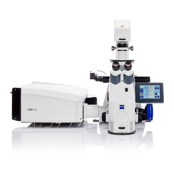
- Page 52 SETUP REQUIREMENTS LSM 710 and LSM 780 Carl Zeiss System Overview LSM 710 and LSM 780 Systems 2.14 System Overview LSM 710 and LSM 780 M60-1-0025 e 02/2010...
- Page 53 LSM 710 and LSM 780 SETUP REQUIREMENTS Systems System Overview LSM 710 and LSM 780 Carl Zeiss 02/2010 M60-1-0025 e...
- Page 54 SETUP REQUIREMENTS LSM 710 and LSM 780 Carl Zeiss System Overview LSM 710 and LSM 780 with ConfoCor 3 Systems 2.15 System Overview LSM 710 and LSM 780 with ConfoCor 3 M60-1-0025 e 02/2010...
- Page 55 LSM 710 and LSM 780 SETUP REQUIREMENTS Systems System Overview LSM 710 and LSM 780 with ConfoCor 3 Carl Zeiss 02/2010 M60-1-0025 e...
- Page 59 Switching off the system..................... 29 Introduction This Quick Guide describes the basic operation of the LSM 710, LSM 780, LSM 710 NLO, LSM 780 NLO and ConfoCor 3 Laser Scanning microscopes with the ZEN 2010 software. The purpose of this document is to guide the user to get started with the system as quick as possible in order to obtain some first images from his samples.
- Page 60 Starting the System Switching on the LSM system • Switch on the main switch (Fig. 1/1) and the safety lock (Fig. 1/2). • When set to ON the power remote switch labeled System/PC provides power to the computer. This allows use of the computer and ZEN software offline •...
- Page 61 Starting the ZEN software • Double click the ZEN 2010 icon on the WINDOWS desktop to start the Carl Zeiss LSM software. The ZEN Main Application window and the LSM 710 / LSM 780 Startup window appear on the screen (Fig. 3) Fig.
- Page 62 Fig. 4 ZEN Main Application Window after Startup with empty image container Fig. 5 ZEN Main Application Window after Startup with several images loaded 02/2010...
- Page 63 Both types of users will appreciate the set of intuitive tools designed to make the use of a confocal microscope from Carl Zeiss easy and fast: The Show all concept ensures that tool panels are never more complex than needed. With Show all de- activated, the most commonly used tools are displayed.
- Page 64 Fig. 7 ZEN Window Layout configuration More features of ZEN 2010 include: − The user can add more columns for tools to the Left Tool Area or detach individual tools to position them anywhere on the monitor. To add a column, drag a tool group by the title bar (e.g., Online Acquisition) to the right and a new tool column automatically opens.
- Page 65 The ZEN software will ask you if you want to save your unsaved images when you try to close the application with unsaved images still open. There is no image database any more like in the earlier Zeiss LSM software versions. Fig. 8...
- Page 66 Advanced data browsing is available through the ZEN File Browser (Ctrl + F or from the File Menu). The ZEN File Browser can be used like the WINDOWS program file browser. Images can be opened by double-click and image acquisition parameters are displayed with the thumbnails (Fig. 9). For more information on data browsing please refer to the detailed operating manual.
- Page 67 Turning on the lasers ZEN 2010 operates all lasers automatically. Whenever they are used (manually or by the Smart Setup function) the lasers are turned on automatically. The Laser Life Extender function of the software shuts all lasers off if ZEN is not used for more than 15 minutes. To manually switch lasers on or off: •...
- Page 68 Setting up the microscope Changing between direct observation and laser scanning mode The Ocular and Acquisition buttons switch between the use of the LSM and the microscope: • Click on the Ocular button to open the controls for the microscope beam path and for direct observation via the eyepieces of the binocular tube, lasers are blocked.
- Page 69 • Use the focusing drive of the microscope to focus the object plane. • Select specimen detail by moving the stage in X and Y using the XY stage fine motion control. Setting the microscope for reflected light • Click on the Reflected Light icon to open the X-Cite 120 Controls and turn it on.
- Page 70 Configuring the beam path and lasers • Click the Acquisition button. Smart Setup The tool Smart Setup is an intuitive, user-friendly interface which can be used for almost all standard applications. It configures all the system hardware for a chosen set of dyes. •...
- Page 71 Fig. 15 Proposals panel of the Smart Setup tool Pressing Apply, automatically sets the hardware parameters in the displayed way for the dyes chosen. Linear Unmixing If the option is selected, the system is set in the lambda mode automatically. Pressing the Auto Exposure button will then optimize the settings of the Gain (Master) and offset for the given laser power and pinhole size.
- Page 72 Setting up a configuration manually Simultaneous scanning of single, double and triple labeling: − Advantage: faster image acquisition − Disadvantage: potential cross-talk between channels Sequential scanning of double and triple labeling; line-by-line or frame-by-frame: − Advantage: Only one detector and one laser are switched on at any one time. This reduces cross- talk.
- Page 73 Settings for track configuration in Channel Mode • Select Channel Mode if necessary (Fig. 17). The Light Path tool displays the selected track configuration which is used for the scan procedure. • You can change the settings of this panel using the following function elements: Fig.
- Page 74 • For storing a new configuration click enter a desired name in the first line of the list box (Fig. 19), then click Ok to store the con- figuration. • For loading an existing configuration click then select it from the list box. •...
- Page 75 Scanning an image Setting the parameters for scanning • Select the Acquisition Mode tool from the Left Tool Area (Fig. 20). • Select the Frame Size as predefined number of pixels or enter your own values (e.g. 300 x 600) in the Acquisition Mode tool.
- Page 76 Setting scan averaging Averaging improves the image by increasing the signal-to-noise ratio. Averaging scans can be carried out line-by-line or frame-by-frame. Frame averaging helps to reduce photo-bleaching, but does not give quite as smooth of an image. • For averaging, select the Line or Frame mode in the Acquisition Mode tool. •...
- Page 77 Image acquisition Once you have set up your parameter as defined in the above section, you can acquire a frame image of your specimen. • Use one of the Auto Exposure, Live, Continuous or Snap buttons to start the scanning procedure to acquire an image. •...
- Page 78 The scanned image appears in a false-color presentation (Fig. 24). If the image is too bright, it appears red on the screen. Red = saturation (maximum). If the image is not bright enough, it appears blue on the screen. Blue = zero (minimum). Adjusting the laser intensity Fig.
- Page 79 Scanning a Z stack • Select Z-Stack in the main tools area. • Open the Z Stack tool in the Left Tool Area. • Select Mode First/Last on the top of the Z-Stack tool. • Click on the button in the Action Button area.
- Page 80 • The WINDOWS Save As window appears. • Enter a file name and choose the appropriate image format. Note: the LSM 5 format is the native Carl Zeiss LSM image data format and contains all available extra information and hardware settings of your experiment.
- Page 81 Using the ConfoCor 3 module • Click on the FCS button. • Use the ConfoCor Tool Group in the Left Tool Area to acquire and analyze FCS data. Fig. 30 ConfoCor Tool Group Setting a configuration • Open the System Configu- ration tool to define the light path, laser lines and pinhole position.
- Page 82 You can change the settings of this panel using the following function elements: Activation / deactivation of the excitation wavelengths (check box) and setting of excitation intensities (slider). Open the Laser Control tool via the Laser icon. Selection of the main dichroic beam splitter (MBS) or secondary dichroic beam splitter (SBS) position through selection from the relevant list box.
- Page 83 Starting a measurement • Select the Acquisition toolbar. The Times, Kinetics and Position panels of the Acquisition toolbar display selected Fig. 34 The ConfoCor 3 Measure Tool: measurement conditions and the positions which are Acquisition used for the FCS experiment. You can change the settings of this panel using the following function elements: Enter the Bleach Time, Measure Time and Repeat Count into the corres- ponding input boxes.
- Page 84 Fig. 35 FCS Correlation diagram You have the following function elements: Activate the FCS Correlation panel to display measured data (Fig. 35). Press the View Options button to define the graph you want to display. Press the Count rate button to display the count rate trace. Press the Correlation button to display the correlation function.
- Page 85 Pressing the Reuse button will set the system configuration to exactly the same values, as used in the experiment. Pressing the Reload button will open the current measurement, if stored raw data are available. This allows you to alter the parameters of your mathematical calculations. Analyzing the data The acquired FCS data is analyzed in the Fit display of the FCS diagram (see figure Fig.
- Page 86 You have the following options: Activate the FCS Fit panel to display fitted data (Fig. 36). Set the red and blue bars to define the start and end points of the curve fit window. Load a predefined model from the Model drop-down menu. You can assemble a model by pressing the Model tool in the ConfoCor tool group.
- Page 87 Switching off the system • Click on the File button in the Main Menu bar and then click on the Exit button to leave the ZEN 2010 software. • If any lasers are still running you should shut them off now in the pop-up window indicating the lasers still in use.
- Page 89 M i c r o s c o p y f r o m C a r l Z e i s s Principles Confocal Laser Scanning Microscopy Optical Image Formation Electronic Signal Processing...
- Page 90 Highlights of Laser Scanning Microscopy 1982 The first Laser Scanning Microscope from Carl Zeiss. The prototype of the LSM 44 series is now on display in the Deutsches Museum in Munich. 1988 The LSM 10 – a confocal system with two fluorescence channels.
- Page 91 Confocal Laser Scanning Microscopy In recent years, the confocal Laser Scanning Micro- scope (LSM) has become widely established as a research instrument. The present brochure aims at giving a scientifically sound survey of the special nature of image forma- tion in a confocal LSM. LSM applications in biology and medicine predomi- nantly employ fluorescence, but it is also possible to use the transmission mode with conventional con-...
- Page 92 Contents Introduction Part 1 Optical Image Formation Point Spread Function Resolution and Confocality Resolution Geometric optic confocality Wave-optical confocality Overview Part 2 Signal Processing Sampling and Digitization Types of A/D conversion Nyquist theorem Pixel size Noise Resolution and shot noise – resolution probability Possibilities to improve SNR Summary...
- Page 93 Following a description of the fundamental diffe- Image generation rences between a conventional and a confocal The complete generation of the two-dimensional microscope, this monograph will set out the object information from the focal plane (object plane) of a confocal LSM essentially comprises special features of the confocal LSM and the capa- bilities resulting from them.
- Page 94 The pinhole is arranged in front of the detector, on a plane conjugate to Carl Zeiss laser scanning microscopes. Configura- the focal plane of the objective. Light coming from planes above or below...
- Page 95 Optical slices With a confocal LSM it is therefore possible to A confocal LSM can therefore be used to advan- exclusively image a thin optical slice out of a thick tage especially where thick specimens (such as specimen (typically, up to 100 µm), a method biological cells in tissue) have to be examined by known as optical sectioning.
- Page 96 Introduction dimension Time series In addition to the possibility to observe a single A field of growing importance is the investigation plane (or slice) of a thick specimen in good con- of living specimens that show dynamic changes trast, optical sectioning allows a great number of even in the range of microseconds.
- Page 97 Point Spread Function In order to understand the optical performance characteristics of a confocal LSM in detail, it is ne- cessary to have a closer look at the fundamental optical phenomena resulting from the geometry of the confocal beam path. As mentioned before, what is most essential about a confocal LSM is that both illumination and observation (detection) are limited to a point.
- Page 98 Optical Image Formation Part 1 Any reference to the PSF in the following discus- is also influenced by all these factors and, sion exclusively refers to the half-maximum area. additionally, by the pinhole size. For reasons of Quantitatively the half-maximum area is described beam path efficiency (see Part 2), the pinhole is in terms of the full width at half maximum never truly a point of infinitely small size and thus...
- Page 99 Resolution and Confocality Wherever quantitative data on the resolving The smaller the pinhole diameter, the more PSF power and depth discrimination of a confocal LSM approaches the order of magnitude of PSF . In the are specified, it is necessary to distinguish clearly limit case (PH <...
- Page 100 Optical Image Formation Part 1 Axial: Resolution 0.88 . Resolution, in case of large pinhole diameters FWHM ill,axial (n- n (PH >1 AU), is meant to express the separate visi- bility, both laterally and axially, of points during = refractive index of immersion liquid, NA = numerical aperture of the microscope objective, the scanning process.
- Page 101 Let the statements made on PSF so far be further Optical axis illustrated by the figure on the left. It shows a sec- tion through the resulting diffraction pattern sur- rounding the focus on the illumination side (PSF ). The lines include areas of equal brightness (isophote presentation).
- Page 102 Optical Image Formation Part 1 Above a pinhole diameter of 1 AU, the influence Geometric optical confocality of diffraction effects is nearly constant and equa- Optical slice thickness (depth discrimination) and tion (4) is a good approximation to describe the stray light suppression (contrast improvement) are depth discrimination.
- Page 103 Thus, equations (2) and (3) for the widths of the Wave-optical confocality axial and lateral half-intensity areas are trans- If the pinhole is closed down to a diameter of formed into: < 0.25 AU (virtually “infinitely small”), the charac- ter of the image changes. Additional diffraction Axial: effects at the pinhole have to be taken into 0.64 .
- Page 104 Optical Image Formation Part 1 From equations (7) and (7a) it is evident that depth It must also be noted that with PH <1 AU, a dis- resolution varies linearly with the refractive index n tinction between optical slice thickness and resolu- of the immersion liquid and with the square of the tion can no longer be made.
- Page 105 Optical slice Fig. 11 (NA = 1.3; n = 1.52; a) Variation of = 496 nm) pinhole diameter Pinhole diameter [AU] 1000 Depth resolution b) Variation of (PH = 0; n = 1.52; numerical aperture = 496 nm) Numerical aperture Depth resolution c) Variation of (PH = 0;...
- Page 106 Optical Image Formation Part 1 Overview Confocal microscopy 1 AU < PH < ∞ Conventional microscopy Confocal microscopy PH < 0.25 AU 1. Optical slice thickness not definable 1. Optical slice thickness 1. Optical slice thickness With a conventional microscope, unlike in con- focal microscopy, sharply defined images of 0.88 .
- Page 107 Part 2 Sampling and Digitization After the optical phenomena have been discussed in Part 1, Part 2 takes a closer look at how the digi- tizing process and system-inherent sources of noise limit the performance of the system . As stated in Part 1, a confocal LSM scans the spec- imen surface point by point.
- Page 108 = time of signal detection (t<<T) tage over point sampling with regard to the signal- to-noise ratio (SNR). Therefore, Carl Zeiss confocal LSM systems operate in the integration mode, as a rule. The absolute integration time can be modi- fied by varying the scanning speed, which also means a change of the pixel time.
- Page 109 If the number of sampling points per feature size is Nyquist theorem smaller than that given by the Nyquist theorem It is known from Part 1 that the information con- (undersampling), part of the information will be tent of the signal is limited by the resolving power lost.
- Page 110 Signal Processing Part 2 For a Carl Zeiss confocal LSM, there is a simple for- Pixel size mula, based on the edge length of the scanned A quantity of decisive importance in this connec- field in the intermediate image: tion is the maximum scanning angle set via the scanning zoom.
- Page 111 Noise The main types of noise important in a confocal With a pinhole diameter <1AU, resolution improves LSM system are detector noise (dark noise, sec- (better point separation thanks to a deeper dip), ondary emission noise), laser noise, and shot noise which is penalized by a drastic loss in energy.
- Page 112 Signal Processing Part 2 Figure 17 (page 22) shows the dependence of the Resolution and shot noise – resolution probability on signal level and pinhole resolution probability diameter by the example of a two-point object If the number of photons detected (N) is below and for different numbers of photoelectrons per 1000, fluorescence emission should be treated as point object.
- Page 113 A resolution probability of 90% is considered ne- The pinhole diameter selected in practice will cessary for resolving the two point images. therefore always be a trade-off between two qual- Accordingly, the two-point object defined above ity parameters: noise (SNR as a function of the can only be resolved if each point produces at least intensity of the detected light) and resolution (or about 25 photoelectrons.
- Page 114 Signal Processing Part 2 averaging method is the lower load on the speci- Possibilities to improve SNR men, as the exposure time per pixel remains con- Pinhole diameters providing a resolution proba- stant. Photon statistics are improved by the addi- bility below 90% may still yield useful images if tion of photons from several scanning runs (SNR = one uses a longer pixel time or employs the signal...
- Page 115 The pictures on the left demonstrate the influence of pixel time and averaging on SNR; object details can be made out much better if the pixel time increases or averaging is employed. Another sizeable factor influencing the SNR of an image is the efficiency of the detection beam path.
- Page 116 Summary This monograph comprehensively deals with the quality parameters of resolution, depth discrimina- tion, noise and digitization, as well as their mutual interaction. The set of equations presented allows in-depth theoretical investigations into the feasibili- ty of carrying out intended experiments with a confocal LSM.
- Page 117 Glossary Aperture angle of a microscope objective Airy unit (diameter of Airy disc) Pixel size in the object plane dpix Full width at half maximum of an intensity FWHM distribution (e.g. optical slice) Refractive index of an immersion liquid Numerical aperture of a microscope objective Pinhole;...
- Page 118 Details To give some further insight into Laser Scanning Microscopy, the following pages treat several aspects of particular importance for practical work with a Laser Scanning Microscope. Pupil Illumination Optical Coordinates Fluorescence Sources of Noise...
- Page 119 Airy distribution causes a certain energy loss (a the intensity curve in the +X direction). Figure 21 (right) decrease in efficiency). [In Carl Zeiss microscope objectives, shows the percentage efficiency as a function of pupil the pupil diameter is implemented by a physical aperture diameter in millimeter, with constant laser beam expansion.
- Page 120 Details Optical Coordinates In order to enable a representation of lateral and axial Thus, when converting a given pinhole diameter into AUs, quantities independent of the objective used, let us intro- we need to consider the system’s total magnification; duce optical coordinates oriented to microscopic imaging. which means that the Airy disk is projected onto the plane of the pinhole (or vice versa).
- Page 121 Details Fluorescence Fluorescence is one of the most important contrasting In principle, the number of photons emitted increases with methods in biological confocal microscopy. the intensity of excitation. However, the limiting parameter Cellular structures can be specifically labeled with dyes is the maximum emission rate of the fluorochrome mole- (fluorescent dyes = fluorochromes or fluorophores) in vari- cule, i.e.
- Page 122 1.29 1.07 8.57 6.43 4.29 2.14 What has been said so far is valid only as long as the mol- ecule is not affected by photobleaching. In an oxygen-rich Laser power [mW] environment, fluorescein bleaches with a quantum efficien- cy of about 2.7·10 –5 .
- Page 123 Details Sources of Noise Sources of noise effective in the LSM exist everywhere in the Dark noise Dark noise is due to the generation of thermal dark electrons signal chain – from the laser unit right up to A/D conversion. , irrespective of whether the sensor is irradiated.
- Page 124 LITERATURE 1. Barton, D.L., Tangyunyong, P., 11. Oldenbourg, R. et al., Scanning Fluorescent Microthermal Imaging, Image sharpness and contrast transfer in coherent Proceedings of 23 Int Symposium for Testing and confocal microscopy, Journal of Microscopy Failure Analysis (10/1997), Santa Clara, California Vol.172, pp.
- Page 125 AUTHORS Stefan Wilhelm, Bernhard Gröbler, Martin Gluch, Hartmut Heinz † (Carl Zeiss Jena GmbH) We gratefully acknowledge the assistance of many other staff members who contributed to this brochure. Carl Zeiss Advanced Imaging Microscopy 07740 Jena GERMANY Phone: ++49-36 41 64 34 00 Telefax: ++49-36 41 64 31 44 E-Mail: micro@zeiss.de...
- Page 126 SNR; several ROIs of any shape can be defined and used simultaneously. The Confocal Laser Scanning Microscope We make it visible. Carl Zeiss · 07740 Jena · Germany · E-mail: micro@zeiss.de · www.zeiss.de/lsm...
- Page 127 M i c r o s c o p y f r o m C a r l Z e i s s Methods Confocal Laser Scanning Microscopy Applications in Research and Teaching. Design, Functions, Methods. We make it visible.
- Page 128 Contents Confocal Laser Scanning Microscopy Having decoded the human genome, biomedical High Resolution in Space and Time research today is focused on exploring the interac- The Confocal Principle tion between cellular components. Scientists want to find out which protein is where, and at what time, Two-Dimensional Images and what other structural and functional modules it interacts with.
- Page 129 High Resolution in Space and Time Confocal laser scanning microscopes (LSMs) are cence lifetimes. With such information it is possi- distinguished by their high spatial and temporal ble to increase the number of fluorescent labels resolving power. They clearly outperform classical used in an experiment, or to use fluorochrome light microscopes especially by their axial resolu- combinations unthinkable with conventional...
- Page 130 Detector Pinhole in the confocal plane Confocal Principle In this chapter, the mode of operation of an LSM Laser light source will be explained using a fluorescence-labeled spe- cimen as an example. Fluorescent dyes, also known as fluorochromes, are used as markers in most bio- Principal dichroic beam splitter medical applications to make the structures of inter-...
- Page 131 Excitation light path Detection light path The laser focused through the objective The only fluorescence that forms a double cone of excitation light reaches the detector is that inside the specimen. While the excitation emitted in the focal plane. intensity is strongest at the center of the Light originating from double cone (in the focal plane), it is suffi- other planes is blocked by...
- Page 132 Two-Dimensional Images For examining flat specimens such as cell culture monolayers, it is usually sufficient to acquire one XY image to obtain the desired information. The same applies if the specimen is a three-dimensional tissue section of which a single optical section is represen- tative.
- Page 133 Confocal section through the cerebellum of a rat. Green: astroglia cells (GFAP labeling); red: superoxide dismutase in neurons. Double labeling of a Drosophila retina. Green: actin; red: Crumbs. Specimen: Dr. O. Baumann, University of Potsdam, Germany.
- Page 134 This can be lengths. In the systems of the Zeiss LSM 510 family, effected by moving either the objective or the every detector is equipped with a separate pin- specimen stage along the Z axis, according to the hole.
- Page 135 Three-dimensional specimen 10µm The orthogonal projection of the three-dimensional image data stack permits the raw data stack to be sectioned anywhere in any of the three mutually perpendicular planes. Bottom left (above): Horizontal section through the center of the pollen grain (XY image). Top: Projection of a vertical section along the horizontal axis in the XY image.
- Page 136 Time Series Within a time series, the LSM 510 permits selec- Dynamic processes in living specimens can be tive, point-accurate illumination of ROIs with laser recorded by means of time series. Data thus light. acquired can be analyzed “off-line”, i.e. after image This function is useful for generating a photo- acquisition, or “on-line”, i.e.
- Page 137 Evaluation of the experiment Selection of ROIs within the specimen. ROI 1: Cytosol ROI 2: Cytoplasmic membrane Investigation of protein movements Time series of PKC-GFP transfected HeLa cells. The stimulation of the cells with PMA at the time t=1 min leads to a redistribution of PKC from the cytosol to the cytoplasmic membrane (times in minutes).
- Page 138 Multifluorescence – The Crosstalk Problem and Its Solution One can distinguish between two kinds of cross- If a specimen is labeled with more than one fluo- talk: emission and excitation crosstalk. rochrome, each image channel should only show the emission signal of one of them. In a pure emission crosstalk between two fluo- If, in a specimen labeled red and green, part of the rochromes A and B, the two emission spectra will...
- Page 139 Emission crosstalk of Alexa Fluor 488 and 546 The excitation efficiency of the two fluorochromes is determined by the point of intersection between the laser line used and the excitation spectrum (dotted line). Accordingly, Alexa Fluor 488 is excited to about 80 %, Alexa Fluor 546 to about 60 %.
- Page 140 Spectral Imaging Whereas the beam paths for conventional and The acquisition of spectral data becomes necessary META detection are identical on the excitation where the overlapping emission signals of multiple- side, the emission beam for spectral detection, labeled specimens have to be separated, or where after having passed the pinhole, hits a reflective the cellular parameter to be measured is coded by grating.
- Page 141 λ Lambda stack All images show the same area, but different spectral windows of the specimen. The marker dyes are represented by different parts of the stack depending on the emission spectrum. By connecting adjacent detector elements (bin- The META Detector is good not only for recording ning), the spectral width of the images can be lambda stacks, but also as a channel detector in extended.
- Page 142 Spectral Imaging With Emission Fingerprinting, autofluorescences Emission Fingerprinting are simply included in the unmixing process. The Emission Fingerprinting is a method for the com- user can subsequently decide between switching the autofluorescence channel off and using it to plete separation (unmixing) of overlapping emission obtain structural information possibly contained in spectra.
- Page 143 Intensity 4000 3000 2000 1000 500 510 520 530 540 550 560 570 Emission wavelength in nm The 3 Steps of Emission Fingerprinting Recording of a lambda stack Definition of reference spectra Linear Unmixing The illustration shows an 8-channel The reference spectra were obtained by Using the reference spectra from the image of a cell culture transfected means of lambda stacks of cells single-...
- Page 144 Spectral Imaging As raw data for unmixing in this case, it is suffi- Channel Unmixing cient to have multichannel images in which one of the marker dyes dominates in each channel. If the emission spectra of fluorescent markers over- Such images can be acquired without the META lap only slightly, the signals can be separated with the Channel Unmixing function.
- Page 145 Here, a reference spectrum is assigned to each Online Fingerprinting image channel before image acquisition starts. The functionality of Online Fingerprinting can be During the experiment proper, lambda stacks are acquired and immediately unmixed in a back- used to separate overlapping emissions even while ground operation.
- Page 146 Spectral Imaging The infrared (IR) light emitted by such lasers can Excitation Fingerprinting penetrate tissues to greater depths than visible By means of tunable excitation lasers such as those light can. Due to its low phototoxicity, IR light is suitable for long-time observation of live samples. used in multiphoton systems, it is possible to detect Usually, the emission wavelength of these lasers also the excitation spectra of fluorochromes.
- Page 147 Excitation Fingerprinting separates widely overlapping emission signals by their excitation spectra. Excitation wavelength in nm Retina of a Drosophila fly, labeled for actin (Alexa Fluor 586 phalloidin); autofluorescence and emission signal can be cleanly separated by Emission Fingerprinting. Specimen: PD Dr. O. Baumann, University of Potsdam, Germany...
- Page 148 ■ Dynamic investigation of living cells – FRAP, FLIP, FRET, Carl Zeiss offers seminars that teach the funda- photoactivation and photoconversion mental theory and explain biomedical research methods, followed by intensive practical hands-on ■ Confocal laser scanning microscopy in training in small groups.
- Page 149 With their capabilities for acquiring, evaluating and presenting experimental data, LSM systems made by Carl Zeiss are tailored to the require- ments of scientists of today and tomorrow.
- Page 150 Handbook of Biological Confocal Microscopy. Plenum Press, New York Selvin P.R. (2000) The renaissance of fluorescence resonance energy transfer. Nat Struct Biol. 7(9):730-4. Review. LINKS Carl Zeiss website on contrasting techniques in light microscopy www.zeiss.de/contrasts EAMNET website on FRAP www.embl-heidelberg.de/eamnet/html/ teaching_modules.html...
- Page 151 LSM 710 / LSM 780 Quasar detector. LSM 710 and LSM 780 While the 'In Tune' laser is busy due to a wavelength change the wavelength...
- Page 152 IMPORTANT NOTES LSM 710 and LSM 780 Carl Zeiss Systems Auto Save, Stich, These functions are described within the context sensitive help in the Focus Speed and software but are not described in the printed manual LSM 7 MP. Parafocal Correction...
- Page 153 HARDWARE ASPECTS ..................9 Optical Diagram of the LSM 710 and LSM 780 Systems (Schematic)......9 Performance and Features of the LSM 710 and LSM 780 Systems ......10 Microscope Equipment of the LSM 710 and LSM 780 Systems........11 Computer Hardware and Software ................13 STARTUP AND SHUTDOWN OF THE SYSTEM ..........
- Page 154 PURPOSE LSM 710 and LSM 780 Carl Zeiss Contents Systems 4.5.2 Maintain ...........................28 4.5.2.1 Set Spline..........................28 4.5.2.2 Camera ..........................29 4.5.2.3 Hardware Administrator ....................29 4.5.2.4 Test Grid ...........................29 4.5.3 Help – About ........................30 Main Toolbar ........................31 Status Bar........................33 Left Tool Area .........................34 4.8.1...
- Page 155 LSM 710 and LSM 780 PURPOSE Systems Contents Carl Zeiss 5.2.8 Ratio Channels ........................81 5.2.9 Tool Group Online Acquisition: Focus Tool ................82 5.2.10 Tool Group Online Acquisition: Stage Tool ................83 5.2.11 Tool Group Online Acquisition: Regions Tool ..............84 5.2.12 Tool Group Multidimensional Acquisition: Z-Stack .............86 5.2.12.1 How to Proceed Setting a Z-Stack Using First/Last Mode............86...
- Page 156 PURPOSE LSM 710 and LSM 780 Carl Zeiss Contents Systems 5.3.14 Adjust ..........................137 5.3.14.1 Burn in Brightness and Contrast ..................137 5.3.14.2 Interpolate Brightness and Contrast.................137 5.3.14.3 Channel Shift ........................139 5.3.14.4 Shading Correction ......................140 Maintain Tab ........................141 5.4.1 Adjust Pinhole and Collimator ..................141 5.4.1.1...
- Page 157 LSM 710 and LSM 780 PURPOSE Systems Contents Carl Zeiss 6.8.6 Clipping Planes........................184 6.8.7 Flying Mode ........................188 6.8.8 3D Rendering Settings in VisArtplus.................188 6.8.9 Series ..........................190 6.8.10 Interactive Measurements....................192 6.8.11 Settings...........................193 6.8.12 Options ...........................194 6.8.13 3D View – Basic.......................195 Histogram View......................196 6.10...
- Page 158 PURPOSE LSM 710 and LSM 780 Carl Zeiss Contents Systems 6.19 Polarization Imaging ....................265 6.19.1 Light Path for Polarization Imaging ..................266 6.19.2 Determination of G-factor ....................267 6.19.3 Obtaining an Anisotropy Image ..................268 6.20 Information View ......................270 RIGHT TOOL AREA, DATA MANAGEMENT AND STORAGE ......272 General..........................272...
- Page 159 The installation of the software for the Laser Scanning Microscope and the basic settings of the equipment components are carried out by Carl Zeiss service staff. This job includes the creation of a customized software configuration in line with the specific hardware components of the customer's microscope system.
- Page 160 PURPOSE LSM 710 and LSM 780 Carl Zeiss Backup Systems Backup System backup − A complete backup of the operating system and application software is available on the enclosed system image CD-ROM. User files backup The following user-generated files should be included in a backup procedure controlled and carried out on a regular basis by the user (keep directory structure): −...
- Page 161 LSM 710 and LSM 780 HARDWARE ASPECTS Systems Optical Diagram of the LSM 710 and LSM 780 Systems (Schematic) Carl Zeiss Hardware Aspects Optical Diagram of the LSM 710 and LSM 780 Systems (Schematic) Halogen Lamp Mercury Vapor Short-Arc Lamp...
- Page 162 HARDWARE ASPECTS LSM 710 and LSM 780 Carl Zeiss Performance and Features of the LSM 710 and LSM 780 Systems Systems Performance and Features of the LSM 710 and LSM 780 Systems Optical and Mechanical Aspects The highly integrated system design makes for the shortest possible optical paths, top-grade optical precision and high stability.
- Page 163 Microscope Equipment of the LSM 710 and LSM 780 Systems The LSM 710 and LSM 780 systems are equipped either with the inverted Axio Observer.Z1 BP or SP microscope or with the upright Axio Imager.Z2, Axio Imager.M2 or Axio Examiner microscopes.
- Page 164 HARDWARE ASPECTS LSM 710 and LSM 780 Carl Zeiss Microscope Equipment of the LSM 710 and LSM 780 Systems Systems Specimen stages and fine focus drives − Mechanical stage The stage with coaxial drive must be mounted on the right side of the stand.
- Page 165 The Camera interface side port can be used with camera adapters 60 N or LSM adapters. Computer Hardware and Software The LSM 710 and LSM 780 systems are controlled via a standard high-end Xeon PC. Linking to the electronic control system is made via Gigabit Ethernet interface. The PC comes with WINDOWS VISTA operating system.
- Page 166 STARTUP AND SHUTDOWN OF THE SYSTEM LSM 710 and LSM 780 Carl Zeiss Startup of the System Systems Startup and Shutdown of the System Startup of the System The LSM system is equipped with a main power switch and two further switches labeled System/PC and Components that are located at the front of the System Electronic Rack.
- Page 167 Maintenance Tool icon With a calibration objective and correct system configuration, the maintenance tool allows convenient self adjustment of the LSM 710 and LSM 780 systems system. The optical beampath, relative pinhole position and scanner adjustment can be set and checked automatically on the LSM 710.
- Page 168 STARTUP AND SHUTDOWN OF THE SYSTEM LSM 710 and LSM 780 Carl Zeiss Startup of the System Systems • First turn the key switch at the power supply of the argon laser from Standby to On. The laser is then held in idle mode for about 5 minutes independent of the run-idle-switch.
- Page 169 LSM 710 and LSM 780 STARTUP AND SHUTDOWN OF THE SYSTEM Systems Startup of the System Carl Zeiss 3.1.2 Starting ZEN The ZEN software is started by double clicking the ZEN icon. The login panel (Fig. 4/a) appears on top of the ZEN Main Application window.
- Page 170 5 minutes after computer shutdown set the power remote switches System/PC and Components or the Main switch on the System Electronic Rack to position OFF. This puts your LSM 710 / LSM 780 microscope system, including the computer, off power. M60-1-0025 e...
- Page 171 LSM 710 and LSM 780 INTRODUCTION TO THE SOFTWARE APPLICATION LAYOUT Systems Overview on the Screen Layout Carl Zeiss Introduction to the Software Application Layout Overview on the Screen Layout Fig. 5 ZEN Main Application window after Startup with empty image container Fig.
- Page 172 Systems Introduction to ZEN ZEN - Efficient Navigation - is the new software for the LSM Family from Carl Zeiss. With the launch of this software in 2007 Carl Zeiss sets new standards in application-friendly software for Laser Scanning Microscopy.
- Page 173 LSM 710 and LSM 780 INTRODUCTION TO THE SOFTWARE APPLICATION LAYOUT Systems Introduction to ZEN Carl Zeiss Fig. 7 Show all mode The Show all concept ensures that tool panels are never more complex than needed. In the basic mode of the tools Show all is deactivated and the tools show only the most relevant functions, covering approximately 80 % of the users application.
- Page 174 INTRODUCTION TO THE SOFTWARE APPLICATION LAYOUT LSM 710 and LSM 780 Carl Zeiss Introduction to ZEN Systems Fig. 8 ZEN window Layout configuration More features of ZEN include: − The user can add more columns to the Left Tool Area or detach individual tools to position them anywhere on the monitor.
- Page 175 LSM 710 and LSM 780 INTRODUCTION TO THE SOFTWARE APPLICATION LAYOUT Systems Function Elements Carl Zeiss Function Elements Function element Description / Explanation Tool group Tool (closed) or tool window (opened) Panel (e.g.: Speed panel) − Field with a subset of tools of a tool window List box or selection box −...
- Page 176 INTRODUCTION TO THE SOFTWARE APPLICATION LAYOUT LSM 710 and LSM 780 Carl Zeiss Function Elements Systems Slider with input ("spin") box and arrows − Setting of numbers in the relevant input box by moving the slider or clicking on the arrow buttons or clicking on the slider and moving via the arrow keys of the keyboard.
- Page 177 LSM 710 and LSM 780 INTRODUCTION TO THE SOFTWARE APPLICATION LAYOUT Systems Application Bar Carl Zeiss Application Bar The application bar includes the following control elements of the ZEN software application window: minimizes the application window switches between one-screen mode and two-screen mode of the window...
- Page 178 INTRODUCTION TO THE SOFTWARE APPLICATION LAYOUT LSM 710 and LSM 780 Carl Zeiss Menu Bar Systems Menu overview File Acquisition Maintain Macro Tools View Window New Acquisition New Acquisition Set Spline Macro VSTA IDE Text View Close Document Document Opens the dialog...
- Page 179 LSM 710 and LSM 780 INTRODUCTION TO THE SOFTWARE APPLICATION LAYOUT Systems Menu Bar Carl Zeiss For detailed information about the image acquisition functions see section Acquisition Tab. The functionalities of the Macro menu are described in CHAPTER 5: MACROS AND VISUAL BASIC of the printed manual.
- Page 180 INTRODUCTION TO THE SOFTWARE APPLICATION LAYOUT LSM 710 and LSM 780 Carl Zeiss Menu Bar Systems 4.5.2 Maintain The Maintain menu from the Menu bar contains additional modules to check and guarantee the interference-free operation of all the software and hardware components of the LSM 710 and LSM 780 systems.
- Page 181 LSM 710 and LSM 780 INTRODUCTION TO THE SOFTWARE APPLICATION LAYOUT Systems Menu Bar Carl Zeiss 4.5.2.2 Camera This function allows the user to adjust the white balance and color balance of a connected camera. • Click on Camera in the Maintain list.
- Page 182 INTRODUCTION TO THE SOFTWARE APPLICATION LAYOUT LSM 710 and LSM 780 Carl Zeiss Menu Bar Systems 4.5.3 Help – About The About window can be accessed via the Help item in the menu bar clicking About opens the following dialog: Fig.
- Page 183 LSM 710 and LSM 780 INTRODUCTION TO THE SOFTWARE APPLICATION LAYOUT Systems Main Toolbar Carl Zeiss Main Toolbar Fig. 16 Main toolbar, left side Fig. 17 Main toolbar, right side Workspace Configuration This function allows loading, saving or deleting a workspace configuration. These workspace...
- Page 184 INTRODUCTION TO THE SOFTWARE APPLICATION LAYOUT LSM 710 and LSM 780 Carl Zeiss Main Toolbar Systems • To load a workspace configuration, click on the button. The following list box will appear: Selecting on of the list entries will load the respective configuration.
- Page 185 LSM 710 and LSM 780 INTRODUCTION TO THE SOFTWARE APPLICATION LAYOUT Systems Status Bar Carl Zeiss Status Bar Fig. 18 Status bar Progress bar Shows an overview of all running processes. If only one process is running the details of this one are shown.
- Page 186 INTRODUCTION TO THE SOFTWARE APPLICATION LAYOUT LSM 710 and LSM 780 Carl Zeiss Left Tool Area Systems Left Tool Area Main Tool tabs Switches between the Ocular, Acquisition, Processing and Maintain main tools to operate the included tool Action buttons...
- Page 187 LSM 710 and LSM 780 INTRODUCTION TO THE SOFTWARE APPLICATION LAYOUT Systems Left Tool Area Carl Zeiss Multidimensional Acquisition: Tools for all multidimensional imaging The tools of this tool group will only appear if selected in the selection panel below the start buttons.
- Page 188 INTRODUCTION TO THE SOFTWARE APPLICATION LAYOUT LSM 710 and LSM 780 Carl Zeiss Left Tool Area Systems 4.8.1 Tool Groups and Tools Multiple Columns Layout of tool groups This function allows moving one or several tool groups in a second or third column of the Left Tool Area.
- Page 189 LSM 710 and LSM 780 INTRODUCTION TO THE SOFTWARE APPLICATION LAYOUT Systems Left Tool Area Carl Zeiss 4.8.2 Context Menu of the Left Tool Area There are 2 different context menus in the Left Tool Area with different functionalities available.
- Page 190 INTRODUCTION TO THE SOFTWARE APPLICATION LAYOUT LSM 710 and LSM 780 Carl Zeiss Center Screen Area Systems Center Screen Area The Center Screen Area is used for displaying scanned images or to show images in the available image views. Using the context menu of the Center Screen Area by clicking the background of a container, the view of this area can be varied individually.
- Page 191 LSM 710 and LSM 780 INTRODUCTION TO THE SOFTWARE APPLICATION LAYOUT Systems Center Screen Area Carl Zeiss Image tabs For each opened image one image tab is shown in the header of the actual container. Three modes can be selected using the context menu of the Center Screen Area (see also section Fig.
- Page 192 INTRODUCTION TO THE SOFTWARE APPLICATION LAYOUT LSM 710 and LSM 780 Carl Zeiss Center Screen Area Systems Arrow down button Clicking on this button shows a list with all opened images for fast image selection. Using the drag and drop function, one or several images can be moved into another opened container.
- Page 193 LSM 710 and LSM 780 INTRODUCTION TO THE SOFTWARE APPLICATION LAYOUT Systems Right Tool Area Carl Zeiss 4.10 Right Tool Area The Right Tool Area is used for displaying and handling the opened images, e.g. save or close. The view of this area can be varied individually, e.g.
- Page 194 INTRODUCTION TO THE SOFTWARE APPLICATION LAYOUT LSM 710 and LSM 780 Carl Zeiss Right Tool Area Systems Save Status icon This icon appears in the image that is not saved yet. Image information Displays the name, type and file size of the image.
- Page 195 LSM 710 and LSM 780 LEFT TOOL AREA AND HARDWARE CONTROL TOOLS Systems Ocular Tab Carl Zeiss Left Tool Area and Hardware Control Tools The Left Tool Area contains all tools for system operation, image acquisition, image processing and maintenance. The functions are organized in the three Main tool tabs Ocular, Acquisition, Processing and Maintain.
- Page 196 LEFT TOOL AREA AND HARDWARE CONTROL TOOLS LSM 710 and LSM 780 Carl Zeiss Ocular Tab Systems 5.1.1.1 Controls for Axio Imager.Z2 • Click on Ocular tab in the Left tool area. Click Online if needed. • The currently set light path of the microscope is displayed.
- Page 197 LSM 710 and LSM 780 LEFT TOOL AREA AND HARDWARE CONTROL TOOLS Systems Ocular Tab Carl Zeiss Microscope settings on Axio Imager for transmitted-light observation • Set the reflector turret position to None and click the On button for transmitted light.
- Page 198 Ocular Tab Systems Fig. 33 LSM 710 / LSM 780 with Axio Imager.Z2 • Via the Objective button, select the required objective as follows: − Open the graphical pop-up menu by clicking on the Objective button. − Click on the objective you want to select.
- Page 199 LSM 710 and LSM 780 LEFT TOOL AREA AND HARDWARE CONTROL TOOLS Systems Ocular Tab Carl Zeiss Microscope settings on Axio Imager for reflected-light observation (Epi-fluorescence) • Turn on the HBO 100 W or X-Cite power supply with switch (Fig. 33/8) •...
- Page 200 LEFT TOOL AREA AND HARDWARE CONTROL TOOLS LSM 710 and LSM 780 Carl Zeiss Ocular Tab Systems 5.1.1.2 Controls for Axio Observer.Z1 • Click on Ocular tab in the Left Tool Area. • The currently set light path of the microscope is displayed.
- Page 201 LSM 710 and LSM 780 LEFT TOOL AREA AND HARDWARE CONTROL TOOLS Systems Ocular Tab Carl Zeiss Conventional setting of the microscope Axio Observer.Z1 The Recording of microscope settings works as described for the microscope Axio Imager. conventional setting Axio Observer.Z1, proceed as follows: •...
- Page 202 Ocular Tab Systems Fig. 35 LSM 710 / LSM 780 with Axio Observer.Z1 Microscope settings on Axio Observer for transmitted-light observation • Click on the Transmitted light button. Click the On button in the Transmitted Light panel and set the transmitted light intensity via the slider or click on 3200 K.
- Page 203 LSM 710 and LSM 780 LEFT TOOL AREA AND HARDWARE CONTROL TOOLS Systems Ocular Tab Carl Zeiss Microscope settings on Axio Observer for reflected-light observation (Epi-fluorescence) • Turn on the HBO 100 power supply switch (Fig. 35/1). • Click on the Reflected Light button and set the shutter to Open.
- Page 204 LEFT TOOL AREA AND HARDWARE CONTROL TOOLS LSM 710 and LSM 780 Carl Zeiss Acquisition Tab Systems Acquisition Tab The Acquisition tool tab hosts all tools for image acquisition. The content of this tab is specific for the hardware of the microscopy system.
- Page 205 LSM 710 and LSM 780 LEFT TOOL AREA AND HARDWARE CONTROL TOOLS Systems Acquisition Tab Carl Zeiss To stop an Experiment: a) The action buttons turn into stop buttons and can be used to stop the system. b) When performing a multidimensional experiment, the graphical representation of the experiment type (see above) turns into a stop button.
- Page 206 Gain (Master) and offset for the given laser power and pinhole size. Further image optimization from this point can be done easily. Fig. 41 Smart Setup menu For questions or suggestions send an e-mail to smartsetup@zeiss.de. M60-1-0025 e 02/2010...
- Page 207 LSM 710 and LSM 780 LEFT TOOL AREA AND HARDWARE CONTROL TOOLS Systems Acquisition Tab Carl Zeiss 5.2.3 Tool Group Setup Manager: Laser Tool The tools Laser, Imaging Setup and Light Path are not displayed by default in Fig. 42 Show manual tools function the software.
- Page 208 Acquisition Tab Systems Diode lasers The LSM 710 and LSM 780 systems can be equipped with Diode 440 and / or Diode 405 lasers that can be used in continuous wavelength (cw) mode or in pulsed (ps) mode (Fig. 44).
- Page 209 LSM 710 and LSM 780 LEFT TOOL AREA AND HARDWARE CONTROL TOOLS Systems Acquisition Tab Carl Zeiss 5.2.4 Tool Group Setup Manager: Imaging Setup Tool Fig. 46 Imaging Setup Tool The Tool group Setup Manager is only visible when Show manual tools is ticked.
- Page 210 LEFT TOOL AREA AND HARDWARE CONTROL TOOLS LSM 710 and LSM 780 Carl Zeiss Acquisition Tab Systems Within the Imaging Setup tool (Fig. information on the laser line and detection band is displayed arrow left activation/deactivation tick box of each track is clicked.
- Page 211 LSM 710 and LSM 780 LEFT TOOL AREA AND HARDWARE CONTROL TOOLS Systems Acquisition Tab Carl Zeiss Online Fingerprinting: imaging mode Online Fingerprinting (Fig. 49) is based on the acquisition of a Lambda stack. It requires using already available emission spectra from the dyes which are used as markers in the imaged specimen.
- Page 212 The configuration can be stored in the Imaging Setup tool. The Channel Mode tab (Fig. 50) displays the current hardware settings within the LSM 710 and LSM 780 system's scan head for an imaging track. The display changes in accordance with the selection of a track in the Imaging Setup tool.
- Page 213 The filter wheel FW1 is the reflector turret of the microscope that holds push-and-click filter cubes and fixed beam combiner cubes. When using the LSM 710 and LSM 780 systems for imaging the filter wheel is automatically set to an empty position if not otherwise defined or set by loading an imaging configuration.
- Page 214 LEFT TOOL AREA AND HARDWARE CONTROL TOOLS LSM 710 and LSM 780 Carl Zeiss Acquisition Tab Systems 5.2.5.4 Setting of Laser Line and Laser Attenuation The Laser icon provides access to the control of the laser lines and their attenuation (Fig. 52). Activate a laser line by checking the according box.
- Page 215 LSM 710 and LSM 780 LEFT TOOL AREA AND HARDWARE CONTROL TOOLS Systems Acquisition Tab Carl Zeiss 5.2.5.6 Ratio Imaging Configuration In the Show all mode of the Light Path tool two additional ratio imaging channels R1 and R2 can be defined.
- Page 216 LEFT TOOL AREA AND HARDWARE CONTROL TOOLS LSM 710 and LSM 780 Carl Zeiss Acquisition Tab Systems 5.2.5.7 Imaging in Lambda Mode (to be selected in the Imaging Setup tool or in the Light Path tool). The Lambda Mode is recording the overall...
- Page 217 LSM 710 and LSM 780 LEFT TOOL AREA AND HARDWARE CONTROL TOOLS Systems Acquisition Tab Carl Zeiss QUASAR detector settings Use the two sliders to define the spectral detection range of the QUASAR detector. Resolution: Step size can be 3.2, 4.9, 9.7, 19.4, 23.3 or 38.9 nm (slightly different values for GaAsP QUASAR).
- Page 218 HBO (XCite 120). It also provides access to the condensor aperture and filters and the tube lens. The use of this function permits the use of a Zeiss AxioCam camera (various models, see ANNEX for description) as an alternative external detector.
- Page 219 Spot − scanning of a spot (Spot + Time Series) The availability of the modes is also dependent on the imaging device used e.g. LSM 710 and LSM 780 systems, LSM 7 LIVE or camera. Depending on the selected scan mode the additional parameters for image acquisition will change.
- Page 220 LEFT TOOL AREA AND HARDWARE CONTROL TOOLS LSM 710 and LSM 780 Carl Zeiss Acquisition Tab Systems 5.2.6.1 Frame Mode for LSM When the scan mode Frame is active, a frame of variable size is scanned pixel by pixel and line by line.
- Page 221 LSM 710 and LSM 780 LEFT TOOL AREA AND HARDWARE CONTROL TOOLS Systems Acquisition Tab Carl Zeiss • Select 8 Bit, 12 Bit or 16 Bit Data Depth, i.e. 256, 4096 or 65536 gray values. • Select the Unidirectional or Bi-directional Scan Direction.
- Page 222 LEFT TOOL AREA AND HARDWARE CONTROL TOOLS LSM 710 and LSM 780 Carl Zeiss Acquisition Tab Systems ROI HDR The HDR function is located under the field Averaging of the Acquisition Mode tool (Fig. 58). HDR is active when the checkbox is ticked.
- Page 223 LSM 710 and LSM 780 LEFT TOOL AREA AND HARDWARE CONTROL TOOLS Systems Acquisition Tab Carl Zeiss As a result of the HDR process, the grey scale is independent from the initial bit depth, but covers a range from 0-1.
- Page 224 LEFT TOOL AREA AND HARDWARE CONTROL TOOLS LSM 710 and LSM 780 Carl Zeiss Acquisition Tab Systems After acquisition, the grey dynamics of the image are automatically compressed (see description above) to match the range of the output display. This step visibly intensifies weak signals (Fig. 62).
- Page 225 LSM 710 and LSM 780 LEFT TOOL AREA AND HARDWARE CONTROL TOOLS Systems Acquisition Tab Carl Zeiss To use the HDR Illumination mode, select Illumination.Tthe number Frames automatically set to 3 (Fig. 63). Fig. 63 HDR Illumination mode The Illumination mode changes the laser power (Fig.
- Page 226 LEFT TOOL AREA AND HARDWARE CONTROL TOOLS LSM 710 and LSM 780 Carl Zeiss Acquisition Tab Systems After acquisition, the grey dynamics of the resulting image are not automatically compressed to match the range of the output display. Use the Gamma and Contrast sliders of the image display (Fig. 66) to adjust the image.
- Page 227 LSM 710 and LSM 780 LEFT TOOL AREA AND HARDWARE CONTROL TOOLS Systems Acquisition Tab Carl Zeiss Scan Area In this panel, the scan field is set for zoom, rotation and offset in relation to the field of view of the microscope.
- Page 228 LEFT TOOL AREA AND HARDWARE CONTROL TOOLS LSM 710 and LSM 780 Carl Zeiss Acquisition Tab Systems 5.2.6.2 Frame Mode for Camera When images are acquired with the camera only Frame mode is available. The following additional parameters and be set:...
- Page 229 LSM 710 and LSM 780 LEFT TOOL AREA AND HARDWARE CONTROL TOOLS Systems Acquisition Tab Carl Zeiss 5.2.6.3 Line Mode In Line mode, fluorescent or reflected light along a straight or freely definable line is displayed in the form of an intensity profile. The line is scanned pixel by pixel.
- Page 230 LEFT TOOL AREA AND HARDWARE CONTROL TOOLS LSM 710 and LSM 780 Carl Zeiss Acquisition Tab Systems If the defined free shape curve becomes too complicated or the selected Scan Speed is too high, the following message appears in the status bar: Maximum scanner acceleration exceeded! •...
- Page 231 LSM 710 and LSM 780 LEFT TOOL AREA AND HARDWARE CONTROL TOOLS Systems Acquisition Tab Carl Zeiss 5.2.7 Tool Group Online Acquisition: Channels Tool The Channels tool provides the control of the parameters of the individual detection channels. It also allows to activate and de-activate tracks by...
- Page 232 LEFT TOOL AREA AND HARDWARE CONTROL TOOLS LSM 710 and LSM 780 Carl Zeiss Acquisition Tab Systems • The slider next to Pinhole enables you to change the pinhole diameter of the relevant channel. − The pinhole diameter is indicated in µm, Optical Slice and Airy Units. The Airy value depends on the aperture of the objective, excitations and the emission wavelength.
- Page 233 LSM 710 and LSM 780 LEFT TOOL AREA AND HARDWARE CONTROL TOOLS Systems Acquisition Tab Carl Zeiss 5.2.8 Ratio Channels The parameters for ratio metric imaging can be set when a ratio channel is selected. • Click on the button of a ratio channel (e.g.
- Page 234 LEFT TOOL AREA AND HARDWARE CONTROL TOOLS LSM 710 and LSM 780 Carl Zeiss Acquisition Tab Systems 5.2.9 Tool Group Online Acquisition: Focus Tool The Focus tool controls the position on Z of the specimen. It controls the internal Z drive of the...
- Page 235 The following software description applies to systems which are equipped with a motorized scanning stage. If an LSM 710 / LSM 780 scan head is attached to the side port of an inverted microscope (Axio Observer) take care not to catch your fingers when moving the motorized XY scanning stage to the maximum position left position.
- Page 236 LEFT TOOL AREA AND HARDWARE CONTROL TOOLS LSM 710 and LSM 780 Carl Zeiss Acquisition Tab Systems 5.2.11 Tool Group Online Acquisition: Regions Tool The Regions tool allows the user to define Regions of Interest (ROI) which are used for image acquisition, sample manipulation (bleaching) and image analysis (Fig.
- Page 237 LSM 710 and LSM 780 LEFT TOOL AREA AND HARDWARE CONTROL TOOLS Systems Acquisition Tab Carl Zeiss A region drawn in the image will be listed in the regions tool. The regions are assigned with numbers. By checking the box next to Hide the regions will not be shown in the image. When highlighting a ROI in the list, the numerical parameters for Center X, Center Y, Width and Height will be shown below.
- Page 238 LEFT TOOL AREA AND HARDWARE CONTROL TOOLS LSM 710 and LSM 780 Carl Zeiss Acquisition Tab Systems 5.2.12 Tool Group Multidimensional Acquisition: Z-Stack To activate the Multidimensional Acquisition tools the check boxes below the action buttons have to be checked. Once checked the tools are active and will be used for acquisition.
- Page 239 LSM 710 and LSM 780 LEFT TOOL AREA AND HARDWARE CONTROL TOOLS Systems Acquisition Tab Carl Zeiss Fast Z Line performs a fast XZ scan for overviews using a continuous movement of the microscope focus (only in Line scan mode and not with the Piezo). The stack size is retained; the interval is adapted depending on the scan speed.
- Page 240 LEFT TOOL AREA AND HARDWARE CONTROL TOOLS LSM 710 and LSM 780 Carl Zeiss Acquisition Tab Systems that position. A Z-Stack is then acquired at the new position when starting the experiment. Hitting Center moves the Center position back to the actual Focus Position setting the Offset again to zero.
- Page 241 LSM 710 and LSM 780 LEFT TOOL AREA AND HARDWARE CONTROL TOOLS Systems Acquisition Tab Carl Zeiss Range Select produces a XZ scan as described above. Dragging the green line moves the whole stack updating the Focus Position accordingly. Dragging a red line changes the Number of Slices and therefore the Range of the stack keeping the Focus Position unchanged.
- Page 242 LEFT TOOL AREA AND HARDWARE CONTROL TOOLS LSM 710 and LSM 780 Carl Zeiss Acquisition Tab Systems 5.2.12.5 Correction The Refractive Index Correction considers the different refractive indices between the immersion medium of the objective (n') and the embedding medium of the specimen (n), which can be set between 0.5 and 3.
- Page 243 Multidimensional Acquisition: Bleaching The use of this function permits setting the bleach parameters using the LSM 710 and LSM 780 systems for bleaching in spot, line or frame mode. The Bleach Settings can be saved under a name and reloaded for later use. Existing settings can also be deleted from the list.
- Page 244 LEFT TOOL AREA AND HARDWARE CONTROL TOOLS LSM 710 and LSM 780 Carl Zeiss Acquisition Tab Systems Use one out of four triggers with Trigger In to trigger the bleaching process. In addition, one out of four triggers can be used as Trigger Out signal to control an external device. (For a detailed description on the trigger use and interface see printed manual CHAPTER 7 ANNEX).
- Page 245 LSM 710 and LSM 780 LEFT TOOL AREA AND HARDWARE CONTROL TOOLS Systems Acquisition Tab Carl Zeiss 5.2.14 Tool Group Multidimensional Acquisition: Time Series In the Time Series tool the parameters for the sequential acquisition of image frames and lines as time series are defined.
- Page 246 LEFT TOOL AREA AND HARDWARE CONTROL TOOLS LSM 710 and LSM 780 Carl Zeiss Acquisition Tab Systems The markers visible in the image series, have different colors with the following meaning: • red: manually set marker with time indication and comments •...
- Page 247 LSM 710 and LSM 780 LEFT TOOL AREA AND HARDWARE CONTROL TOOLS Systems Acquisition Tab Carl Zeiss 5.2.15 Tool Group Multidimensional Acquisition: Tile Scan This function permits a frame to be created as an overview image of the specimen. The size of the final tiled image depends on the settings for the Fig.
- Page 248 LEFT TOOL AREA AND HARDWARE CONTROL TOOLS LSM 710 and LSM 780 Carl Zeiss Acquisition Tab Systems Bounding grid: Positions that should be part of the tile scan have to be marked using Add. Using these positions a bounding grid is created, which finally defines the dimensions of the tile scan.
- Page 249 LSM 710 and LSM 780 LEFT TOOL AREA AND HARDWARE CONTROL TOOLS Systems Acquisition Tab Carl Zeiss 5.2.16 Tool Group Multidimensional Acquisition: Information Experiment This tool displays the information of the current experiments in terms of the acquisition mode and type of data produced.
- Page 250 LEFT TOOL AREA AND HARDWARE CONTROL TOOLS LSM 710 and LSM 780 Carl Zeiss Acquisition Tab Systems Scan overview image opens a new interactive window (Fig. 90) to define the number of Tiles, the Objective (chose from the drop down menu) and a potential Zoom factor with which to scan an overview image.
- Page 251 LSM 710 and LSM 780 LEFT TOOL AREA AND HARDWARE CONTROL TOOLS Systems Acquisition Tab Carl Zeiss this application and additional method for Auto-Focus is available: Auto-Focus Map (Fig. 93). When this method is chosen, click Record z-Map for the system to define the auto-focus position for each of the positions in the sample carrier.
- Page 252 LEFT TOOL AREA AND HARDWARE CONTROL TOOLS LSM 710 and LSM 780 Carl Zeiss Acquisition Tab Systems 5.2.19 Image Optimization This section describes an example how to acquire an image, using an excitation wavelength of 561 nm and a fluorescence emission range above 570 nm. Use the MBS 488/561 as the main dichroic beam splitter.
- Page 253 LSM 710 and LSM 780 LEFT TOOL AREA AND HARDWARE CONTROL TOOLS Systems Acquisition Tab Carl Zeiss • Activate channel 1 via the check box. You may want to assign a different color to the channel which will be taken over for the initial display of the image.
- Page 254 LEFT TOOL AREA AND HARDWARE CONTROL TOOLS LSM 710 and LSM 780 Carl Zeiss Acquisition Tab Systems • The Imaging Setup tool now shows the active channel and the detection range for the signal. You may want to save these settings as a specific imaging configuration (Fig.
- Page 255 LSM 710 and LSM 780 LEFT TOOL AREA AND HARDWARE CONTROL TOOLS Systems Acquisition Tab Carl Zeiss 5.2.19.3 Defining the Detection Parameters • Activate the Channels tool (Fig. 101). • Set the Pinhole to one Airy Unit clicking 1 AU.
- Page 256 LEFT TOOL AREA AND HARDWARE CONTROL TOOLS LSM 710 and LSM 780 Carl Zeiss Acquisition Tab Systems • To adjust the black level (background), use the Digital Offset slider so that areas without picture content just show a trace of blue.
- Page 257 LSM 710 and LSM 780 LEFT TOOL AREA AND HARDWARE CONTROL TOOLS Systems Processing Tab Carl Zeiss Processing Tab 5.3.1 General Structure of the Processing Tab The functions of the Processing tab are designed to cover a large range of Image Processing methods to process and analyze already stored as well as just scanned images including mathematical operators and algorithms.
- Page 258 LEFT TOOL AREA AND HARDWARE CONTROL TOOLS LSM 710 and LSM 780 Carl Zeiss Processing Tab Systems The Processing tool tab is structured as follows (see Fig. 105): On the top, under the Main tool tab, is the Processing tab header with the button.
- Page 259 LSM 710 and LSM 780 LEFT TOOL AREA AND HARDWARE CONTROL TOOLS Systems Processing Tab Carl Zeiss 5.3.2 Maximum Intensity Projection The Maximum Intensity Projection function generates a maximum intensity projection image along the z-, time- or channel dimension of a multidimensional image data set (Fig.
- Page 260 LEFT TOOL AREA AND HARDWARE CONTROL TOOLS LSM 710 and LSM 780 Carl Zeiss Processing Tab Systems 5.3.3 Image Calculator The Image Calculator tool provides a calculator- style interface to apply arithmetic operators to the selected image. • To open the Image Calculator tool (Fig. 107), click on the Image Calculator in the list of methods in the Processing main tool tab.
- Page 261 LSM 710 and LSM 780 LEFT TOOL AREA AND HARDWARE CONTROL TOOLS Systems Processing Tab Carl Zeiss All actions performed in the Image Calculator have immediate effect on the previews. After pressing the Apply button on the top of the Processing main tab, a new image document with the resulting image is crated.
- Page 262 LEFT TOOL AREA AND HARDWARE CONTROL TOOLS LSM 710 and LSM 780 Carl Zeiss Processing Tab Systems 5.3.4 Average The Average processing function will calculate an image that can be averaged in pixel and time. • Activate Average in the Processing tool.
- Page 263 LSM 710 and LSM 780 LEFT TOOL AREA AND HARDWARE CONTROL TOOLS Systems Processing Tab Carl Zeiss 5.3.5 Filter The filter function permits the processing of scanned images with a Median, Smooth, Sharpen or Bandpass filter. • To open the Filter tool, select it from the list in the Processing main tab (Fig.
- Page 264 LEFT TOOL AREA AND HARDWARE CONTROL TOOLS LSM 710 and LSM 780 Carl Zeiss Processing Tab Systems Smooth Filter The smooth filter is a lowpass filter with Gaussian characteristic. The gray value of each center pixel is replaced with the weighted average value of the surrounding neighbor pixels.
- Page 265 LSM 710 and LSM 780 LEFT TOOL AREA AND HARDWARE CONTROL TOOLS Systems Processing Tab Carl Zeiss 5.3.6 Correlation • Activate Correlation in the Processing tool. The Correlation menu will appear (Fig. 116). The Correlation processing function will display the spatial correlation of an image or image stack.
- Page 266 LEFT TOOL AREA AND HARDWARE CONTROL TOOLS LSM 710 and LSM 780 Carl Zeiss Processing Tab Systems 5.3.7 Modify Series This function allows to modify dimensions of images or image series (Fig. 117). Depending on the input image (for example...
- Page 267 LSM 710 and LSM 780 LEFT TOOL AREA AND HARDWARE CONTROL TOOLS Systems Processing Tab Carl Zeiss 5.3.8 • Activate ICS in the Processing tool by clicking on the arrow. The ICS menu will appear with three sub- Fig. 119...
- Page 268 LEFT TOOL AREA AND HARDWARE CONTROL TOOLS LSM 710 and LSM 780 Carl Zeiss Processing Tab Systems If Immobile structures (+Stack) is selected, the average frame of the stack will be computed and subtracted from each original frame. This creates a new average subtracted stack of the same size. An average value of all pixels from all frames is computed, which is a scalar, and added to each pixel of the single frames of the average subtracted stack to avoid negative numbers.
- Page 269 LSM 710 and LSM 780 LEFT TOOL AREA AND HARDWARE CONTROL TOOLS Systems Processing Tab Carl Zeiss The output image will be a correlation stack. • If the Merge correlations check box is ticked, the output will be an average correlation Fig.
- Page 270 LEFT TOOL AREA AND HARDWARE CONTROL TOOLS LSM 710 and LSM 780 Carl Zeiss Processing Tab Systems 5.3.8.3 Map Filter • Activate Map filter in the ICS tool. The Map filter menu will appear (Fig. 125). • To select an image map press the Select button and click into the image map in the container.
- Page 271 − or have a look at http://zeiss-campus.magnet.fsu.edu/ and the respective brochures from Carl Zeiss MicroImaging GmbH In brief, with the knowledge of the spectral characteristic of individual components of a multi-component sample, even heavily overlapping individual spectral characteristics can be mathematically extracted from experimental multi-channel data.
- Page 272 LEFT TOOL AREA AND HARDWARE CONTROL TOOLS LSM 710 and LSM 780 Carl Zeiss Processing Tab Systems • To open the Linear Unmixing tool, click on Linear Unmixing in the Processing tool list in the Processing tab (Fig. 126). • Pressing the...
- Page 273 LSM 710 and LSM 780 LEFT TOOL AREA AND HARDWARE CONTROL TOOLS Systems Processing Tab Carl Zeiss The following additional settings and functions are available for linear unmixing (see Fig. 127): Auto scale balances the intensity of the unmixed channels to equal levels.
- Page 274 LEFT TOOL AREA AND HARDWARE CONTROL TOOLS LSM 710 and LSM 780 Carl Zeiss Processing Tab Systems Display channels with statistical confidence: This option displays an additional channel per unmixed component which shows the relative statistical error in each unmixed component channel. This statistical uncertainty of the pixel intensity in the unmixed channel is calculated based on the (Poisson-) noise of the acquired (input-) channels, the bandwidth and –position and the quality of the reference spectra.
- Page 275 LSM 710 and LSM 780 LEFT TOOL AREA AND HARDWARE CONTROL TOOLS Systems Processing Tab Carl Zeiss Fig. 130 Image Display window after unmixing 02/2010 M60-1-0025 e...
- Page 276 LEFT TOOL AREA AND HARDWARE CONTROL TOOLS LSM 710 and LSM 780 Carl Zeiss Processing Tab Systems 5.3.10 Ion Concentration This Processing function permits the calibration of ion concentrations in physiological experiments. To open the Ion Concentration tools click Ion concentration (Fig.
- Page 277 LSM 710 and LSM 780 LEFT TOOL AREA AND HARDWARE CONTROL TOOLS Systems Processing Tab Carl Zeiss From these pull downs, the following combinations of parameters can be chosen: Dye: single wavelength vs. ratiometric Available methods for single wavelength: Titration or equation (both, in vitro and in situ calibration).
- Page 278 LEFT TOOL AREA AND HARDWARE CONTROL TOOLS LSM 710 and LSM 780 Carl Zeiss Processing Tab Systems 5.3.10.1 Single Wavelength Dyes – Offline Calibration • Subtract background/autofluorescence image from raw images to obtain better raw data to start with (Fig. 134).
- Page 279 LSM 710 and LSM 780 LEFT TOOL AREA AND HARDWARE CONTROL TOOLS Systems Processing Tab Carl Zeiss 5.3.10.2 Ratiometric Dyes • Fura-2, Indo-, SNARF, Cameleon, Ratiometric Pericam, Phluorin. • Display fluorescence ratio R over time • Display fluorescence ratio R corrected for background/autofluorescence over time •...
- Page 280 LEFT TOOL AREA AND HARDWARE CONTROL TOOLS LSM 710 and LSM 780 Carl Zeiss Processing Tab Systems • Perform equation- or titration calibration (compare R with a calibration curve → titration calibration or put R values in calibration formula). Fig. 136...
- Page 281 LSM 710 and LSM 780 LEFT TOOL AREA AND HARDWARE CONTROL TOOLS Systems Processing Tab Carl Zeiss Ratiometric Dyes Equation Calibration (Grynkiewicz) (Fig. 137) Fura-2, Indo-1,.. (dissociation constant) taken from literature : derived from ion-free state of the dye (e.g. 0 R max: derived from ion-bound state of the dye (e.g.
- Page 282 LEFT TOOL AREA AND HARDWARE CONTROL TOOLS LSM 710 and LSM 780 Carl Zeiss Processing Tab Systems 5.3.11 Stitch With this function tiled images acquired with Versions of ZEN 2010 and the tiling function can be stitched together in 2D and 3D.
- Page 283 LSM 710 and LSM 780 LEFT TOOL AREA AND HARDWARE CONTROL TOOLS Systems Processing Tab Carl Zeiss 5.3.12 Deconvolution The 3D Deconvolution option is used for the resolution enhancement of fluorescence image stacks. When a three-dimensional object is reproduced by an optical system the resulting image of the object does not correspond exactly to the object's actual form.
- Page 284 LEFT TOOL AREA AND HARDWARE CONTROL TOOLS LSM 710 and LSM 780 Carl Zeiss Processing Tab Systems As the resolution of an optical system is significantly lower in the axial direction than in the lateral (X/Y-)direction, the greatest improvement in resolution can be achieved in the Z-direction.
- Page 285 LSM 710 and LSM 780 LEFT TOOL AREA AND HARDWARE CONTROL TOOLS Systems Processing Tab Carl Zeiss open Deconvolution tools click Deconvolution (Fig. 142). 5.3.12.1 Methods The Method pull down menu (Fig. 142) permits the selection between the calculation methods Nearest Neighbour, Inverse and Iterative.
- Page 286 LEFT TOOL AREA AND HARDWARE CONTROL TOOLS LSM 710 and LSM 780 Carl Zeiss Processing Tab Systems 5.3.12.2 PSF Tab In the PSF settings section of the Deconvolution tool, a theoretical point spread function (PSF) is calculated from the systems settings (objective data, wavelengths, pinhole diameter).
- Page 287 LSM 710 and LSM 780 LEFT TOOL AREA AND HARDWARE CONTROL TOOLS Systems Processing Tab Carl Zeiss 5.3.13 Copy The Copy function provides means to copy or duplicate images. Copy Channel (Fig. 144) produces a copy of the selected image.
- Page 288 LEFT TOOL AREA AND HARDWARE CONTROL TOOLS LSM 710 and LSM 780 Carl Zeiss Processing Tab Systems Subset The Subset function is a set of tools to truncate a multidimensional data set in all available coordinates to the desired size (Fig. 145).
- Page 289 LSM 710 and LSM 780 LEFT TOOL AREA AND HARDWARE CONTROL TOOLS Systems Processing Tab Carl Zeiss 5.3.14 Adjust 5.3.14.1 Burn in Brightness and Contrast The Burn in brightness and contrast function creates a new image document in which the...
- Page 290 LEFT TOOL AREA AND HARDWARE CONTROL TOOLS LSM 710 and LSM 780 Carl Zeiss Processing Tab Systems In the Interpolation panel (Fig. 150) the parameters for the interpolation procedure are set. • In the Coordinate pull down, select the dimension for the interpolation. Depending on the image data type, x, y, z and time are the possible options.
- Page 291 LSM 710 and LSM 780 LEFT TOOL AREA AND HARDWARE CONTROL TOOLS Systems Processing Tab Carl Zeiss 5.3.14.3 Channel Shift Channel Shift is used to produce a congruent image with relation to the pixels of the various channels (Fig. 151).
- Page 292 LEFT TOOL AREA AND HARDWARE CONTROL TOOLS LSM 710 and LSM 780 Carl Zeiss Processing Tab Systems 5.3.14.4 Shading Correction Shading Correction is a tool to manipulate images which are unevenly illuminated over the field of view. This works with single images, time series, Z-Stacks or tiled images.
- Page 293 LSM 710 and LSM 780 LEFT TOOL AREA AND HARDWARE CONTROL TOOLS Systems Maintain Tab Carl Zeiss Maintain Tab The Maintain tool group on the Maintain tab provides functions to adjust hardware components of the system. 5.4.1 Adjust Pinhole and Collimator...
- Page 294 LEFT TOOL AREA AND HARDWARE CONTROL TOOLS LSM 710 and LSM 780 Carl Zeiss Maintain Tab Systems 5.4.1.1 Pinholes Section Panel The Pinholes Section panel has the following options: Description field: Display of the relevant active channel. Setting of diameter, X-, Y- and Z-position of the pinhole in relation to Diameter [µm] slider...
- Page 295 LSM 710 and LSM 780 LEFT TOOL AREA AND HARDWARE CONTROL TOOLS Systems Maintain Tab Carl Zeiss 5.4.1.2 Collimators Section Panel The Collimators Section panel has the following options (Fig. 154): Name drop down menu Selection of the collimator (IR / VIS or UV / VIS) to be adjusted via the drop down menu.
- Page 296 LEFT TOOL AREA AND HARDWARE CONTROL TOOLS LSM 710 and LSM 780 Carl Zeiss Maintain Tab Systems 5.4.1.3 How to Adjust the Pinhole Adjustment pinholes performed manually or automatically. Temporary optimization of the adjustment: The position of the pinhole relative to the detector...
- Page 297 • Click the Stop action button to stop the continuous scan. Automatic pinhole adjustment: The automatic adjustment allows the LSM 710 and LSM 780 systems pinholes to be used with any combination of beam splitters and more than one active channel.
- Page 298 LEFT TOOL AREA AND HARDWARE CONTROL TOOLS LSM 710 and LSM 780 Carl Zeiss Maintain Tab Systems A change of the pinhole diameter made manually in the Pinholes section panel will update the pinhole diameter in the Channels tool and vice versa.
- Page 299 LSM 710 and LSM 780 LEFT TOOL AREA AND HARDWARE CONTROL TOOLS Systems Maintain Tab Carl Zeiss 5.4.2 Objectives This function permits changed objectives to be activated and the parfocality to be set without having to exit the software. The function can be called up in the Maintain panel in the Objectives tool of the Maintain tool group (see Fig.
- Page 300 Add Objective window – Potential The new objective is stored in the database in the Objectives objective directory under User defined Objective. If you have activated the Non Zeiss check box, objectives from other manufacturers can also be included in the database. Fig. 164...
- Page 301 LSM 710 and LSM 780 LEFT TOOL AREA AND HARDWARE CONTROL TOOLS Systems Maintain Tab Carl Zeiss 5.4.2.3 Remove Objective You can remove user-defined objectives. Hence, all objectives in the User Defined Objectives directory can be deleted (see Fig. 165).
- Page 302 LEFT TOOL AREA AND HARDWARE CONTROL TOOLS LSM 710 and LSM 780 Carl Zeiss Maintain Tab Systems 5.4.2.5 Focus Speed The focus Speed can be changed for every objective present in the nosepiece via the Focus Speed tab in the Objectives Tool. Use the speed...
- Page 303 LSM 710 and LSM 780 LEFT TOOL AREA AND HARDWARE CONTROL TOOLS Systems Maintain Tab Carl Zeiss 5.4.3 LSM Options In the LSM Options tool the following user-accessible program Settings of the ZEN software are available (see Fig. 169): − Load configuration −...
- Page 304 LEFT TOOL AREA AND HARDWARE CONTROL TOOLS LSM 710 and LSM 780 Carl Zeiss Maintain Tab Systems Hardware Press the Load configuration register to open the panel. The Hardware tab (see Fig. 171) allows you to set several hardware defaults.
- Page 305 LSM 710 and LSM 780 CENTER SCREEN AREA / IMAGE CONTAINERS - … Systems Structure and Functional Concept of the … Carl Zeiss Center Screen Area / Image Containers - Display and Image Analysis Structure and Functional Concept of the Center Screen Area and the Image Display Container 6.1.1...
- Page 306 CENTER SCREEN AREA / IMAGE CONTAINERS - … LSM 710 and LSM 780 Carl Zeiss Structure and Functional Concept of the … Systems Fig. 174 Image Display window; Select - Overlay View tabs: The View tabs make all viewing options and image analysis functions directly available from the main view.
- Page 307 LSM 710 and LSM 780 CENTER SCREEN AREA / IMAGE CONTAINERS - … Systems Structure and Functional Concept of the … Carl Zeiss Fig. 175 Image Display window; Select - Overlay View Option control tabs: These tabs allow individual activation / deactivation of the available View Option control blocks by clicking on the tabs.
- Page 308 CENTER SCREEN AREA / IMAGE CONTAINERS - … LSM 710 and LSM 780 Carl Zeiss Structure and Functional Concept of the … Systems In order to reset the tab layout – use the entry Reset Tab Layout in the context menu or click on the triangle to access the View Options Area menu.
- Page 309 LSM 710 and LSM 780 CENTER SCREEN AREA / IMAGE CONTAINERS - … Systems Structure and Functional Concept of the … Carl Zeiss The Image Display: The Image Display contains and displays the image data or –depending on the active view type – a combination of image data, overlays, graphs and tables.
- Page 310 CENTER SCREEN AREA / IMAGE CONTAINERS - … LSM 710 and LSM 780 Carl Zeiss 2D View Systems 2D View This function allows to − display a single image in frame mode, − display multiple channel images in superimposed mode.
- Page 311 LSM 710 and LSM 780 CENTER SCREEN AREA / IMAGE CONTAINERS - … Systems 2D View Carl Zeiss The functions of the individual buttons are: Zoom Normal: The image is fitted automatically to size of the Image Display window which is always the originally displayed size.
- Page 312 CENTER SCREEN AREA / IMAGE CONTAINERS - … LSM 710 and LSM 780 Carl Zeiss 2D View Systems Dimensions – Channels The Channel(s) buttons are designed to switch on/off channels or the display of the merged image as well as to assign color look-up tables (LUTs) to...
- Page 313 LSM 710 and LSM 780 CENTER SCREEN AREA / IMAGE CONTAINERS - … Systems 2D View Carl Zeiss Reuse Clicking the button transfers ALL acquisition parameters (exception: objective and collimator, see below) from the stored image data to the Microscope Hardware Settings / Control tools and applies those parameters directly to the system.
- Page 314 CENTER SCREEN AREA / IMAGE CONTAINERS - … LSM 710 and LSM 780 Carl Zeiss 2D View Systems Dimensions – Positions When clicking Positions a crosshair will be displayed at the cursor position in the image. With a mouse click the position of the crosshair in the image will be added to the Positions list in the Stage tool and the Positions tool.
- Page 315 LSM 710 and LSM 780 CENTER SCREEN AREA / IMAGE CONTAINERS - … Systems 2D View Carl Zeiss The current position slider is displayed in the View Option control block Dimensions and can also me moved manually or by entering the slice number in the input box. The slider can be accessed only, when the player animation is off.
- Page 316 CENTER SCREEN AREA / IMAGE CONTAINERS - … LSM 710 and LSM 780 Carl Zeiss 2D View Systems Functional Description The overlay function uses a plane separate from the image plane (the graphics plane) and therefore does not change the content of the image(s).
- Page 317 LSM 710 and LSM 780 CENTER SCREEN AREA / IMAGE CONTAINERS - … Systems 2D View Carl Zeiss Closed polyline tool: Creation of a closed polyline figure in the Image Display. The first click sets the starting point, each additional click adds a further line, a click with the right mouse button closes the figure and ends the procedure.
- Page 318 CENTER SCREEN AREA / IMAGE CONTAINERS - … LSM 710 and LSM 780 Carl Zeiss 2D View Systems Cut Region tool: The region of a Z-Stack or 4D-image surrounded by an Overlay element is extracted and displayed separately in a new Image Display. This function is only active if the active Overlay element defines a closed contour or volume.
- Page 319 LSM 710 and LSM 780 CENTER SCREEN AREA / IMAGE CONTAINERS - … Systems 2D View Carl Zeiss Activation of the tick box keeps all overlays of the image document hidden. Activation of the tick box displays the number of every overlay element in the image display To load / save overlays from / to a file use the Load / Save buttons in this View Options control block.
- Page 320 CENTER SCREEN AREA / IMAGE CONTAINERS - … LSM 710 and LSM 780 Carl Zeiss Split View Systems Split View This function allows to − display the individual channels of a multi channel image as well as the superimposed image.
- Page 321 LSM 710 and LSM 780 CENTER SCREEN AREA / IMAGE CONTAINERS - … Systems Gallery View Carl Zeiss Gallery View This function allows to: − display images (Z-Stack, time series, combination of both) side by side in a tiled fashion, −...
- Page 322 CENTER SCREEN AREA / IMAGE CONTAINERS - … LSM 710 and LSM 780 Carl Zeiss Ortho View Systems Ortho View This function allows to − display a Z-Stack of images in an orthogonal view − measure distances in three dimensions The settings of the Dimensions, Display, Player and Overlay view options control blocks apply.
- Page 323 LSM 710 and LSM 780 CENTER SCREEN AREA / IMAGE CONTAINERS - … Systems Ortho View Carl Zeiss 6.5.1 Ortho - Select Function • By changing the parameters X, Y and Z in the Ortho View Option control block, the section plane can be positioned at any XYZ coordinate of the Z-Stack.
- Page 324 CENTER SCREEN AREA / IMAGE CONTAINERS - … LSM 710 and LSM 780 Carl Zeiss Ortho View Systems 6.5.2 Ortho - Distance Function • Activating the tick box permits length measurements in 3D space. • Click on the button to set the first XYZ-point for the measurement of the spatial distance.
- Page 325 LSM 710 and LSM 780 CENTER SCREEN AREA / IMAGE CONTAINERS - … Systems Cut View Carl Zeiss Cut View This function allows to display a user defined section plane (= cut plane) of a Z-Stack. It automatically improves the image of the section plane by trilinear interpolation.
- Page 326 CENTER SCREEN AREA / IMAGE CONTAINERS - … LSM 710 and LSM 780 Carl Zeiss 2.5 D View Systems 2.5 D View This function allows to − display the two-dimensional intensity distribution of an image in an pseudo 3D mode, −...
- Page 327 LSM 710 and LSM 780 CENTER SCREEN AREA / IMAGE CONTAINERS - … Systems 2.5 D View Carl Zeiss Fig. 196 2.5 D Image Display The 2.5 D view control tab contains the following function elements: Profile button Profile display (vertical polygon display). Setting of the Profile Distance between 1 and 20 using the slider.
- Page 328 CENTER SCREEN AREA / IMAGE CONTAINERS - … LSM 710 and LSM 780 Carl Zeiss 3D View (Image VisArtplus) Systems 3D View (Image VisArtplus) With Image VisArtplus the Z-Stack of images generated by using the LSM systems can be displayed in three dimensions.
- Page 329 LSM 710 and LSM 780 CENTER SCREEN AREA / IMAGE CONTAINERS - … Systems 3D View (Image VisArtplus) Carl Zeiss On the 3D View control panel the different render modes are accessible: Shadow, Transparent, Maximum, Surface and Mixed. The rendering quality can be adjusted by using slider.
- Page 330 CENTER SCREEN AREA / IMAGE CONTAINERS - … LSM 710 and LSM 780 Carl Zeiss 3D View (Image VisArtplus) Systems Series: 3D Data can be visualized online by simply grabbing and turning the data stack with the mouse. In addition to this interactive way of interacting with the data VisArtplus is able to perform render series where the dataset is turned around one of the axis, along a freely defined position list or a freely definable start and end point.
- Page 331 LSM 710 and LSM 780 CENTER SCREEN AREA / IMAGE CONTAINERS - … Systems 3D View (Image VisArtplus) Carl Zeiss Fig. 200 Image Display window, 3D display, Shadow projection, Front view The zoom wheel to the left of the Image Display window allows continuous zooming of the 3D reconstructed image.
- Page 332 CENTER SCREEN AREA / IMAGE CONTAINERS - … LSM 710 and LSM 780 Carl Zeiss 3D View (Image VisArtplus) Systems The following additional buttons are available in the Any View shadow projection mode: • After activation of the Frame button (below the image), a bounding box is drawn around the 3D reconstructed image.
- Page 333 LSM 710 and LSM 780 CENTER SCREEN AREA / IMAGE CONTAINERS - … Systems 3D View (Image VisArtplus) Carl Zeiss 6.8.2 Transparency Render Mode Principle: In the Transparent mode a three-dimensional image with a transparent effect is calculated. At least...
- Page 334 CENTER SCREEN AREA / IMAGE CONTAINERS - … LSM 710 and LSM 780 Carl Zeiss 3D View (Image VisArtplus) Systems By means of the Depth Coding function (Fig. 204), the Z-information contained in a sequence can be colored with the colors of the rainbow, in which case "blue"...
- Page 335 LSM 710 and LSM 780 CENTER SCREEN AREA / IMAGE CONTAINERS - … Systems 3D View (Image VisArtplus) Carl Zeiss Surface rendering (voxel based) Basic button Surface rendering (triangle based) Advanced button Full Resolution button High accuracy surface rendering (triangle based) 6.8.5...
- Page 336 CENTER SCREEN AREA / IMAGE CONTAINERS - … LSM 710 and LSM 780 Carl Zeiss 3D View (Image VisArtplus) Systems 6.8.6 Clipping Planes Fig. 208 Image Display window, 3D display, Clipping planes VisArtplus can display up to three so called Clipping planes. These planes can be used to cut the volume open in order to visualize structures which are within a given volume.
- Page 337 LSM 710 and LSM 780 CENTER SCREEN AREA / IMAGE CONTAINERS - … Systems 3D View (Image VisArtplus) Carl Zeiss All these functions can be used for creating a render series in order to show them and to make spectacular movies for presentations.
- Page 338 CENTER SCREEN AREA / IMAGE CONTAINERS - … LSM 710 and LSM 780 Carl Zeiss 3D View (Image VisArtplus) Systems These adjustments can be made individually for each plane. The clipping planes can be chosen by selecting the respective button.
- Page 339 LSM 710 and LSM 780 CENTER SCREEN AREA / IMAGE CONTAINERS - … Systems 3D View (Image VisArtplus) Carl Zeiss Clipping Planes Settings Activate or deactivate the clipping functionality globally by the check box Enable clipping. displays the three clipping planes in the default mode (opaque surface, colored outlines, clipping planes active, no clipping).
- Page 340 CENTER SCREEN AREA / IMAGE CONTAINERS - … LSM 710 and LSM 780 Carl Zeiss 3D View (Image VisArtplus) Systems 6.8.7 Flying Mode The Flying mode is an interactive way to explore three-dimensional data. By pressing the mouse wheel you can fly into and through a dataset. Moving the mouse left / right and up / down controls the flying direction.
- Page 341 LSM 710 and LSM 780 CENTER SCREEN AREA / IMAGE CONTAINERS - … Systems 3D View (Image VisArtplus) Carl Zeiss − Roughness (for Shadow mode only): You can also influence the roughness used to display the surface of the image structures. This changes the plasticity of the display (relative scale between 0 and 1). This setting quickly can produce artifacts if pixels reach saturation.
- Page 342 CENTER SCREEN AREA / IMAGE CONTAINERS - … LSM 710 and LSM 780 Carl Zeiss 3D View (Image VisArtplus) Systems 6.8.9 Series The Series tab (Fig. 217) displays the Render Fig. 217 Series tab Series control panel. This panel allows settings for the axis to be used for rotation of the 3D reconstructed images.
- Page 343 LSM 710 and LSM 780 CENTER SCREEN AREA / IMAGE CONTAINERS - … Systems 3D View (Image VisArtplus) Carl Zeiss Position List mode In the Position List mode, the image is reconstructed between any required number of interim positions to be determined individually. Just...
- Page 344 CENTER SCREEN AREA / IMAGE CONTAINERS - … LSM 710 and LSM 780 Carl Zeiss 3D View (Image VisArtplus) Systems 6.8.10 Interactive Measurements VisArt plus features three tools for interactive measurements in Euclidian space. Measurements can be performed in all graphic-card accelerated modes (Transparency, Surface, Maximum and Mixed). The measured tools are shown as shapes in the rendered volume and can be changed there interactively.
- Page 345 LSM 710 and LSM 780 CENTER SCREEN AREA / IMAGE CONTAINERS - … Systems 3D View (Image VisArtplus) Carl Zeiss 6.8.11 Settings Flying mode Ticking the tick box Flying mode activates the mode, unticking deactivates it. To show or hide an overview image tick the Show overlay image box.
- Page 346 Renderer: OpenGL Software is rendering the 3D data with the CPU; OpenGL Hardware uses the Graphics card to do this. Especially when having trouble with non Zeiss-certified graphics cards it is sometimes useful to switch to a pure CPU based rendering.
- Page 347 LSM 710 and LSM 780 CENTER SCREEN AREA / IMAGE CONTAINERS - … Systems 3D View (Image VisArtplus) Carl Zeiss Intensity Alpha textures: This feature optimizes the usage of the graphic ram with single channel images. Display Lists activates a batch processing of the operations the graphics card is doing. It results in faster rendering.
- Page 348 CENTER SCREEN AREA / IMAGE CONTAINERS - … LSM 710 and LSM 780 Carl Zeiss Histogram View Systems Histogram View The Histogram function (Histo View) allows to − display a histogram (distribution of pixel intensities) of an image or Region of Interest, −...
- Page 349 LSM 710 and LSM 780 CENTER SCREEN AREA / IMAGE CONTAINERS - … Systems Histogram View Carl Zeiss Fig. 229 Image Display, Histogram view The Histo button can also be used online during scanning. 02/2010 M60-1-0025 e...
- Page 350 CENTER SCREEN AREA / IMAGE CONTAINERS - … LSM 710 and LSM 780 Carl Zeiss Histogram View Systems Histogram functions: Skip Black and Skip White tick boxes: Ignore black pixels (gray value 0) and Ignore white pixels (gray value 255 or 4096) in the histogram.
- Page 351 LSM 710 and LSM 780 CENTER SCREEN AREA / IMAGE CONTAINERS - … Systems Colocalization View Carl Zeiss 6.10 Colocalization View The Colocalization function permits interactive analysis of two channels of an image by computing a scatter diagram (co-localization). The settings of the Dimensions, Display, Player and Overlay view options control blocks apply.
- Page 352 CENTER SCREEN AREA / IMAGE CONTAINERS - … LSM 710 and LSM 780 Carl Zeiss Colocalization View Systems Fig. 232 Image Display, Colocalization view Fig. 233 Scatter diagram and threshold with crosshair M60-1-0025 e 02/2010...
- Page 353 LSM 710 and LSM 780 CENTER SCREEN AREA / IMAGE CONTAINERS - … Systems Colocalization View Carl Zeiss 6.10.1 How a Scatter Diagram is Generated All pixels having the same positions in both images are considered a pair. Of every pair of pixels (P1, P2) from the two source images, the intensity level of pixel P1 is interpreted as X coordinate, and that of pixel P2 as Y coordinate of the scatter diagram.
- Page 354 CENTER SCREEN AREA / IMAGE CONTAINERS - … LSM 710 and LSM 780 Carl Zeiss Colocalization View Systems 6.10.2 Quantitative Colocalization Parameters Shown in the Data Table − No. of pixels in image ROI or scatter region − Area / relative area of image ROI or scatter region −...
- Page 355 LSM 710 and LSM 780 CENTER SCREEN AREA / IMAGE CONTAINERS - … Systems Profile View Carl Zeiss 6.10.4 Overlap Coefficient, Overlap Coefficient after Manders (Manders, Verbeek and Aten, J. Microscopy 169:375-382, 1993) ∑ ∑ ∑ − Another parameter used to quantify colocalization in image pairs −...
- Page 356 CENTER SCREEN AREA / IMAGE CONTAINERS - … LSM 710 and LSM 780 Carl Zeiss Profile View Systems Fig. 235 Image Display - Profile View, Line Profile with markers Fig. 236 Image Display - Profile View, Profile displayed in Image...
- Page 357 LSM 710 and LSM 780 CENTER SCREEN AREA / IMAGE CONTAINERS - … Systems Profile View Carl Zeiss Control block tools The Profile toolbar contains the following tools: Select button: Activates the mouse button for selection, resizing or movement of the profile line in the Image Display window.
- Page 358 CENTER SCREEN AREA / IMAGE CONTAINERS - … LSM 710 and LSM 780 Carl Zeiss Topography View Systems 6.12 Topography View This optional function allows to − process, display and measure topographic information. − use frame Z-Stacks − and frame Z-Stacks over time The Topography function is mainly used for applications in material research and industry.
- Page 359 LSM 710 and LSM 780 CENTER SCREEN AREA / IMAGE CONTAINERS - … Systems Topography View Carl Zeiss The Topography view contains the following image view control blocks: Set 2D or 3D, kind of generation, thresholds, filters and fits Display Set options of different 3D views.
- Page 360 CENTER SCREEN AREA / IMAGE CONTAINERS - … LSM 710 and LSM 780 Carl Zeiss Topography View Systems First / Last maximum • Click on the First maximum button to calculate the topography surface by using the first maximum coming from the top.
- Page 361 LSM 710 and LSM 780 CENTER SCREEN AREA / IMAGE CONTAINERS - … Systems Topography View Carl Zeiss • Chose a Filter: − None − Median − Low pass − High pass In case of Low and High pass, Gauss and Butterworth FFT filters can be chosen below. This function performs a Fast Fourier Transformation (FFT) in the frequency range, applies highpass or lowpass filtering in the frequency range and performs the inverse FFT.
- Page 362 CENTER SCREEN AREA / IMAGE CONTAINERS - … LSM 710 and LSM 780 Carl Zeiss Topography View Systems 6.12.2 Control Block: 3D If 3D is chosen within the Display block, following settings can be applied in the 3D control block (Fig.
- Page 363 LSM 710 and LSM 780 CENTER SCREEN AREA / IMAGE CONTAINERS - … Systems Topography View Carl Zeiss Additionally the shading model can be adjusted using the Surface, Light and Projection tabs: Ambient/Specular: Material properties; many % of the light component are projected by the material into which spectral ranges.
- Page 364 CENTER SCREEN AREA / IMAGE CONTAINERS - … LSM 710 and LSM 780 Carl Zeiss Topography View Systems Setting via scrollbars: see section 3D View (Image VisArtplus). You can set the x, y and z scales to an identical ratio by opening a context menu in the Image with a click of the right mouse button and selecting the Metric equal ratio function.
- Page 365 LSM 710 and LSM 780 CENTER SCREEN AREA / IMAGE CONTAINERS - … Systems Topography View Carl Zeiss Also these check boxes can be ticked, to perform the measurements: • Roughness: Calculation of the roughness parameters • Volume: Calculation of the volume parameters.
- Page 366 CENTER SCREEN AREA / IMAGE CONTAINERS - … LSM 710 and LSM 780 Carl Zeiss Topography View Systems These tools are available for measurements within the profile diagram: Move the markers. Zoom within the diagram by clicking the left mouse button, drawing a rectangle around the interesting area, release the left mouse button.
- Page 367 LSM 710 and LSM 780 CENTER SCREEN AREA / IMAGE CONTAINERS - … Systems Topography View Carl Zeiss Clicking on the Color button opens a color selection box where the color of the drawing element can be selected with a click of the mouse.
- Page 368 CENTER SCREEN AREA / IMAGE CONTAINERS - … LSM 710 and LSM 780 Carl Zeiss Topography View Systems 2D Amplitude parameters (Profile Roughness): Mean height z Dispersion Arithmetic mean deviation Root mean square deviation Asymmetry Skewness Sharpness Kurtosis Extremes Highest peak...
- Page 369 LSM 710 and LSM 780 CENTER SCREEN AREA / IMAGE CONTAINERS - … Systems Topography View Carl Zeiss − Kurtosis of the distribution of all profile height values R ∑ ⋅ ⋅ ⋅ − Maximum peak height R − − Maximum valley depth R −...
- Page 370 CENTER SCREEN AREA / IMAGE CONTAINERS - … LSM 710 and LSM 780 Carl Zeiss Topography View Systems 3D Amplitude parameters (Topography Roughness): Mean height z Dispersion Arithmetic mean deviation Root mean square deviation Asymmetry Skewness RSsk PSsk WSsk Sharpness...
- Page 371 LSM 710 and LSM 780 CENTER SCREEN AREA / IMAGE CONTAINERS - … Systems Topography View Carl Zeiss − Maximum peak height RS − − Maximum valley depth S − − Maximum roughness depth RS (= Peak to Valley / PV) −...
- Page 372 CENTER SCREEN AREA / IMAGE CONTAINERS - … LSM 710 and LSM 780 Carl Zeiss Topography View Systems 6.12.3.5 Volume Measurement in 3D • Tick the check box within the Measure line: − The volume parameters are calculated and displayed to the right of the image.
- Page 373 LSM 710 and LSM 780 CENTER SCREEN AREA / IMAGE CONTAINERS - … Systems Topography View Carl Zeiss true surface = sum of all triangles formed by adjacent data points of the surface Sda : reconstruction developed surface area ratio: Sdr : Σ...
- Page 374 CENTER SCREEN AREA / IMAGE CONTAINERS - … LSM 710 and LSM 780 Carl Zeiss Mean of ROI: Additional View Type for Time Series Systems 6.13 Mean of ROI: Additional View Type for Time Series The Mean of ROI View Type allows to −...
- Page 375 LSM 710 and LSM 780 CENTER SCREEN AREA / IMAGE CONTAINERS - … Systems Mean of ROI: Additional View Type for Time Series Carl Zeiss Fig. 250 Image Display window, Mean ROI display for time series in single plane Fig. 251...
- Page 376 CENTER SCREEN AREA / IMAGE CONTAINERS - … LSM 710 and LSM 780 Carl Zeiss Mean of ROI: Additional View Type for Time Series Systems The Mean of ROI view options control block contains the following function elements: ROIs can be managed in the Overlay View Option control block.
- Page 377 LSM 710 and LSM 780 CENTER SCREEN AREA / IMAGE CONTAINERS - … Systems Mean of ROI: Additional View Type for Time Series Carl Zeiss Buttons for diagram display options: Mode pull down: choose between area and mean mode Area: Display of the area of the ROI in the intensity time diagram, depending on the set threshold values.
- Page 378 CENTER SCREEN AREA / IMAGE CONTAINERS - … LSM 710 and LSM 780 Carl Zeiss Kinetic / FRAP View: Additional View Type … Systems 6.14 Kinetic / FRAP View: Additional View Type for Time Series The FRAP function permits interactive analysis of bleaching experiments, including −...
- Page 379 LSM 710 and LSM 780 CENTER SCREEN AREA / IMAGE CONTAINERS - … Systems Kinetic / FRAP View: Additional View Type … Carl Zeiss Available tools in the FRAP View: Channels Select single channels or all channels for analysis in the general Dimensions view options control box.
- Page 380 CENTER SCREEN AREA / IMAGE CONTAINERS - … LSM 710 and LSM 780 Carl Zeiss Kinetic / FRAP View: Additional View Type … Systems Example: FRAP Performed in a Nucleus Expressing GFP Labeled Proteins Display of the image series in the Mean ROI display mode: The drawing tools are used to define the ROI to be analyzed (ROI 1), the background ROI (ROI 2), and the reference ROI (ROI 3) (see Fig.
- Page 381 LSM 710 and LSM 780 CENTER SCREEN AREA / IMAGE CONTAINERS - … Systems Kinetic / FRAP View: Additional View Type … Carl Zeiss • Switch to the FRAP View tab. • Mark the check boxes for background and reference region.
- Page 382 CENTER SCREEN AREA / IMAGE CONTAINERS - … LSM 710 and LSM 780 Carl Zeiss Kinetic / FRAP View: Additional View Type … Systems Fig. 255 Image window displaying the analysis of FRAP data using a mono exponential fit Fig. 256...
- Page 383 A more general introduction to FRAP experiments can be found in the "Methods" brochure "Photomanipulation with Laser Scanning Microscopes" by Carl Zeiss Microimaging GmbH (order no. 45- 0060 e/09.06) and the references cited therein.
- Page 384 CENTER SCREEN AREA / IMAGE CONTAINERS - … LSM 710 and LSM 780 Carl Zeiss Lambda Coded: Additional View Types for … Systems 6.15 Lambda Coded: Additional View Types for Lambda Mode The Lambda Coded View displays a Lambda Stack in a wavelength-coded color view.
- Page 385 LSM 710 and LSM 780 CENTER SCREEN AREA / IMAGE CONTAINERS - … Systems Lambda Coded: Additional View Types for … Carl Zeiss Fig. 259 Dimensions view option control block for Lambda Coded View 02/2010 M60-1-0025 e...
- Page 386 CENTER SCREEN AREA / IMAGE CONTAINERS - … LSM 710 and LSM 780 Carl Zeiss FRET View Systems 6.16 FRET View In the FRET View type (Fig. 260), FRET data sets can be analyzed: − Data gained performing Acceptor Photobleaching −...
- Page 387 LSM 710 and LSM 780 CENTER SCREEN AREA / IMAGE CONTAINERS - … Systems FRET View Carl Zeiss 6.16.1 Tools in the FRET View Options Control Block Acceptor Photobleaching FRET tab (Fig. 261): Within this tab the overlay regions present in the...
- Page 388 CENTER SCREEN AREA / IMAGE CONTAINERS - … LSM 710 and LSM 780 Carl Zeiss FRET View Systems Settings tab (Fig. 264): This tab provides access to the definition of the parameters used for the image analysis. The General tab applies to both methods, Acceptor Photobleaching and Sensitized Emission.
- Page 389 LSM 710 and LSM 780 CENTER SCREEN AREA / IMAGE CONTAINERS - … Systems FRET View Carl Zeiss Fc or Youvan method: Displays the Fc image with intensities converted from the FRET index calculated for each pixel using the Youvan method. This method assumes that the signal recorded in the FRET channel is the sum of real FRET signal overlaid by donor crosstalk and acceptor signal induced by direct (donor) excitation.
- Page 390 CENTER SCREEN AREA / IMAGE CONTAINERS - … LSM 710 and LSM 780 Carl Zeiss FRET View Systems Thresholds tab (Fig. 268): The threshold for the image analysis can be set manually using the slider or editing text box next to Donor , Acceptor , FRET or All , where All moves all sliders to the same value.
- Page 391 This includes all LSM 710, LSM 780, LSM 710 NLO, LSM 780 NLO, LSM 7 MP (if NDD detectors calibrated) and LSM 700 systems. Detector calibration is a service task. This calibration allows that reference spectra can be...
- Page 392 CENTER SCREEN AREA / IMAGE CONTAINERS - … LSM 710 and LSM 780 Carl Zeiss Unmixing View Systems Fig. 272 Unmixing Display of a Lambda stack acquired on an LSM 710 / LSM 780 M60-1-0025 e 02/2010...
- Page 393 LSM 710 and LSM 780 CENTER SCREEN AREA / IMAGE CONTAINERS - … Systems Unmixing View Carl Zeiss Fig. 273 Unmixing Display of a Lambda stack with a spectral dataset acquired on an LSM 700 with a variable secondary dichroic...
- Page 394 CENTER SCREEN AREA / IMAGE CONTAINERS - … LSM 710 and LSM 780 Carl Zeiss Unmixing View Systems Tools in the Unmixing View Options Control Block − Tools for display customization and drawing: Drawing tools Drawing tools work the same as in the Overlay View Option control...
- Page 395 LSM 710 and LSM 780 CENTER SCREEN AREA / IMAGE CONTAINERS - … Systems Unmixing View Carl Zeiss Fig. 274 Save to Spectra Database: database interface with subfolder structure 02/2010 M60-1-0025 e...
- Page 396 CENTER SCREEN AREA / IMAGE CONTAINERS - … LSM 710 and LSM 780 Carl Zeiss Unmixing View Systems 6.17.1 Automatic Component Extraction The use of this function permits the automatic search for the individual reference spectra in a Lambda stack.
- Page 397 Tools in the Unmixing - Extract View Options Control Block: These tools (Unmixing – Extract) are only available for Lambda stacks on an LSM 710 and LSM 780 system with 34 channel Quasar detector. Hence, they are not available for LSM 700 or LSM 7 LIVE Hardware (tool tab grayed).
- Page 398 CENTER SCREEN AREA / IMAGE CONTAINERS - … LSM 710 and LSM 780 Carl Zeiss Raster Scanning Image Correlation Spectroscopy Systems 6.18 Raster Scanning Image Correlation Spectroscopy (RICS) RICS is an image analysis tool that allows retrieving dynamic information from intensity images or image stacks.
- Page 399 LSM 710 and LSM 780 CENTER SCREEN AREA / IMAGE CONTAINERS - … Systems Raster Scanning Image Correlation Spectroscopy Carl Zeiss 6.18.1 Access to RICS If you have taken an image or an image series or have opened an old image or image stack, a click on the RICS tab of the View tab list will make the RICS functionality available.
- Page 400 CENTER SCREEN AREA / IMAGE CONTAINERS - … LSM 710 and LSM 780 Carl Zeiss Raster Scanning Image Correlation Spectroscopy Systems 6.18.3 Correlation Functionalities Activate Correlation under the RICS view. The Correlation options will be displayed in the View control window (Fig.
- Page 401 LSM 710 and LSM 780 CENTER SCREEN AREA / IMAGE CONTAINERS - … Systems Raster Scanning Image Correlation Spectroscopy Carl Zeiss Fig. 279 Image window with two selected regions of interest in the intensity image Correlation allows you to select the type of correlation analysis you want to perform.
- Page 402 CENTER SCREEN AREA / IMAGE CONTAINERS - … LSM 710 and LSM 780 Carl Zeiss Raster Scanning Image Correlation Spectroscopy Systems For a two channel cross-correlation setup, two drop-down menus will be displayed (Fig. 282). The upper channel will be the one that will be cross- correlated against the lower channel.
- Page 403 LSM 710 and LSM 780 CENTER SCREEN AREA / IMAGE CONTAINERS - … Systems Raster Scanning Image Correlation Spectroscopy Carl Zeiss When Immobile structures (+Stack) is selected, the average image from the stack (pixel by pixel) is subtracted from every image and a scalar is added to avoid negative pixels. The scalar is computed from the average of all pixels from all frames of the stack.
- Page 404 CENTER SCREEN AREA / IMAGE CONTAINERS - … LSM 710 and LSM 780 Carl Zeiss Raster Scanning Image Correlation Spectroscopy Systems Fig. 289 Correlation displayed clockwise as Diagram, Image, Table and 2.5 D Note, that for different representations different options will become available.
- Page 405 LSM 710 and LSM 780 CENTER SCREEN AREA / IMAGE CONTAINERS - … Systems Raster Scanning Image Correlation Spectroscopy Carl Zeiss For the Diagram and Table, the X and Y pull down menu and the Draw symmetric curve check box will be additionally available.
- Page 406 CENTER SCREEN AREA / IMAGE CONTAINERS - … LSM 710 and LSM 780 Carl Zeiss Raster Scanning Image Correlation Spectroscopy Systems If Table , Image or Diagram was selected from the Representation pull down menu, the 2D image will be exported. If 2.5 D was selected, a pseudo 3D image will be stored. If the Show residuals box is checked, the Residuals will be displayed in a format as selected in the Representation pull down menu (Fig.
- Page 407 LSM 710 and LSM 780 CENTER SCREEN AREA / IMAGE CONTAINERS - … Systems Raster Scanning Image Correlation Spectroscopy Carl Zeiss Fig. 295 The Map settings menu In Map Selection you can determine the map type with the Map type pull down menu (Fig. 296).
- Page 408 CENTER SCREEN AREA / IMAGE CONTAINERS - … LSM 710 and LSM 780 Carl Zeiss Raster Scanning Image Correlation Spectroscopy Systems minimum and maximum will be displayed as black in the maps. This option is helpful in eliminating extreme outliers.
- Page 409 LSM 710 and LSM 780 CENTER SCREEN AREA / IMAGE CONTAINERS - … Systems Raster Scanning Image Correlation Spectroscopy Carl Zeiss The map size in terms of region numbers is displayed in the info area of the Settings menu. By pressing Generate a map is created with region and shift sizes as determined in the settings.
- Page 410 CENTER SCREEN AREA / IMAGE CONTAINERS - … LSM 710 and LSM 780 Carl Zeiss Raster Scanning Image Correlation Spectroscopy Systems 6.18.5 Fitting Functionalities Activate Fitting under the RICS view. The Fit options will be displayed in the View control window (Fig.
- Page 411 LSM 710 and LSM 780 CENTER SCREEN AREA / IMAGE CONTAINERS - … Systems Raster Scanning Image Correlation Spectroscopy Carl Zeiss You can select a model from the Model pull down menu in the Fit table. Every stored model will be listed (Fig.
- Page 412 CENTER SCREEN AREA / IMAGE CONTAINERS - … LSM 710 and LSM 780 Carl Zeiss Raster Scanning Image Correlation Spectroscopy Systems By clicking with the right mouse button into the Result table the table’s menu box will pop up (Fig. 305). You have the option to copy the table to the clipboard by selecting Copy, or by choosing save to save the data as a text file.
- Page 413 LSM 710 and LSM 780 CENTER SCREEN AREA / IMAGE CONTAINERS - … Systems Raster Scanning Image Correlation Spectroscopy Carl Zeiss 6.18.6 Model Functionalities Activate Model under the RICS view. The Model options will be displayed in the View control window (Fig.
- Page 414 CENTER SCREEN AREA / IMAGE CONTAINERS - … LSM 710 and LSM 780 Carl Zeiss Raster Scanning Image Correlation Spectroscopy Systems For three of the terms (G , A and G ) settings menus are available. To open a settings menu, press the corresponding Settings button.
- Page 415 LSM 710 and LSM 780 CENTER SCREEN AREA / IMAGE CONTAINERS - … Systems Raster Scanning Image Correlation Spectroscopy Carl Zeiss Translation G (ξ,ψ) describes the translational diffusion term of the model function. To open the Translation settings press the Settings button (Fig. 314).
- Page 416 CENTER SCREEN AREA / IMAGE CONTAINERS - … LSM 710 and LSM 780 Carl Zeiss Raster Scanning Image Correlation Spectroscopy Systems Components allow selecting 1 , 2 or 3 components by pressing the respective button. Free / anomalous allow via pull- down menus to select between free Fig.
- Page 417 G can be measured using horizontally polarized excitation light and is defined as However, since in the LSM 710 and LSM 780 systems the polarization of the excitation light can not be changed easily from vertical to horizontal, G has to be determined with an isotropic fluorescent dye...
- Page 418 CENTER SCREEN AREA / IMAGE CONTAINERS - … LSM 710 and LSM 780 Carl Zeiss Polarization Imaging Systems 6.19.1 Light Path for Polarization Imaging In case emission polarizers (analyzers) are available, you have a Notch filter cascade selection button available situated on top of the MBS button (see Fig.
- Page 419 6.19.2 Determination of G-factor In order to determine the G-factor for the LSM 710 and LSM 780 systems, you need an isotropic medium like a fluorescent dye at a 1 mM concentration, for example fluorescein. Use the multi track configuration with P and S analyzers selected and with a beam path and channel settings appropriate to your dye.
- Page 420 CENTER SCREEN AREA / IMAGE CONTAINERS - … LSM 710 and LSM 780 Carl Zeiss Polarization Imaging Systems 6.19.3 Obtaining an Anisotropy Image Set up a multitrack configuration with alternating analyzers: P in track 1, S in track 2, leaving all other settings equal (see Fig.
- Page 421 LSM 710 and LSM 780 CENTER SCREEN AREA / IMAGE CONTAINERS - … Systems Polarization Imaging Carl Zeiss In the Ratio1 channel choose the Ratio Type 3 formula. With Ch1-T1 and Ch1-T2 corresponding to I and Is this formula corresponds to the formula for anisotropy r. For the factors put in the value for the G- factor in the numerator and 2 x G in the denominator.
- Page 422 CENTER SCREEN AREA / IMAGE CONTAINERS - … LSM 710 and LSM 780 Carl Zeiss Information View Systems 6.20 Information View The Information View is a view type that shows a summary information sheet of all relevant image acquisition parameters. Parts of it can be edited – like, e.g., the Notes field.
- Page 423 LSM 710 and LSM 780 CENTER SCREEN AREA / IMAGE CONTAINERS - … Systems Information View Carl Zeiss Fig. 323 Information View of a 2 channel Z-Stack 02/2010 M60-1-0025 e...
- Page 424 RIGHT TOOL AREA, DATA MANAGEMENT… LSM 710 and LSM 780 Carl Zeiss General Systems Right Tool Area, Data Management and Storage General Document Type Images are stored as files (*.lsm format) in folders just like in any other Windows application.
- Page 425 LSM 710 and LSM 780 RIGHT TOOL AREA, DATA MANAGEMENT… Systems ZEN File Browser Carl Zeiss ZEN File Browser The ZEN File Browser is a versatile tool to access your image data. • In the menu bar, click on File and select the New File Browser … item to open the ZEN File Browser .
- Page 426 RIGHT TOOL AREA, DATA MANAGEMENT… LSM 710 and LSM 780 Carl Zeiss ZEN File Browser Systems Pressing the Re-use button loads the hardware settings of the .lsm image highlighted (blue) in the browser window. You do not have to open the image to apply the Re-use function.
- Page 427 LSM 710 and LSM 780 RIGHT TOOL AREA, DATA MANAGEMENT… Systems ZEN File Browser Carl Zeiss Opening Images: • To open an image, double click on it. • For opening multiple images at a time, select the desired images and press Load .
- Page 428 RIGHT TOOL AREA, DATA MANAGEMENT… LSM 710 and LSM 780 Carl Zeiss ZEN File Browser Systems Fig. 328 Images with information M60-1-0025 e 02/2010...
- Page 429 LSM 710 and LSM 780 RIGHT TOOL AREA, DATA MANAGEMENT… Systems ZEN File Browser Carl Zeiss 7.2.2 Form View of the ZEN File Browser All images in a given folder can be accessed by using the slider below the Info-Panel or using the stepper on the right side of the slider.
- Page 430 RIGHT TOOL AREA, DATA MANAGEMENT… LSM 710 and LSM 780 Carl Zeiss ZEN File Browser Systems 7.2.3 Table View of the ZEN File Browser The Table View is especially useful in case a lot of files are found in one folder.
- Page 431 LSM 710 and LSM 780 RIGHT TOOL AREA, DATA MANAGEMENT… Systems ZEN File Browser Carl Zeiss The columns displayed in the table are selectable from the Table columns button. The options shown in Fig. 331 are available. The width of the columns is editable and the order of the columns can be freely shifted by dragging the header of the columns.
- Page 432 RIGHT TOOL AREA, DATA MANAGEMENT… LSM 710 and LSM 780 Carl Zeiss Open Images Systems Open Images The Open Images section (Fig. 333) is located in the Right Tool Area of ZEN. It displays all images and data which are open in ZEN. Its purpose is multi-fold: −...
- Page 433 LSM 710 and LSM 780 RIGHT TOOL AREA, DATA MANAGEMENT… Systems Open Images Carl Zeiss − The context menu of the Open Image section allows the following functions: − Selecting one of the view options Textual View , Thumbnail View or Big View −...
- Page 434 RIGHT TOOL AREA, DATA MANAGEMENT… LSM 710 and LSM 780 Carl Zeiss Open Images Systems − A progress bar (Fig. 336) is shown for each image facilitating the overview if e.g. many processing function are running at the same time.
- Page 435 LSM 710 and LSM 780 RIGHT TOOL AREA, DATA MANAGEMENT… Systems Opening of Files via the "Open" Command … Carl Zeiss Opening of Files via the "Open" Command in the File Menu • Click on the File menu of the menu bar and select Open or click on the icon of the main toolbar.
- Page 436 RIGHT TOOL AREA, DATA MANAGEMENT… LSM 710 and LSM 780 Carl Zeiss Export of Images Systems Export of Images The Export function allows the export of acquired images and images loaded • Select the image to be exported. • Click on the Export menu Item in the File menu of the menu bar This opens the Export window (Fig.
- Page 437 LSM 710 and LSM 780 INDEX Systems Carl Zeiss Index Coordinate label ...........166 Correlation ...........113 Crop.............161 2D View ............158 Depth coding..........182 3D distance measurement ......172 Dichroic beam splitters........60 3D View basic..........195 Digital Gain ..........104 3D VisArtplus..........176 Digital Offset ..........104 Maximum ..........182 Mixed ............183...
- Page 438 INDEX LSM 710 and LSM 780 Carl Zeiss Systems Image view controls........39 Select............171 Image view tabs ..........39 Imaging configuration ........35 Imaging setup ..........57 Pinhole adjustment .......141, 144 Information On Experiment ......97 Polarization Imaging ........265 Ion concentration .........124 Processing Brightness ..........137 Channel shift ..........139...
- Page 439 LSM 710 and LSM 780 INDEX Systems Carl Zeiss Statistical confidence ........122 Status bar ............33 View control Stitch............130 Dimensions ..........158 Subset ............136 Display ............162 System startup..........14 Overlay .............163 Player............162 View type Text view ............40 2.5 D ............174 Textual view button ........41 Cut ............173...
- Page 441 LSM 710 and LSM 780 MACROS AND VISUAL BASIC Systems Contents Carl Zeiss CHAPTER 5 MACROS AND VISUAL BASIC CONTENTS Page MACROS AND VISUAL BASIC................2 Macros..........................2 5.1.1 Macro Language ........................2 5.1.2 Macro Control ........................3 5.1.3 Overview of Available Macros ....................8 5.1.4...
- Page 442 The VBA functionality described in this paragraph refers to VBA as implemented in the Carl Zeiss LSM software 4.2. With the introduction of ZEN, the range of functions is and will be extended, while VBA can still be used with the functions described below. The extension of functionality applies mostly to display and access of image data.
- Page 443 LSM 710 and LSM 780 MACROS AND VISUAL BASIC Systems Macros Carl Zeiss 5.1.2 Macro Control To open the Macro Control, choose Macro from the Macro List in the main Menu Bar. This opens the Macro Control window. 5.1.2.1 Edit Macro Function This function allows you to manage project data.
- Page 444 MACROS AND VISUAL BASIC LSM 710 and LSM 780 Carl Zeiss Macros Systems Macros are stored and managed in project files (*.lvb). Before you can record or edit a macro, you have to create a project as follows: • Press the New button to create a project file.
- Page 445 LSM 710 and LSM 780 MACROS AND VISUAL BASIC Systems Macros Carl Zeiss Recording panel Before recording a command sequence, you can enter the name for the macro to be created in the Name input box of the Recording panel.
- Page 446 MACROS AND VISUAL BASIC LSM 710 and LSM 780 Carl Zeiss Macros Systems Proceed as follows to edit a macro: • Select the required macro from the Project list box of the Macros panel. • Click on the Edit button. The Microsoft Visual Basic editing window will be opened.
- Page 447 LSM 710 and LSM 780 MACROS AND VISUAL BASIC Systems Macros Carl Zeiss 5.1.2.3 Editing and Debugging of Macros The Edit in the Edit Macro panel button activates IDE (Integrated Development Environment), which allows macros to be edited and debugged. Under the Help - Macrosoft Visual Basics Help menu item, IDE contains detailed "online"...
- Page 448 MACROS AND VISUAL BASIC LSM 710 and LSM 780 Carl Zeiss Macros Systems 5.1.3 Overview of Available Macros Documentation files (*.rtf, *.doc) of advanced macros will be located in the macro directory. Name Description AOTFfitlin50Monitor.lvb Linearize laser attenuation (AOTF or mechanical) AOTFDriverPower50.lvb...
- Page 449 Sample Macros The ZEN software package includes a set of basic macros free of charge Other macros from the list (previous page) are part of the product range of Carl Zeiss MicroImagniing GmbH. Please contact your sales representative for a quote.
- Page 450 1x Bead Slide 1 µm for dry objectives, # 000000-1371-460, 1x Bead Slide 200 nm for immersion objectives, # 000000-1371-461); to be ordered from Carl Zeiss Jena. Basic Functions of the Macro The KolliMatic macro features two main parts, Basic Calibration and Calibration Matrix: A - Basic Calibration −...
- Page 451 LSM 710 and LSM 780 MACROS AND VISUAL BASIC Systems Macros Carl Zeiss B - Calibration Matrix − Is based on the calibration results of the Basic Calibration − Needs to be performed for each objective separately − Is for calibrating the collimator position of both collimators RGB + V only for the selected Objective...
- Page 452 MACROS AND VISUAL BASIC LSM 710 and LSM 780 Carl Zeiss Macros Systems Preparations for using the Calibration Slides: • Follow the steps shown in the macro Sample and Configuration Setup window. • Use the arrows or the drop down menu in order...
- Page 453 LSM 710 and LSM 780 MACROS AND VISUAL BASIC Systems Macros Carl Zeiss Fig. 11 Beads focused for maximal vertical thinness Fig. 12 Beads optimally focused 02/2010 M60-1-0025 e...
- Page 454 MACROS AND VISUAL BASIC LSM 710 and LSM 780 Carl Zeiss Macros Systems Running the Basic Calibration If you have selected the check box for Basic Calibration, please follow these instructions. In order to run the Calibration Matrix only, move on to Running the Calibration Matrix.
- Page 455 LSM 710 and LSM 780 MACROS AND VISUAL BASIC Systems Macros Carl Zeiss Running the Calibration Matrix If you haven’t selected the check box for Basic Calibration, the Calibration Matrix will be selected automatically. In this case, when the Calibration Matrix is running, the Basic Calibration is disabled.
- Page 456 MACROS AND VISUAL BASIC LSM 710 and LSM 780 Carl Zeiss Macros Systems • In order to follow the calibration process, just simply click into the Message Window and; a scroll bar will appear (Fig. 16). • The Calibration Matrix takes about 25 min for each objective.
- Page 457 LSM 710 and LSM 780 MACROS AND VISUAL BASIC Systems Macros Carl Zeiss Important Information! If the calibration has been completed successfully with the Calibration Matrix, do no longer use the pinhole and collimator functionality software in the Maintain menu.
- Page 458 MACROS AND VISUAL BASIC LSM 710 and LSM 780 Carl Zeiss Macros Systems The calibration results of the Basic Calibration will be saved in a file named BasicCalibrationLog.txt (to be found in the BIN-Folder as well). Changes won’t effect the stored values.
- Page 459 LSM 710 and LSM 780 MACROS AND VISUAL BASIC Systems Macros Carl Zeiss 5.1.6 OptimizeLIVE Macro For temporary optimization of the LSM 7 LIVE beampath, try to avoid a modification of the general adjustment in the pinhole dialogue. Use the Optimize slider instead. The general setting of the confocal aperture might still be correct for other scan parameters.
- Page 460 MACROS AND VISUAL BASIC LSM 710 and LSM 780 Carl Zeiss Macros Systems 5.1.7 Image Match (Scanfield Transformation) Macro Adjusts the Image match between two scanning imaging systems or a manipulation and a scanning imaging system. To adjust the image match, set up a multitracking that creates one channel for each scanning and/or manipulation system.
- Page 461 LSM 710 and LSM 780 MACROS AND VISUAL BASIC Systems Macros Carl Zeiss • Choose split image for the image window display. You will see the scanned areas of both scanning systems and an overlay. Fig. 22 Split image • Choose LSM (710) or DuoScan to adjust the image match.
- Page 462 MACROS AND VISUAL BASIC LSM 710 and LSM 780 Carl Zeiss Macros Systems 5.1.8 Visual Macro Editor The Visual Macro Editor is an optional software module which allows the user to program a variety of scanning procedures, image calculations or image processing routines as well as the combination of these functions.
- Page 463 LSM 710 and LSM 780 MACROS AND VISUAL BASIC Systems Macros Carl Zeiss 5.1.8.2 Function Description Toolbar buttons Starts the macro starting with the first block in the program flow column. Stops the macro at any time. Stop Break Interrupts the macro. Clicking the button again will resume the actual program flow of the Macro.
- Page 464 MACROS AND VISUAL BASIC LSM 710 and LSM 780 Carl Zeiss Macros Systems It is not possible to drag an action block for program flow into the data flow or an action block for data flow into the program flow.
- Page 465 LSM 710 and LSM 780 MACROS AND VISUAL BASIC Systems Macros Carl Zeiss Acquisition Action Blocks Loads a Beam Path Configuration form the Configuration list or uses the current one that is set up in the main program. Use the pull down menu in the Property/Value list to select the Configuration that should be used for imaging.
- Page 466 MACROS AND VISUAL BASIC LSM 710 and LSM 780 Carl Zeiss Macros Systems This block performs the Find function according to the main software. This action block sets the Z-position according to the focusing procedure set with the parameters available. The image generated displays the image or image series aquired to set the focus position.
- Page 467 LSM 710 and LSM 780 MACROS AND VISUAL BASIC Systems Macros Carl Zeiss It is possible to export images choosing a certain file format from the pull down list that appears when highlighting the line File Format in the properties list.
- Page 468 MACROS AND VISUAL BASIC LSM 710 and LSM 780 Carl Zeiss Macros Systems Process Action Blocks This block allows to do image calculation using the pixel intensities of the image. The operators for the formulas that can be used are described in the Ratio Dialogue within the Process Menu.
- Page 469 LSM 710 and LSM 780 MACROS AND VISUAL BASIC Systems Macros Carl Zeiss This action block defines a mask (image region with defined properties) derived from an image window. The image can be a newly acquired or imported image. The properties of the mask based on the image data can be selected according to the following properties: −...
- Page 470 MACROS AND VISUAL BASIC LSM 710 and LSM 780 Carl Zeiss Macros Systems Sub Macro Actions Blocks This block allows to set a sub macro within a main macro. The sup macro is opened by clicking onto the arrow in the block icon and closed clicking the top right of the program flow column.
- Page 471 LSM 710 and LSM 780 MACROS AND VISUAL BASIC Systems Macros Carl Zeiss • Assign the value 1 to a variable named Index (Fig. 25). Fig. 25 Assignment of the variable • Click the single action blocks and assign the...
- Page 472 MACROS AND VISUAL BASIC LSM 710 and LSM 780 Carl Zeiss Macros Systems • The movement of the stage is defined highlighting the Move stage action block, chosing the Mode Relative Position in the Property list and typing in the value 10 for X...
- Page 473 LSM 710 and LSM 780 MACROS AND VISUAL BASIC Systems VBA PROGRAMMING FOR LSM Carl Zeiss VBA PROGRAMMING FOR LSM 5.2.1 VBA Programming 5.2.1.1 General Syntax Object oriented programming Class Modules 5.2.1.2 Working with Files I/O operations, file operations are implemented in VBA...
- Page 474 MACROS AND VISUAL BASIC LSM 710 and LSM 780 Carl Zeiss VBA PROGRAMMING FOR LSM Systems Example: Type CALIBRATION_PARAM lambda As Long angle As Double Translation As Double End Type Function DataFromCalibFile(strGetFile As String, calibArray() As CALIBRATION_PARAM) As Integer Dim hFile As Long...
- Page 475 LSM 710 and LSM 780 MACROS AND VISUAL BASIC Systems VBA PROGRAMMING FOR LSM Carl Zeiss 5.2.1.3 Access to Windows API Function and external DLLs - with Type Libraries - with Declare Statements API Text Viewer searches WIN32API.TXT Fig. 32...
- Page 476 MACROS AND VISUAL BASIC LSM 710 and LSM 780 Carl Zeiss VBA PROGRAMMING FOR LSM Systems Example: Declare Sub Sleep Lib "kernel32" (ByVal dwMilliseconds As Long) Declare Function RegOpenKeyEx _ Lib "advapi32.dll" Alias "RegOpenKeyExA" _ (ByVal hKey As Long, ByVal lpSubKey As String, _...
- Page 477 LSM 710 and LSM 780 MACROS AND VISUAL BASIC Systems VBA PROGRAMMING FOR LSM Carl Zeiss Function FServerFromDescription(strName As String, strPath As String) As Boolean Dim lngResult As Long Dim strTmp As String Dim hKeyServer As Long Dim strBuffer As String...
- Page 478 MACROS AND VISUAL BASIC LSM 710 and LSM 780 Carl Zeiss VBA PROGRAMMING FOR LSM Systems lngResult = RegCloseKey(hKeyServer) hKeyServer = 0 i = Len(strPath) Do Until (i = 0) If (VBA.Mid(strPath, i, 1) = "\") Then strPath = Left(strPath, i - 1)
- Page 479 LSM 710 and LSM 780 MACROS AND VISUAL BASIC Systems VBA PROGRAMMING FOR LSM Carl Zeiss 5.2.2 Programming for LSM 5.2.2.1 Object structure Application Lsm5Application Lsm5 Lsm5VbaProject CpAmplifiers Lsm5Info CpCollimators Lsm5Tools CpFilterSets Lsm5Constants CpFocus CpIntegrators Lsm5Options CpLamps Lsm5Hardware CpLaserLines CpLasers...
- Page 480 MACROS AND VISUAL BASIC LSM 710 and LSM 780 Carl Zeiss VBA PROGRAMMING FOR LSM Systems To search the object structure, use the object browser. Fig. 34 Object Browser window M60-1-0025 e 02/2010...
- Page 481 LSM 710 and LSM 780 MACROS AND VISUAL BASIC Systems VBA PROGRAMMING FOR LSM Carl Zeiss 5.2.2.2 Differences in access to LSM Hardware with LSM 7 Hardware Object / DsRecording Object Object Browser Project / Library LsmVba auswählen und Objekt Struktur untersuchen...
- Page 482 MACROS AND VISUAL BASIC LSM 710 and LSM 780 Carl Zeiss VBA PROGRAMMING FOR LSM Systems For lI = 0 To TD.DetectionChannelCount - 1 Set DetS = TS.DetectionChannelObjectByIndex(lI, success) Set DetD = TD.DetectionChannelObjectByIndex(lI, success) DetD.Filter1 = DetS.Filter1 DetD.Filter2 = DetS.Filter2 DetD.DetectorGain = DetS.DetectorGain...
- Page 483 LSM 710 and LSM 780 MACROS AND VISUAL BASIC Systems VBA PROGRAMMING FOR LSM Carl Zeiss - Access with Lsm5Hardware Object Status will be updated at Scan Start to current recording parameters Private Sub FillPinholeList() Dim count As Long Dim i As Long...
- Page 484 MACROS AND VISUAL BASIC LSM 710 and LSM 780 Carl Zeiss VBA PROGRAMMING FOR LSM Systems 5.2.2.3 Access to hidden Interface - When do I need to use the hidden Interface? - How do I find the desired Object / Method? Open Tool / References Browse CP.dll search, open...
- Page 485 LSM 710 and LSM 780 MACROS AND VISUAL BASIC Systems VBA PROGRAMMING FOR LSM Carl Zeiss 5.2.2.4 Access to scanned pictures Example: Function ChannelIndexFromChannelName(ChannelName As String, ChannelIndex As Long) Dim num As Long, num1 As Long, num2 As Long Dim i As Long...
- Page 486 MACROS AND VISUAL BASIC LSM 710 and LSM 780 Carl Zeiss VBA PROGRAMMING FOR LSM Systems Function GetAveragePixel(ScanDoc As DsRecordingDoc, ChannelName As String, mean As Double) Dim line As Variant Dim x As Long, xmax As Long Dim y As Long, ymax As Long...
- Page 487 LSM 710 and LSM 780 MACROS AND VISUAL BASIC Systems VBA PROGRAMMING FOR LSM Carl Zeiss 5.2.2.5 Backup Recording Dim Recording As DsRecording Dim BackupRecording As DsRecording Dim ScanDoc As DsRecordingDoc Set Recording = Lsm5.DsRecording 'create a backup recording Set BackupRecording = Lsm5.CreateBackupRecording Success = BackupRecording.Copy(Recording)
- Page 488 MACROS AND VISUAL BASIC LSM 710 and LSM 780 Carl Zeiss VBA PROGRAMMING FOR LSM Systems 5.2.2.6 Events - What are Events good for? - Mouse Events - Scan state events - Hardware Events Private Sub Lsm5VbaDoc_SystemEvent(ByVal EventNr As Long, ByVal Param As Variant)
- Page 489 LSM 710 and LSM 780 TOOLS, ADDITIONAL SOFTWARE Systems Contents Carl Zeiss CHAPTER 6 TOOLS, ADDITIONAL SOFTWARE CONTENTS Page TOOLS, ADDITIONAL SOFTWARE ................. 2 3D for LSM ..........................2 6.1.1 Overview and Explanations....................2 6.1.2 User Interface........................3 6.1.3 Functions ..........................11 Change Filters........................60 Stand Select ........................62...
- Page 490 6.1.1.1 The Image Sequence (0, 0, 0) Voxel The "3D for LSM" handles image sequences generated by the Zeiss LSM software. This can be Single slice with three-dimensional image data or a time sequence single channel of two-dimensional images (slices). Each slice (as well as the sequence) can consist of up to eight channels.
- Page 491 LSM 710 and LSM 780 TOOLS, ADDITIONAL SOFTWARE Systems 3D for LSM Carl Zeiss 6.1.1.2 The Image Properties Every image sequence has its own set of properties. They contain the scaling and the scaling units. The scaling and its units are required for 3D reconstruction and measurement. If a sequence of LSM-TIFF images is read in, the image properties are loaded automatically from the file header and allocated to the image properties of the new image sequence.
- Page 492 TOOLS, ADDITIONAL SOFTWARE LSM 710 and LSM 780 Carl Zeiss 3D for LSM Systems Display window This window is used to display image sequences. Display window Fig. 3 Display window Dialog boxes All dialog boxes provide three buttons. Pressing the OK button executes the function with the defined parameters and closes the dialog window.
- Page 493 LSM 710 and LSM 780 TOOLS, ADDITIONAL SOFTWARE Systems 3D for LSM Carl Zeiss File Menu Open Image Opens a file selector dialog to load an image sequence. Save Image As Opens a file selector to save an image or image sequence.
- Page 494 TOOLS, ADDITIONAL SOFTWARE LSM 710 and LSM 780 Carl Zeiss 3D for LSM Systems Scrap Selects or deletes objects of a defined size. Fill Holes Fills holes in objects. View Menu Set Channel Colour The colour and the weight of the single channels can be defined.
- Page 495 LSM 710 and LSM 780 TOOLS, ADDITIONAL SOFTWARE Systems 3D for LSM Carl Zeiss Gallery The Gallery is used as an overview of the images available in memory and their contents. It is located just below the Tool bar. Each small image represents a sequence. The middle slice of each image sequence is shown.
- Page 496 TOOLS, ADDITIONAL SOFTWARE LSM 710 and LSM 780 Carl Zeiss 3D for LSM Systems Player This function plays back the sequential images of an image sequence. Fig. 4 The image sequence is displayed in the Display window. The display process is working as a background task;...
- Page 497 LSM 710 and LSM 780 TOOLS, ADDITIONAL SOFTWARE Systems 3D for LSM Carl Zeiss Control Element of the Player The three arrow shaped controls on the scale show the start slice and the currently displayed sequential image. The values (positions) can be changed using the mouse. Press and hold the left mouse button and move the pointer to the desired position.
- Page 498 TOOLS, ADDITIONAL SOFTWARE LSM 710 and LSM 780 Carl Zeiss 3D for LSM Systems Set Channel Colour This function sets the colour and weight for the channels. Fig. 5 Each image sequence can get its own colour definitions. All functions will inherit the colour definition from the Input sequence to the Output sequence.
- Page 499 Fig. 6 The individual files of a Zeiss TIFF image sequence are read and saved as an image sequence in image memory. In addition, the image properties are read out of the TIFF files and allocated to the image sequence Input.
- Page 500 3D for LSM Systems Carl Zeiss Vision image sequences must have a number digit at the end of the base filename. They are used to indicate the different channels in a multichannel sequence. The numbering starts with zero (0). If a sequence is saved in the Carl Zeiss Vision format the numbers are generated automatically.
- Page 501 By choosing the Carl Zeiss Vision file format "KE Images (*0.img)", two files per channel are saved. On one hand the Carl Zeiss Vision type image sequence file, on the other hand the file with the image properties. One pair of files is written per channel. They are numbered automatically, starting with zero.
- Page 502 TOOLS, ADDITIONAL SOFTWARE LSM 710 and LSM 780 Carl Zeiss 3D for LSM Systems Print This function prints the current Display window contents. The standard Windows print dialog is opened. Before the execution of this function any image or image sequence can be selected to be displayed. From a multichannel sequence any channel status (on or off) combination can be defined.
- Page 503 LSM 710 and LSM 780 TOOLS, ADDITIONAL SOFTWARE Systems 3D for LSM Carl Zeiss Edit Channels This function allows to add or to remove channels to a single or multichannel image. On the Add Channel tab sheet the channels of (different) Input sequences can be defined to add (combine) channels to an Output sequence.
- Page 504 TOOLS, ADDITIONAL SOFTWARE LSM 710 and LSM 780 Carl Zeiss 3D for LSM Systems On the Delete Channel tab sheet channels of the Input 1 image sequence can be selected to delete channels. Fig. 10 This operation might save time and memory for further processing if not all channels are needed.
- Page 505 LSM 710 and LSM 780 TOOLS, ADDITIONAL SOFTWARE Systems 3D for LSM Carl Zeiss 6.1.3.3 Functions in the Process Menu Arithmetics - Add This function adds two image sequences. Fig. 11 The Add tab sheet of the Arithmetics dialog window must be selected.
- Page 506 TOOLS, ADDITIONAL SOFTWARE LSM 710 and LSM 780 Carl Zeiss 3D for LSM Systems Arithmetics - Subtract This function subtracts two image sequences. Fig. 12 The Subtract tab sheet of the Arithmetics dialog window must be selected. If one or both input sequences are multichannel sequence, any number or combination can be selected.
- Page 507 LSM 710 and LSM 780 TOOLS, ADDITIONAL SOFTWARE Systems 3D for LSM Carl Zeiss Contrast - Interactive This function allows interactive changes of the contrast of an image sequence. Fig. 13 The Interactive tab sheet of the Contrast dialog window must be selected.
- Page 508 TOOLS, ADDITIONAL SOFTWARE LSM 710 and LSM 780 Carl Zeiss 3D for LSM Systems The Output histogram shows the resulting histogram. The horizontal axis represents the grey values from 0 to the maximum, which is either 255 or 4095, depending whether the input is 8 bit or 12 bit. The vertical axis represents the pixel count.
- Page 509 LSM 710 and LSM 780 TOOLS, ADDITIONAL SOFTWARE Systems 3D for LSM Carl Zeiss Contrast - Automatic This function scales the grey values of an image sequence to the maximum possible range. Fig. 14 The Automatic tab sheet of the Contrast dialog window must be selected.
- Page 510 TOOLS, ADDITIONAL SOFTWARE LSM 710 and LSM 780 Carl Zeiss 3D for LSM Systems The Output histogram shows the resulting histogram. They are not editable. The horizontal axis represents the grey values from 0 to the maximum, which is either 255 or 4095, depending whether the input is 8 bit or 12 bit.
- Page 511 LSM 710 and LSM 780 TOOLS, ADDITIONAL SOFTWARE Systems 3D for LSM Carl Zeiss Contrast – Linearize This function scales a range of grey values of an image sequence to equal area fractions in the histogram. Fig. 15 The Linearize tab sheet of the Contrast dialog window must be selected.
- Page 512 TOOLS, ADDITIONAL SOFTWARE LSM 710 and LSM 780 Carl Zeiss 3D for LSM Systems Parameters: Image Input image sequence Output Output image sequence SkipBlack 0 - Grey value black is ignored 1 - Grey value black is taken into account...
- Page 513 LSM 710 and LSM 780 TOOLS, ADDITIONAL SOFTWARE Systems 3D for LSM Carl Zeiss Morphology The following four functions perform basic operations of mathematical morphology on image sequences. Fig. 17 As generalization of the morphology of two-dimensional images to three dimensions the structural elements are small volumina.
- Page 514 TOOLS, ADDITIONAL SOFTWARE LSM 710 and LSM 780 Carl Zeiss 3D for LSM Systems The input image sequence is analyzed voxel by voxel with a selected shape (Shape). The voxel to be analyzed is always the central voxel of the shape. The shape type determines which neighboring voxels are used to compute the resulting voxel.
- Page 515 LSM 710 and LSM 780 TOOLS, ADDITIONAL SOFTWARE Systems 3D for LSM Carl Zeiss Sequential image: Volume view: Cube cross shape: created through application of "cube" and "cross" one after the other. For regions (voxels) that are at the edge of the image sequence, it assumed for erosion that there are white voxels with a grey value of 255 (4095) outside the edge.
- Page 516 TOOLS, ADDITIONAL SOFTWARE LSM 710 and LSM 780 Carl Zeiss 3D for LSM Systems The result of erosion and dilation is called opening. On the one hand, this maintains to some extent the original size of the regions while not losing the smoothing effect of erosion on the image. This name stands for the operation of reducing convex bulges in the contour of the region.
- Page 517 LSM 710 and LSM 780 TOOLS, ADDITIONAL SOFTWARE Systems 3D for LSM Carl Zeiss Morphology - Erode This function erodes structures in an image sequence. Fig. 20 In the Morphology dialog window, the tab sheet Erode must be selected. Erosion makes bright regions smaller on a dark background. It also results in separation of thin connections between regions.
- Page 518 TOOLS, ADDITIONAL SOFTWARE LSM 710 and LSM 780 Carl Zeiss 3D for LSM Systems Morphology - Dilate This function dilates structures in an image sequence. Fig. 21 In the Morphology dialog window, the tab sheet Dilate must be selected. Dilation makes bright regions larger on a dark background. It also results in the filling of gaps and smoothing of small contour details.
- Page 519 LSM 710 and LSM 780 TOOLS, ADDITIONAL SOFTWARE Systems 3D for LSM Carl Zeiss Morphology - Open This function carries out an opening. Fig. 22 In the Morphology dialog window, the tab sheet Open must be selected. This function carries out an erosion followed by a dilation. For the most part, the opening maintains the original size of the regions.
- Page 520 TOOLS, ADDITIONAL SOFTWARE LSM 710 and LSM 780 Carl Zeiss 3D for LSM Systems Morphology - Close This function carries out a closing. Fig. 23 In the Morphology dialog window, the tab sheet Close must be selected. This function carries out a dilation followed by an erosion. For the most part, the closing maintains the original size of the regions.
- Page 521 LSM 710 and LSM 780 TOOLS, ADDITIONAL SOFTWARE Systems 3D for LSM Carl Zeiss Segment - Interactive This function carries out a grey value segmentation by means of thresholding. Fig. 24 The Interactive tab sheet of Segment dialog window must be selected.
- Page 522 TOOLS, ADDITIONAL SOFTWARE LSM 710 and LSM 780 Carl Zeiss 3D for LSM Systems If Blue/Red is selected, the voxels with grey values within the interval Low, High remain unchanged. Voxels with grey values less than Low are highlighted in blue; those with grey values higher than High are highlighted in red.
- Page 523 LSM 710 and LSM 780 TOOLS, ADDITIONAL SOFTWARE Systems 3D for LSM Carl Zeiss Segment - Automatic The function carries out an automatic grey value segmentation by means of thresholding. Fig. 25 The Automatic tab sheet of the Segment dialog window must be selected. Segmentation is especially used to generate binary regions.
- Page 524 TOOLS, ADDITIONAL SOFTWARE LSM 710 and LSM 780 Carl Zeiss 3D for LSM Systems Parameters: Input Input image sequence Output Resulting image sequence Colour Green - Selected interval is displayed in green Blue/Red - Grey values below the selected interval are displayed in blue, grey...
- Page 525 LSM 710 and LSM 780 TOOLS, ADDITIONAL SOFTWARE Systems 3D for LSM Carl Zeiss Boolean - And This function carries out a bit-by-bit And calculation for the image sequences Input 1 and Input 2. Fig. 26 The And tab sheet of the Boolean dialog window must be selected.
- Page 526 TOOLS, ADDITIONAL SOFTWARE LSM 710 and LSM 780 Carl Zeiss 3D for LSM Systems If one or both input sequences are multichannel sequences, any number or combination can be selected. The number of selected channels for Input 1 and Input 2 must be the same. They will be combined from left to right.
- Page 527 LSM 710 and LSM 780 TOOLS, ADDITIONAL SOFTWARE Systems 3D for LSM Carl Zeiss Boolean - Not This function carries out a bit-by-bit negation of an image. Fig. 29 The Not tab sheet of the Boolean dialog window must be selected.
- Page 528 TOOLS, ADDITIONAL SOFTWARE LSM 710 and LSM 780 Carl Zeiss 3D for LSM Systems Boolean - Mask This function masks a grey value image sequence. Fig. 30 The Mask tab sheet of the Boolean dialog window must be selected. This function modifies the Output image sequence depending on the mask image sequence used.
- Page 529 LSM 710 and LSM 780 TOOLS, ADDITIONAL SOFTWARE Systems 3D for LSM Carl Zeiss Scrap This function deletes or selects objects in a specified size range. Fig. 31 The operation deletes or selects objects on the basis of their total volume in voxels. Objects with a volume within the range MinVolume to MaxVolume are effected.
- Page 530 TOOLS, ADDITIONAL SOFTWARE LSM 710 and LSM 780 Carl Zeiss 3D for LSM Systems Fill Holes This function fills holes in all objects. Fig. 32 All holes in objects are filled by this operation. Holes are structures, which have a grey value of 0 and are surrounded completely by voxels with a grey value not equal to 0.
- Page 531 LSM 710 and LSM 780 TOOLS, ADDITIONAL SOFTWARE Systems 3D for LSM Carl Zeiss 6.1.3.4 Functions in the View Menu Render - Surface This function displays an image sequence according to the gradient shading method. Fig. 33 The Surface tab sheet of the Render dialog window must be selected.
- Page 532 TOOLS, ADDITIONAL SOFTWARE LSM 710 and LSM 780 Carl Zeiss 3D for LSM Systems Method The Input sequence defines the data to be reconstructed. If it is a multichannel sequence one or all channels can be selected for the reconstruction.
- Page 533 LSM 710 and LSM 780 TOOLS, ADDITIONAL SOFTWARE Systems 3D for LSM Carl Zeiss Grey High High grey value threshold of the region to be displayed Aperture Measure of the extent of the highlights Reflection Weight of the defuse brightness components in comparison to the highlights...
- Page 534 TOOLS, ADDITIONAL SOFTWARE LSM 710 and LSM 780 Carl Zeiss 3D for LSM Systems Render - Alpha This function displays an image sequence according to the alpha rendering method. Fig. 34 The Alpha tab sheet of the Render dialog window must be selected.
- Page 535 LSM 710 and LSM 780 TOOLS, ADDITIONAL SOFTWARE Systems 3D for LSM Carl Zeiss Method The Input sequence defines the data to be reconstructed. If it is a multichannel sequence one or all channels can be selected for the reconstruction.
- Page 536 TOOLS, ADDITIONAL SOFTWARE LSM 710 and LSM 780 Carl Zeiss 3D for LSM Systems Parameters: Input Input image sequence Output Resulting image sequence Number of Views Number of reconstructions to be calculated Angle X Angle of rotation on the X-axis, start position...
- Page 537 LSM 710 and LSM 780 TOOLS, ADDITIONAL SOFTWARE Systems 3D for LSM Carl Zeiss Render - Alpha: Method Description Each Output pixel is a weighted sum of the Input voxels along a ray in view direction through the Input sequence. Each Input voxel has an opacity value, dependent only on its grey value. The opacity values are defined by the parameters Threshold, Ramp, and Max.
- Page 538 TOOLS, ADDITIONAL SOFTWARE LSM 710 and LSM 780 Carl Zeiss 3D for LSM Systems 6.1.3.5 Functions in the Measurement Menu Measurement Concept Measurement is based on regions (objects) in three-dimensional space. Segmenting an image sequence generates these. The image segmentation process produces a mask image that defines the region.
- Page 539 LSM 710 and LSM 780 TOOLS, ADDITIONAL SOFTWARE Systems 3D for LSM Carl Zeiss Measurement Process The measurement process consists of three steps: region definition, checking of the validity of the regions, and feature calculation. Region definition: - Automatically from the mask image...
- Page 540 TOOLS, ADDITIONAL SOFTWARE LSM 710 and LSM 780 Carl Zeiss 3D for LSM Systems Automatic Object Measurement – Object Features A measurement feature describes a region characterized by a number (e.g. volume, area or a densitometrical statistic). The features can be selected on the Object Features and Volume Features tab sheets.
- Page 541 LSM 710 and LSM 780 TOOLS, ADDITIONAL SOFTWARE Systems 3D for LSM Carl Zeiss The dialog shows two lists. One shows the Available Features as groups (on the left). The other one shows the Selected Features. Double-clicking on items of the left list will add the Selected Features to the right list.
- Page 542 TOOLS, ADDITIONAL SOFTWARE LSM 710 and LSM 780 Carl Zeiss 3D for LSM Systems Object Features (geometric) If Object Features are selected, one set of measurement data is calculated for each object. Group Name Name Description Volume Volume Volume of the object.
- Page 543 LSM 710 and LSM 780 TOOLS, ADDITIONAL SOFTWARE Systems 3D for LSM Carl Zeiss Object Features (densitometric) Group Name Name Description Mean Densitometric MeanD Densitometric mean value of an object. Standard Deviation StdD Standard deviation of the densitometric values of an object.
- Page 544 TOOLS, ADDITIONAL SOFTWARE LSM 710 and LSM 780 Carl Zeiss 3D for LSM Systems The dialog shows two lists. One shows the Available Features as groups (on the left). The other one shows the Selected Features. Double-clicking on items of the left list will add the Selected Features to the right list.
- Page 545 LSM 710 and LSM 780 TOOLS, ADDITIONAL SOFTWARE Systems 3D for LSM Carl Zeiss Automatic Object Measurement - Condition The measurement conditions are used to limit the objects to be evaluated (e.g. only objects with defined minimum value). All objects are tested against the defined conditions. If the conditions are fulfilled the feature values are written to the data table.
- Page 546 TOOLS, ADDITIONAL SOFTWARE LSM 710 and LSM 780 Carl Zeiss 3D for LSM Systems Automatic Object Measurement - General This function carries out an automatic measurement and labeling. Measured Object Features Measured Volume Features Fig. 40 The regions must be defined by an image sequence Mask Image (the objects must be separated from one another by black voxels with the grey value 0).
- Page 547 LSM 710 and LSM 780 TOOLS, ADDITIONAL SOFTWARE Systems 3D for LSM Carl Zeiss A single object of interest can be visualized. Clicking on a specific row in the data grid chooses the object. By selecting a row in the data grid a new image is created with the object of interest visualized.
- Page 548 TOOLS, ADDITIONAL SOFTWARE LSM 710 and LSM 780 Carl Zeiss Change Filters Systems Change Filters The Change Filters tool is used to update the filter data in the software after a change of filters in the reflector turret. • Close the ZEN software program.
- Page 549 Fig. 42 Edit Filter/Beam Splitter List window If you have activated the Non Zeiss check box, filter sets from other manufacturers can also be included in the database. • To remove an new filter set from the database, select it with a click of the mouse in the New Filter Cubes Stand panel and then click on Remove.
- Page 550 TOOLS, ADDITIONAL SOFTWARE LSM 710 and LSM 780 Carl Zeiss Stand Select Systems Stand Select The Stand Select tool permits a new or updated database to be assigned to the ZEN software program. This function should preferably be Fig. 43 Select Stand Database window performed by authorized service personnel.
- Page 551 With a calibration objective and correct system configuration, the maintenance tool allows convenient self adjustment of the LSM 710 and LSM 780 systems. The optical beampath, relative pinhole position and scanner adjustment can be set and checked automatically on the LSM 710 and LSM 780 systems. 6.4.1...
- Page 552 − If red is indicated, repeat the test one more time; if it still fails, contact your local LSM service technician. On LSM 7 DUO systems, the maintenance tool calibrates only the components of the LSM 710 scanhead, not the LSM 7 LIVE scanhead.
- Page 553 Allows to automatically adjust the optimal beampath between the MBS, the scanning mirrors and the master pinhole on all LSM 710 and LSM 780 systems. On LSM 710 and LSM 780 systems with additional 405, 440 or IR (NLO) lasers, make sure the respective lasers are checked.
- Page 554 TOOLS, ADDITIONAL SOFTWARE LSM 710 and LSM 780 Carl Zeiss Maintenance Tool Systems Scanner linearity test (Scan field): => Test tool: calibration objective (turret will be set automatically) Subset of task scan field checks the scanner linearity and runs much faster than scanner calibration.
- Page 555 Acquisition conditions for RICS....................9 Recommendations for Excitation Laser Lines and Emission Filters of Dyes ....10 Changing the Filter Wheel in the LSM 710 and LSM 780 Systems .......11 Changing Filters in the LSM 7 LIVE Scanning Module ...........12 Mounting and Dismounting the LSM 710 / LSM 780 Scan Head ........13 Detaching / Attaching the LSM 7 LIVE Scanning Module from / to Microscope Stands ......................14...
- Page 556 ANNEX LSM 710 and LSM 780 Carl Zeiss Software and Hardware Options Systems ANNEX Software and Hardware Options This section describes optional software and hardware configurations. Depending on your configuration, the content of dialogue and function may differ. 7.1.1 Software The following software packages for ZEN 2010 are available: −...
- Page 557 LSM 710 and LSM 780 ANNEX Systems Software and Hardware Options Carl Zeiss If your configuration does not include the "Visual Macro Editor" software package, the following functions are not available: − VME button in the Macro Menu If your configuration does not include the "Topography" software package, the following functions are not available: −...
- Page 558 ANNEX LSM 710 and LSM 780 Carl Zeiss Software and Hardware Options Systems 7.1.2 Hardware Depending on whether the following hardware components are available or not, the content of the screens may differ: − Piezo objective focusing device / Z-Piezo stage −...
- Page 559 Carl Zeiss Courses on "How to Operate the System in an Optimized Way" Carl Zeiss is offering training courses on how to operate the system in an optimized way. − Courses are held in our application center in Jena, Germany.
- Page 560 Carl Zeiss Beam path Configuration Guide for … Systems Beam path Configuration Guide for Fluorescence Microscopy with the LSM 710 and LSM 780 Systems 7.3.1 Optical elements in the Configuration Control window All wavelength values given in Nanometer [nm]. 7.3.1.1 Main Dichroic Beam Splitter (MBS) •...
- Page 561 LSM 710 and LSM 780 ANNEX Systems Beam path Configuration Guide for … Carl Zeiss Fig. 1 Light Path tool, Imaging Setup tool • Select proper emission bands front channels activate channels. Example: select > 575 nm in front of Ch 3, and a band of 500 – 550 nm in front of Ch 2.
- Page 562 ANNEX LSM 710 and LSM 780 Carl Zeiss Beam path Configuration Guide for … Systems b) Multiple tracks – sequential acquisition of channels Multi-tracking is the method of choice for multi- fluorescence imaging. It allows avoiding artifacts from emission crosstalk that can occur in simultaneous excitation and detection of multiple dyes.
- Page 563 LSM 710 and LSM 780 ANNEX Systems Beam path Configuration Guide for … Carl Zeiss Line and Frame Mode of Multitracking • Settings can be used for Line or Frame wise multi-tracking acquisition. • In Line mode the lines are scanned in turns for all tracks with the corresponding laser lines turned on exclusively.
- Page 564 ANNEX LSM 710 and LSM 780 Carl Zeiss Recommendations for excitation Laser Lines … Systems Recommendations for Excitation Laser Lines and Emission Filters of Dyes Laser line/MBS Emission bands DAPI > 385/420, max. at 461 EBFP > 385/420, max. at 447 Hoechst >...
- Page 565 You can remove and replace filter wheels by push and click. When opening the cover, the safety control will switch off the laser light. To exchange the filter wheel of the LSM 710 and LSM 780 systems proceed as follows: • Loosen the four fastening screws ( /1).
- Page 566 ANNEX LSM 710 and LSM 780 Carl Zeiss Changing Filters in the LSM 7 LIVE … Systems Changing Filters in the LSM 7 LIVE Scanning Module For optimum investigation of specimens it is useful to employ filter wheels permitting the motor- controlled change between different filters for narrow-band or broad-band detection depending on the wavelength.
- Page 567 Moving the scan heads between Axio Observer.Z1, Axio Examiner and Axio Imager.Z2: • Loosen the three fastening screws of the LSM 710 / LSM 780 (Fig. 6/1 and Fig. 7/1). • Slowly pull the scanning module (Fig. 7/2) away from the microscope port or the tube (Fig. 7/3).
- Page 568 ANNEX LSM 710 and LSM 780 Carl Zeiss Detaching / Attaching the LSM 7 LIVE … Systems Detaching / Attaching the LSM 7 LIVE Scanning Module from / to Microscope Stands Tool needed: 3 mm Allen key The user can remove the Scanning Module from one microscope and attach it to another within a few minutes.
- Page 569 LSM 710 and LSM 780 ANNEX Systems Detaching / Attaching the LSM 7 LIVE … Carl Zeiss Fig. 8 Change-over of the Scanning Module 02/2010 M60-1-0025 e...
- Page 570 The number of filters is limited by the capacity of the filter wheel. The change of the filter wheel as a whole involves complete readjustment by trained Zeiss service personel. The filter wheels of channel 1 (vertical filter wheel) and 2 (horizontal filter wheel) of the Scanning Module have 7 positions in which a filter, including its mount, can be changed in a reproducible position without requiring readjustment.
- Page 571 7.10.3 Additional Information on the Operation The piezo fine focusing stage is a high-precision, sensitive accessory for the LSM systems from Carl Zeiss and must therefore be treated carefully. High mechanical stress, such as the use of specimens weighing more than 100 g or the application of pressure or knocks on the movable stage tongue, can result in damage and therefore in failure of the stage function.
- Page 572 ANNEX LSM 710 and LSM 780 Carl Zeiss The Use of the Piezo Fine Focusing Stage Systems Fine focusing is performed mechanically via an inclined position of the stage tongue. Therefore, the lifting range Z at the location of the image field depends on the position of the piezo stage in relation to the optical axis.
- Page 573 LSM 710 and LSM 780 ANNEX Systems Piezo Objective Focusing Device Carl Zeiss 7.11 Piezo Objective Focusing Device For Axio Imager.Z2, Axio Imager.M2 and Axio Observer.Z1 ≥ 200 µm Range: Minimum step size: 15 nm Speed: Piezo objective focussing device Slices Step size [µm]...
- Page 574 ANNEX LSM 710 and LSM 780 Carl Zeiss Z-Piezo Focussing Insert for Scanning Stages Systems 7.12 Z-Piezo Focussing Insert for Scanning Stages For Axio Imager.Z2, Axio Imager.M2 and Axio Observer.Z1 ≥ 200 µm Range: Minimum step size: 5 nm Speed:...
- Page 575 Specifications of Trigger-Interface Application: With LSM 710, LSM 780, LSM 710 NLO, LSM 780 NLO and LSM 7 MP you can control various actions externally using Trigger-In or force external devices to work at a defined time depending on an action using Trigger-Out during time series.
- Page 576 ANNEX LSM 710 and LSM 780 Carl Zeiss Specifications of Trigger-Interface Systems 15-point high density D-Type plug: Plug "A" Signal name Description Software interface SyncOUT0 synch output Synchronous output Trigger 1 out SyncOUT1 Synchronous output Trigger 2 out SyncOUT2 Synchronous output...
- Page 577 LSM 710 and LSM 780 ANNEX Systems Specifications of Trigger-Interface Carl Zeiss Timing of the synchronization signals Stack out, Line out and Frame out These signals are directly generated by the hardware and cannot be influenced by the software. They are also not seen by the user software and cannot be used to generate any markers within the image acquistion.
- Page 578 ANNEX LSM 710 and LSM 780 Carl Zeiss AxioCam High Resolution Digital Cameras Systems 7.14 AxioCam High Resolution Digital Cameras 7.14.1 High Resolution Microscopy Camera AxioCam HRm Rev.3 FireWire Cat. No 426511-0000-000, incl. digital interface and cable High Range Monochrome Number of Pixels: 1388 (H) x 1040 (V) = 1.4 Mega pixel...
- Page 579 LSM 710 and LSM 780 ANNEX Systems AxioCam High Resolution Digital Cameras Carl Zeiss Integration Time: 1 ms to several minutes Cooling: One stage Peltier cooling Optical Interface: C-Mount Size: about 11 cm x 8 cm x 6.5 cm (2.3" x 3.2" x 2.6")
- Page 580 ANNEX LSM 710 and LSM 780 Carl Zeiss AxioCam High Resolution Digital Cameras Systems 7.14.4 Microscope camera ports and camera port adapters for the AxioCam In order to mount a Camera onto the AxioObserver.Z1, the following parts are needed To use the front port of the microscope: Adapter Video V200 C 2/3"...
- Page 581 WITH LSM FAMILY SYSTEMS CONTENTS Page MULTIPHOTON LASER SCANNING MICROSCOPY WITH LSM FAMILY SYSTEMS ...2 NLO (Non Linear Optics) Laser for LSM 710 NLO, LSM 780 NLO and LSM 7 MP .....2 8.1.1 Laser Control tool .........................3 8.1.2 Imaging Setup and Light Path tool ..................4 8.1.3...
- Page 582 These lasers are not operated via the ZEN software. Refer to the operator manual of the manufacturer for using these lasers in combination with the LSM 710 NLO, LSM 780 NLO or LSM 7 MP. However it is needed to indicate the tuned wavelength in the software to ensure the appropriate setting of the AOM frequency (as described below in section 8.1.1).
- Page 583 LSM 710 and LSM 780 MULTIPHOTON LASER SCANNING MICROSCOPY … Systems NLO (Non Linear Optics) Laser for LSM … Carl Zeiss 8.1.1 Laser Control tool The Laser Control tool shows the NLO laser as an additional laser of the system.
- Page 584 MULTIPHOTON LASER SCANNING MICROSCOPY … LSM 710 and LSM 780 Carl Zeiss NLO (Non Linear Optics) Laser for LSM … Systems 8.1.2 Imaging Setup and Light Path tool Application of the NLO laser requires special main dichroic beam splitters to be activated in the Light Path tool.
- Page 585 Carl Zeiss Non Descanned Detection (NDD) The use of Non-Descanned Detectors with the LSM 710, LSM 780 NLO and LSM 7 MP is only useful in combination with the NLO laser. The Non-Descanned Detection modules can be used on the reflected or transmitted-light beam path (transmission NDDs not available for Axio Imager) or simultaneously on both beam paths.
- Page 586 MULTIPHOTON LASER SCANNING MICROSCOPY … LSM 710 and LSM 780 Carl Zeiss Non Descanned Detection (NDD) Systems 8.2.1 Mounting Dismounting NDD modules from the NDD port To take off a NDD module from its port or from the NDD module before loosen the screws (Fig. 3/1 und 2) on the port connection.
- Page 587 Systems Non Descanned Detection (NDD) Carl Zeiss The NDD detectors need to be connected to the systems in the following way: LSM 710 NLO, LSM 780 NLO LSM 7 MP LVDS A (710 / 780 module) GaAsP nosepiece NDD Detector...
- Page 588 This is done in the same way for all LSM 710 NLO and LSM 780 NLO systems with NDDs independent of the microscope used. The first NDD module, which is attached to the NDD port, is always a two channel module (Fig.
- Page 589 The arrangement of the filters inside the NDD modules depends on the orientation of the modules on the microscope. For LSM 710 NLO and LSM 780 NLO systems on Axio Observer the NDDs are attached to the sideport (up to 5 channels are possible) or the transmission port (maximum of two channels is possible). All dichroics offered by Zeiss for this modules, are long pass dichroics.
- Page 590 MULTIPHOTON LASER SCANNING MICROSCOPY … LSM 710 and LSM 780 Carl Zeiss Non Descanned Detection (NDD) Systems Fig. 9 Potential filter equipment for a three channel NDD set up on Axio Examiner starting with an NDD module type A or B Adding NDD channel 3, 4 or 5 to an existing set up does require rearranging and swapping of individual filters within and between single P&C cubes for the systems with Axio Examiner or Axio Imager.
- Page 591 LSM 710 and LSM 780 MULTIPHOTON LASER SCANNING MICROSCOPY … Systems Non Descanned Detection (NDD) Carl Zeiss The filter cube with the emission filter for the green signal is then placed in front of channel three. The new additional filter cube for the red signal is placed into the two-channel NDD module in front of channel one.
- Page 592 MULTIPHOTON LASER SCANNING MICROSCOPY … LSM 710 and LSM 780 Carl Zeiss Non Descanned Detection (NDD) Systems For Axio Examiner:LSM Detector BiG with GaAsP at Transmitted Light NDD Port BiG NDD as channel 1+2 NDD module type B, 2 channels for upright...
- Page 593 LSM 710 and LSM 780 MULTIPHOTON LASER SCANNING MICROSCOPY … Systems Non Descanned Detection (NDD) Carl Zeiss LSM Detector BiG with GaAsP at Transmitted Light NDD Port External detection module BiG DC/transmitted light BiG NDD as channel 1+2; no additional NDD modules can be attached Once the NDD modules are connected and the filters are placed in front of the detectors, the hardware needs to be indicated to the software.
- Page 594 MULTIPHOTON LASER SCANNING MICROSCOPY … LSM 710 and LSM 780 Carl Zeiss Non Descanned Detection (NDD) Systems In this tab the type (Fig. 13) and number of NDD modules (Fig. 14) can be set. From a drop down list the type of filter placed in front of each channel can be selected (Fig.
- Page 595 LSM 710 and LSM 780 MULTIPHOTON LASER SCANNING MICROSCOPY … Systems Non Descanned Detection (NDD) Carl Zeiss Fig. 15 Configuration tool with NDD tab active. The drop down list shows the available filters. The one positioned in front of the corresponding channel needs to be selected.
- Page 596 MULTIPHOTON LASER SCANNING MICROSCOPY … LSM 710 and LSM 780 Carl Zeiss Non Descanned Detection (NDD) Systems In addition indicate the presence of the GaAsP nosepiece NDD detector in the Accessories tab (Fig. 17). The GaAsP nosepiece NDD detector sits inside the objective holder.
- Page 597 LSM 710 and LSM 780 MULTIPHOTON LASER SCANNING MICROSCOPY … Systems Non Descanned Detection (NDD) Carl Zeiss How to Use the GaAsP nosepiece NDD Detector: A. General Instructions for Use Before using this detector for image acquisition, proceed as follows: •...
- Page 599 Principle of Fluorescence Correlation Spectroscopy............. 4 9.1.2 Optical Diagram of the ConfoCor 3 and the LSM 710 and LSM 780 Systems (Schematic) ... 5 9.1.3 Performance Features of the ConfoCor 3 and the LSM 710 and LSM 780 Systems ..... 6 9.1.3.1...
-
Page 600: Table Of Contents
CONFOCOR 3 Carl Zeiss Contents ConfoCor 3 9.9.4 PCH..........................78 9.9.5 Formula ........................... 79 9.10 X-Y-Z Scan........................80 9.11 Save Info ......................... 81 9.12 Action Buttons......................... 82 9.12.1 Count Rate ........................83 9.12.2 New ..........................85 9.12.2.1 Loading Stored Data Files....................87 9.12.2.2 FCS Correlation ....................... -
Page 601: Confocor
CONFOCOR 3 ConfoCor 3 Introduction to Laser Scanning Microscopy and Fluorescence … Carl Zeiss CONFOCOR 3 Principle of Laser Scanning Microscopy To yield information on their inner structure by conventional transmitted-light microscopy, specimens have to be very thin and translucent; otherwise image definition will be poor. In many cases it is a problem to satisfy these requirements. - Page 602 CONFOCOR 3 Carl Zeiss Introduction to Laser Scanning Microscopy and Fluorescence ... ConfoCor 3 9.1.1 Principle of Fluorescence Correlation Spectroscopy Fluorescence Correlation Spectroscopy (FCS) analyzes the diffusion time of molecules and their differences if they have bound together. This is done by fluctuation analysis of fluorescence-labeled molecules within a well defined volume element.
- Page 603 Zoom Optics Infrared Blocking Filter The diagram above is a schematic representation of the LSM 710 / LSM 780-ConfoCor 3 system. Laser light is focused onto the specimen through an objective in a diffraction-limited mode. Light emitted at the focal plane and at planes below and above it is directed via an XY scanner onto a main dichroic beam splitter (MDBS), which separates the emissions from the excitation light.
- Page 604 The compact scanning module can be fitted to an inverted microscope in less than three minutes. The ConfoCor 3 can only be mounted in combination with the LSM 710 / LSM 780 scanning module to the side port of the Axio Observer.Z1 microscope stand.
- Page 605 9.1.3.2 Microscope Equipment of the ConfoCor 3 and the LSM 710 and LSM 780 Systems The LSM 710 and LSM 780 systems are equipped either with the inverted Axio Observer.Z1 SP microscope required in combination with the ConfoCor 3. Only the differences from the delivered operating manual "Axio Observer.Z1" will be explained here.
- Page 606 Photo equipment The stand does not feature an integrated SLR-port, but microscope cameras as described in the Axio Observer.Z1 and LSM 710 and LSM 780 systems operation manual can be used. TV adaptation The TV port at the side and the tubes can be used as described in the Axio Observer.Z1 operation manual.
- Page 607 The software of the LSM 710 and LSM 780 systems and ConfoCor 3 has two levels. On the simple operator interface level, a result will be achieved after a few prompts; graphical prompting of the user in conjunction with automatic setting of many parameters is an ideal tool for daily routine jobs.
- Page 608 Starting the Software ConfoCor 3 Starting the Software • To start the software double click the ZEN icon on your desktop or select ZEN in Carl-Zeiss ZEN from the Windows Programs menu. − The "Login Zeiss Imaging" switchboard will appear (Fig. 4).
- Page 609 CONFOCOR 3 ConfoCor 3 Main Menu for ConfoCor Carl Zeiss Main Menu for ConfoCor The major functions can be selected from the Toolgroup ConfoCor (see Fig. 6) in the tab Acquisition. For opening saved files the File button from the Menu Tab has to be used.
- Page 610 CONFOCOR 3 Carl Zeiss Main Menu for ConfoCor ConfoCor 3 Note this button only appears if the Start ConfoCor Experiment button was pressed and an active measurement was triggered. Will end an active measurement. Data will be available to store...
- Page 611 CONFOCOR 3 ConfoCor 3 Methods Carl Zeiss Methods The Methods tool lists all existing measure methods for selection or deletion. • Click on the Methods tool button. − This opens the Method tool box (see Fig. 7). Another click will close the window again. Scroll tabs will allow to view all the contents.
- Page 612 CONFOCOR 3 Carl Zeiss Carrier Position ConfoCor 3 Carrier Position The Carrier position tool allows the definition and orientation of a sample carrier, so that single wells Fig. 9 Carrier Position tool box can be approached automatically. It also allows the z-focus to be positioned into the solution.
- Page 613 CONFOCOR 3 ConfoCor 3 Carrier Position Carl Zeiss • Now you can move any chamber into the measurement position by clicking the appropriate field on the sample carrier. − The selected chamber is then automatically approached via the motorized microscope stage. When the defined position has been reached, the position of the chamber is displayed in the lower cleft corner in the Positions panel and is highlighted in blue.
- Page 614 CONFOCOR 3 Carl Zeiss Carrier Position ConfoCor 3 9.5.2 Z (Focus) Control The functions of the Z (Focus) tool allows you to position the focus in Z direction. • Press the Z (Focus) tool tab to open the Z (Focus) tool box (Fig. 13).
- Page 615 CONFOCOR 3 ConfoCor 3 Carrier Position Carl Zeiss Proceed as follows: • Make sure that the front lens of the water immersion objective is wetted by a drop of water. Use fluorescence-free double-distilled water, since otherwise the immersion water will cause background fluorescence and deteriorate the correlation signal.
- Page 616 CONFOCOR 3 Carl Zeiss Carrier Position ConfoCor 3 • Set up scan mode in the LSM Acquisition Mode tool (Fig. 15). Choose Line scan, highest Scan speed, 512 pixels and Zoom 1 for convenience. • Set up scan control in the LSM Channels tool (Fig.
- Page 617 CONFOCOR 3 ConfoCor 3 Carrier Position Carl Zeiss • Press Continuous to start a continuous scan. Press the Line Scan register (Fig. 17). • The diagram (Fig. 18) shows a line with a bump, which is the result of reflected light on the...
- Page 618 CONFOCOR 3 Carl Zeiss Carrier Position ConfoCor 3 • Continue cautiously moving upwards. The line position will reach its maximum (Fig. 20). Fig. 20 Line diagram at lower cover slip surface • Having cautiously moved over a short distance the line position will decline again to a minimum and than rise again to a second maximum (Fig.
- Page 619 CONFOCOR 3 ConfoCor 3 System Configuration Carl Zeiss System Configuration In the System Configuration tab you will be able to define the beam path of your experiment (setting dichroic beam splitters and filters as well as laser lines), to adjust the pinhole size and its optimal position.
- Page 620 CONFOCOR 3 Carl Zeiss System Configuration ConfoCor 3 Loading a predefined beam path You can select a saved beam path configuration by the drop down menu. The selected beam path will than be loaded and shown with its settings in the displayed diagram. You can alter the beam path if required.
- Page 621 CONFOCOR 3 ConfoCor 3 System Configuration Carl Zeiss Selection of dichroics and filters Main dichroic beam splitter (MBS): The MBS reflects the specified laser lines and allows the resulting fluorescence spectrum to pass through. Note that the MBS is from the LSM module for the ConfoCor 3, but within the ConfoCor unit in the ConfoCor 2.
- Page 622 CONFOCOR 3 Carl Zeiss System Configuration ConfoCor 3 Infrared blocking filter (IBF): The IBF is used to block any IR excitation light in NLO applications. The IBF is positioned in a 90 angle to the incident beam. • Click on the IBF icon to open a list of block filters (Fig.
- Page 623 CONFOCOR 3 ConfoCor 3 System Configuration Carl Zeiss Secondary dichotic beam splitter (SBS or NFT): The SBS splits the fluorescence spectrum onto the various detection channels. The longpass (LP) filter lets wavelengths pass above the specified border wavelength and reflects wavelengths below. The...
- Page 624 CONFOCOR 3 Carl Zeiss System Configuration ConfoCor 3 Emission filter 1 and 2 (EF1 and EF2): The EF is used to narrow the fluorescence spec- trum. The bandpass (BP) filter allows the range within the specified border wavelengths to pass through.
- Page 625 CONFOCOR 3 ConfoCor 3 System Configuration Carl Zeiss Selection of laser lines • Press the Laser button to display the Laser control expansion submenu (Fig. 28). Alternatively open the Laser Lines by clicking on the triangle. They provide settings for: −...
- Page 626 CONFOCOR 3 Carl Zeiss System Configuration ConfoCor 3 Measurement / Pinhole Adjustment • Press the Measurement / Pinhole Adjustment tab to display the laser control panel used for measurements and pinhole adjustments (Fig. 30). Press again to close the panel.
- Page 627 CONFOCOR 3 ConfoCor 3 System Configuration Carl Zeiss Procedure for setting the laser power • Open the Laser tool by clicking the Laser tab in the Setup Manager tool group (Fig. 33). Close the tool box by clicking again. The selections On the laser to be switched on, if it is controlled by the software.
- Page 628 CONFOCOR 3 Carl Zeiss System Configuration ConfoCor 3 Procedure for defining a new beam path configuration • First tick the laser or lasers (in case of cross -correlation) you need for excitation of your sample. • Then choose a major dichroic beam splitter which should correspond to the chosen laser(s).
- Page 629 CONFOCOR 3 ConfoCor 3 System Configuration Carl Zeiss pupil filling will automatically be set to 100 %. The last entered value will be taken when switching back to FCS mode. • It is recommended to set the pinhole diameter to a size corresponding to the used excitation wavelength that corresponds to 1 AIRY unit: −...
- Page 630 CONFOCOR 3 Carl Zeiss System Configuration ConfoCor 3 Please note that there is only 1 pinhole for both channels for the ConfoCor 3. You can adjust the pinhole to either channel. The values for the pinhole alignment done with the last channel will be stored and used.
- Page 631 CONFOCOR 3 ConfoCor 3 System Configuration Carl Zeiss Fig. 37 Automatic Pinhole Adjustment in x coarse (left) and x fine (right) • You can also alter the position by typing into the input boxes or using the sliders or scroll arrows. If both coordinates are changed, the system will regard this as a new alignment.
- Page 632 CONFOCOR 3 Carl Zeiss System Configuration ConfoCor 3 Procedure to Adjust Pinholes • Mount a carrier with a suitable dye solution and orient the sample stage. Select the test sample. • Select the beam path or the method with attached beam path you want to use.
- Page 633 CONFOCOR 3 ConfoCor 3 Acquisition Carl Zeiss Acquisition The Acquisition panel allows you to define the times and positions for your measurement. • Click on the Acquisition register to open the Fig. 38 Acquisition panel Acquisition panel (Fig. 38). Click on any other register to leave the panel.
- Page 634 CONFOCOR 3 Carl Zeiss Acquisition ConfoCor 3 Repeat Count The Repeat Count value determines the number of measurements, i.e. how often Measure Time is to be performed in a row, or in other words the repetitions. Enter a number in the input box or use the scroll arrows.
- Page 635 CONFOCOR 3 ConfoCor 3 Acquisition Carl Zeiss 9.7.1.2 Kinetics In the Kinetics panel you can set kinetics parameters. • Click on the Kinetics tab to open the Kinetics panel (Fig. 40). Click again to close the panel. Fig. 40 Kinetics panel •...
- Page 636 CONFOCOR 3 Carl Zeiss Acquisition ConfoCor 3 Summand / Factor Summand allows to define the linear increase in the spacing between data points if linear was chosen in Shape. Factor allows to define the exponential increase in the spacing between data points if exp was chosen in Shape.
- Page 637 CONFOCOR 3 ConfoCor 3 Acquisition Carl Zeiss The system will display in the Calculated Duration box the total time of the experiment taken into account measurement times, kinetic data points and the time the system needs for mechanical switches. The start of the single data points can be retrieved from the Save Info panel.
- Page 638 CONFOCOR 3 Carl Zeiss Acquisition ConfoCor 3 If well positions are changed the system will automatically activate the XY Stage tab. The X,Y Stage tab will only be available for scanning stages. It is deactivated for manual stages. Even if the Scanner button is activated, selecting a well will automatically deactivate the Scanner and activates the X,Y-Stage mode.
- Page 639 CONFOCOR 3 ConfoCor 3 Acquisition Carl Zeiss Current Position In Current Position you can select a well from a carrier where you can perform a measurement. You can also control the stage. When using this method, FCS measurements are performed on a fixed position with parked scanners and without automatic table movement.
- Page 640 CONFOCOR 3 Carl Zeiss Acquisition ConfoCor 3 • Bleach a whole in a suitable sample like a dried dye layer and record coordinates. • Image your sample. • Press the Cross Hair button. This will display a cross hair in your scan image. Click again to remove the cross hair.
- Page 641 CONFOCOR 3 ConfoCor 3 Acquisition Carl Zeiss Sample Carrier The Sample Carrier is used to approach multiple wells sequentially in an automatic mode. • Click on the Sample Carrier tab to open the Sample Carrier panel (Fig. 45). Note, the scheme of the carrier is only visible, if it was adjusted in the Carrier Position panel.
- Page 642 CONFOCOR 3 Carl Zeiss Acquisition ConfoCor 3 You can also use the arrow buttons or the joy stick to move the microscope stage, but in those cases, the orientation gets lost. The selected chamber will not any more be highlighted in blue in these case.
- Page 643 CONFOCOR 3 ConfoCor 3 Acquisition Carl Zeiss LSM Image The LSM image panel can be used to define several measurement positions in a scanned image, which will be approached sequentially. You can approach this positions either with the scanning stage is the XY stage button is activated, or by the mirrors, if the Scanning button is activated.
- Page 644 CONFOCOR 3 Carl Zeiss Acquisition ConfoCor 3 • Move the cursor to the desired position in the scan image (drag and drop). Press the Add Pos button to add the position marked with the crossline to the Positions list (Fig. 48). A crossline is set at the selected cursor position in the image (Fig.
- Page 645 CONFOCOR 3 ConfoCor 3 Processing Carl Zeiss The X-, Y- and Z- coordinates of the selected positions are only stored in the Positions table and not in the scanned image (stack). Accordingly, all the marked positions in all the Z-planes are displayed in the scanned image (Z-stack), no matter in which Z-plane they lie.
- Page 646 CONFOCOR 3 Carl Zeiss Processing ConfoCor 3 • Click on the arrow of the Model drop down menu to display a list of pre-defined models and select one of them. This will load the model for this channel (Fig. 52). The model name will...
- Page 647 CONFOCOR 3 ConfoCor 3 Processing Carl Zeiss 9.8.2 Data Handling The Data Handling panel allows you to specify the way data will be recorded. • Click on the Data handling bar to open the Data handling panel (Fig. 53). Click again to close the panel.
- Page 648 CONFOCOR 3 Carl Zeiss Processing ConfoCor 3 Count rate The Count rate panel allows you to specify the binning time used for the count rate trace. • Click on the Count rate register to open the Count rate panel (Fig. 54). Click any other register to leave the panel.
- Page 649 CONFOCOR 3 ConfoCor 3 Processing Carl Zeiss Dust filter The Dust filter panel allows you to activate an electronic dust filter that will be active in operation during the measurement. You can define a threshold in the Count rate intensity that, if...
- Page 650 CONFOCOR 3 Carl Zeiss Models ConfoCor 3 Models The Models tool is used to define model equations to which measured data can be fitted. • Click on the Models tab to open the Models definition panel (Fig. 57). Click the tab again to close the panel.
- Page 651 CONFOCOR 3 ConfoCor 3 Models Carl Zeiss 9.9.2 Model Equations In the following, the mathematical equations of the correlation functions and the fit equations will be more detailed. The acquired correlation functions must be fitted to models in order to retrieve meaningful parameters.
- Page 652 CONFOCOR 3 Carl Zeiss Models ConfoCor 3 Definition of the cross-correlation function The formalism for the cross-correlation function is identical to the auto-correlation function, with the exception that the signal in one channel is not compared to itself, but to a signal in a second channel.
- Page 653 CONFOCOR 3 ConfoCor 3 Models Carl Zeiss A. Amplitudes The Amplitude of the correlation function is influenced by the offset, background and the number of particles in dependence of the geometric factor. The amplitude is also influenced by the process of correlation.
- Page 654 CONFOCOR 3 Carl Zeiss Models ConfoCor 3 Amplitude A γ ( − (5a) where γ is the geometric factor accounting for the point spread function (PSF) and N the mean number of particles. In the ConfoCor software γ can be a fit or a predefined fixed value. In case γ is a fit value N must be fixed in the fit procedure.
- Page 655 CONFOCOR 3 ConfoCor 3 Models Carl Zeiss The following relations exist: ω τ for 1 photon excitation (5e) ⋅ ω τ for 2 photon excitation (5f) ⋅ ω (5g) ω Equations 5e or 5f, dependent on the excitation source, can be used to retrieve ω...
- Page 656 CONFOCOR 3 Carl Zeiss Models ConfoCor 3 B. Anti-bunching Terms Anti-bunching is the phenomenon that a molecule cannot produce emitted photons as long as it stays in the excited state. Hence during the transition time required to drop back to the ground state, which...
- Page 657 CONFOCOR 3 ConfoCor 3 Models Carl Zeiss and τ is a fixed parameter and must be user defined; κ are fit parameters; k is either a fit parameter or can be fixed. and κ Note, fixing k to "1" result s in a simple anti-bunching term.
- Page 658 CONFOCOR 3 Carl Zeiss Models ConfoCor 3 C. Bunching Terms Bunching is the phenomenon of a burst of photons during a certain time interval, the duration of which is determined by photo-physical processes including triplet, blinking, flickering and protonation. These terms are exponential decay functions.
- Page 659 CONFOCOR 3 ConfoCor 3 Models Carl Zeiss Blinking is based on the phenomenon that the electron distribution over conjugated systems can change in dependence on the local environment, for example changes in the pH, which will lead to molecules in a bright and dim or dark state.
- Page 660 CONFOCOR 3 Carl Zeiss Models ConfoCor 3 C.4 IndDependent triplet and blinking In this case the terms are just representatives for two dependent bunching terms that are linked by multiplication. Note that the triplet fraction, if present, could be fitted to either of the terms.
- Page 661 CONFOCOR 3 ConfoCor 3 Models Carl Zeiss C.6 Double stretched exponential - bunching This is a double exponential function, where the exponentials are added. τ τ κ κ − ⋅ − ⋅ τ τ − ⋅ − ⋅ not normalized (7p) τ...
- Page 662 CONFOCOR 3 Carl Zeiss Models ConfoCor 3 D. Diffusion Terms Diffusion is driven by Brownian motion. We can distinguish translational, rotational and flow diffusion. D.1 Rotational diffusion In the most general form, rotation can be described as the sum of 5 exponential terms τ...
- Page 663 CONFOCOR 3 ConfoCor 3 Models Carl Zeiss τ τ − − τ τ ⋅ ⋅ ⋅ τ normalized (8f) − frequency factors and τ and τ with R being the amplitude, c and c relative amplitudes, r and r rotational diffusion times.
- Page 664 CONFOCOR 3 Carl Zeiss Models ConfoCor 3 D.2 Translational diffusion In its general form, translational diffusion is defined as: ⎞ ⎛ ⎜ ⎟ ⎜ ⎟ ⎜ ⎟ ⎜ ⎟ Φ ∑ ∑ τ with the constraint Φ (8g) ⎜ ⎟...
- Page 665 CONFOCOR 3 ConfoCor 3 Models Carl Zeiss τ are fitted parameters. They can be converted to diffusion coefficients D using formulas 5e or 5f. The values, but in this case the lateral radius ω ConfoCor software allows you to directly fit to D has to be specified as a fixed value.
- Page 666 CONFOCOR 3 Carl Zeiss Models ConfoCor 3 Note that the brightness contributes as the square to the correlation function, in other words a double as bright molecule will contribute 4 fold more. Therefore, the fitted number of molecules must be corrected to obtain the real number N of diffusing particles;...
- Page 667 CONFOCOR 3 ConfoCor 3 Models Carl Zeiss D.3 Flow Flow signifies active transport either via cytoplasmic movement or directed transport. If flow occurs in the absence of translational diffusion, the term is defined as follows: ⎛ ⎞ τ − ⎜ ⎜...
- Page 668 CONFOCOR 3 Carl Zeiss Models ConfoCor 3 9.9.3 Definition of a Model • To define a model press the Term tab of the term you want to be part of your final correlation equation. The tab will be highlighted in blue. Press the tab again to remove the term from the final equation.
- Page 669 CONFOCOR 3 ConfoCor 3 Models Carl Zeiss Background • You can set an offset by setting a value in the Background selection box (Fig. 60). Either type in a value or use the arrows. The description box gives you information on the background.
- Page 670 CONFOCOR 3 Carl Zeiss Models ConfoCor 3 Antibunching You can select the anti-bunching term in its normalized form by checking the Normalized check box or in its non-normalized form by de- checking the box (Fig. 62). You can select the Dependent antibunching, rotation and translation form by checking the respective box.
- Page 671 CONFOCOR 3 ConfoCor 3 Models Carl Zeiss Triplet The triplet represents bunching terms that are exponential decay functions. You can select the triplet term in their normalized form by checking the Normalized check box or in their non-normalized form by de-checking the box (Fig.
- Page 672 CONFOCOR 3 Carl Zeiss Models ConfoCor 3 Rotation You can select between normalized and non- normalized rotation functions by selecting / dese- lecting the Normalized check box (Fig. 66). You can select the Dependend antibunching, rotation and translation by checking the respective box.
- Page 673 CONFOCOR 3 ConfoCor 3 Models Carl Zeiss You can select between symmetric rotation (1 exponential term) and asymmetric rotation (double exponential term) by selecting / deselecting the Asymmetric check box (Fig. 68). If the Asymmetric box is checked, you can define...
- Page 674 CONFOCOR 3 Carl Zeiss Models ConfoCor 3 Translation You have the following possibilities for setting parameters (Fig. 69): • You can either fit to fractions (used normally when no brightness differences are observed between different components) or to fractional intensities,...
- Page 675 CONFOCOR 3 ConfoCor 3 Models Carl Zeiss Flow You can determine, if you want to fit to the diffusion time or to the velocity directly by un- ticking or ticking the Velocity check box (Fig. 70). In the latter case you have to provide the radial dimension of the confocal volume in the ω...
-
Page 676: Pch
CONFOCOR 3 Carl Zeiss Models ConfoCor 3 9.9.4 In the PCH (Photon Counting Histogram) panel you can determine concentrations and the molecular brightness of molecules. • Click on the PCH tab to open the PCH control panel (Fig. 72). Click on any other register to leave the panel. -
Page 677: Formula
CONFOCOR 3 ConfoCor 3 Models Carl Zeiss 9.9.5 Formula The Formula panel allows you to define your own fit equation. • Press the Formula register to open the Formula panel (Fig. 73). Click any other register to leave the panel. -
Page 678: X-Y-Z Scan
CONFOCOR 3 Carl Zeiss X-Y-Z Scan ConfoCor 3 9.10 X-Y-Z Scan The X-Y-Z Scan panel allows you to perform scans in the x, y and z directions and combinations thereof. Especially in the z-direction, it helps positioning the focus to the correct position, e.g. in a cell membrane. -
Page 679: Save Info
CONFOCOR 3 ConfoCor 3 Save Info Carl Zeiss 9.11 Save Info The Save Info panel displays all information about the experiment. It allows you to save all data into a method. • Click on the Save Info tab to open the Save Fig. -
Page 680: Action Buttons
CONFOCOR 3 Carl Zeiss Action Buttons ConfoCor 3 9.12 Action Buttons Action buttons used trigger measurements and to adjust the measurement conditions. The following buttons are available Fig. 77 Action buttons (Fig. 77): − New: opens up a new Image FCS Diagram within a container. -
Page 681: Count Rate
CONFOCOR 3 ConfoCor 3 Action Buttons Carl Zeiss 9.12.1 Count Rate • Click on the Count rate button to open the Count rate window (Fig. 78). Display Select one of the display modes Count rate, Correlation or Counts / molecule by pressing the corresponding button. - Page 682 CONFOCOR 3 Carl Zeiss Action Buttons ConfoCor 3 • Click on the Correlation Display button to display the correlation amplitude (G(0)) of each activated physical Software channel (Fig. 80). This amplitude is constantly updated and calculated each 0.5 ms if Averaging is switched Off.
-
Page 683: New
CONFOCOR 3 ConfoCor 3 Action Buttons Carl Zeiss 9.12.2 • Click on the New Document button to open a new FCS Diagram in an Image Container. New diagrams per session will be numbered starting with 0 (Fig. 82). • Click the Close button (cross symbol) in the tab to close the FCS Diagram. The Close Image window will appear and prompt you to acknowledge closing the diagram if anything on the data was altered. - Page 684 CONFOCOR 3 Carl Zeiss Action Buttons ConfoCor 3 Fig. 83 View, Data and Info tab The FCS diagram contains four layers, which can be selected by pressing the corresponding tab: − FCS Correlation: Displays the count race trace, correlation function, photon counting histogram and pulse density histogram.
-
Page 685: Loading Stored Data Files
CONFOCOR 3 ConfoCor 3 Action Buttons Carl Zeiss 9.12.2.1 Loading Stored Data Files To load stored data files you have to use the File menu from the Main Toolbar (Fig. 85). Fig. 85 File Menu • Go to File and than choose Open. This will... -
Page 686: Fcs Correlation
CONFOCOR 3 Carl Zeiss Action Buttons ConfoCor 3 • This will open the data files or images in new Image FCS Diagrams or new Image, respectively, in a new Container. Note that the FCS and image will be loaded into different diagrams. - Page 687 CONFOCOR 3 ConfoCor 3 Action Buttons Carl Zeiss Fig. 87 Image Container with FCS Diagram containing loaded data and FCS Correlation selected Result Table The Result Table below the diagrams displays the measuring results. The width of the columns can also be changed by moving the border lines.
- Page 688 CONFOCOR 3 Carl Zeiss Action Buttons ConfoCor 3 Selection of lines and properties of the table can be defined by pressing the right mouse button, when the cursor is within the table. This will open the Result Table context menu (Fig. 88) offering different options.
- Page 689 CONFOCOR 3 ConfoCor 3 Action Buttons Carl Zeiss The Table properties windows (Fig. 89) chosen from the Result context menu, which is opened by a right mouse click within the table, displays several options: − If the Show standard deviation box is...
- Page 690 CONFOCOR 3 Carl Zeiss Action Buttons ConfoCor 3 Count Rate Trace In the Count Rate diagram the count rate(s) (CR) in kHz is plotted vs. running time. If a cross correlation set-up is used, the count rate trace for each channel is displayed. If you have set an electronic dust filter in the Measurement tool in Processing, loaded Raw data files with an electronic dust filter activated or placed cut limits manually the cut off regions will be displayed as matted areas.
- Page 691 CONFOCOR 3 ConfoCor 3 Action Buttons Carl Zeiss Correlation G(τ) The diagram shows the correlation functions for each activated channel. The zoom function can be used to display certain diagram areas of interest in an enlarged form: Use the left mouse button to click on the margin of the area of interest in the diagram, keep the mouse button pressed and draw a rectangular above the area of interest in any required direction.
- Page 692 CONFOCOR 3 Carl Zeiss Action Buttons ConfoCor 3 Fig. 93 The Correlation Curve window in normal (upper graph) and normalized (lower graph) display Photon Counting Histogram This diagram (also called Photon Counting Histogram) shows the frequency plotted against the photon number in a certain time bin.
- Page 693 CONFOCOR 3 ConfoCor 3 Action Buttons Carl Zeiss By clicking the right mouse button you can choose between different options in the Pulse density Histogram context menu (Fig. 94). • If you have zoomed in, you can go to the original image size by choosing the Reset diagram zoom option.
- Page 694 CONFOCOR 3 Carl Zeiss Action Buttons ConfoCor 3 Options / Correlation Diagrams In the Options / Correlation Diagrams field you can handle files and displayed graphs. Three submenus are available: − View / View Options: Select / Deselect graphs − Data / Data Options: File handling −...
- Page 695 CONFOCOR 3 ConfoCor 3 Action Buttons Carl Zeiss Data • Save allows you to save the whole data set (curves, fitting results, fit parameters, raw data) to a disk. If Save is pressed you will have the choice between FCS data files (.fcs) without raw data or FCS files with raw data (.fcs)
- Page 696 CONFOCOR 3 Carl Zeiss Action Buttons ConfoCor 3 Reuse With a click on the Reuse button, the configuration used to record the selected measurement will be loaded. Reload With Reload the current active measurement will be reopened. If raw data have been stored and attached to the measurement the Correlate window will appear (Fig.
- Page 697 ConfoCor 3 Action Buttons Carl Zeiss Note, that calculation of an average will be performed at the beginning of the measurement. If peak count rates will come at the beginning, this kind of dust filter does not work. Also, due to the necessity to average signals over a certain integration time, more than only the peak area will be discarded.
- Page 698 CONFOCOR 3 Carl Zeiss Action Buttons ConfoCor 3 Info • Click on the Info tab to open the Info Options menu (Fig. 102). • Open the Sample Comment field by clicking the arrow to enter a comment in the comment box.
-
Page 699: Fcs Coincidence
CONFOCOR 3 ConfoCor 3 Action Buttons Carl Zeiss 9.12.2.3 FCS Coincidence The coincidence analysis generates a two-dimensional color-coded count rate histogram (Fig. 103) in this diagram. During the measurement the count rates in channel 1 (red) and channel 2 (blue) are registered in small time windows and the respective frequencies are calculated. -
Page 700: Fcs Fit
CONFOCOR 3 Carl Zeiss Action Buttons ConfoCor 3 9.12.2.4 FCS Fit • Click on the FCS Fit tab to activate the Fit display mode (Fig. 104). In general this menu allows you to work with already measured and probably also fitted correlation curves. - Page 701 ConfoCor 3 Action Buttons Carl Zeiss For fitting to a PCH model the upper graph shows the PCH Fit diagram curves (frequency against count rate per bin) with the overlaid fit graph. If an experiment is loaded for which no fit has been conducted yet, only the measured curve is shown and no fit curve.
- Page 702 CONFOCOR 3 Carl Zeiss Action Buttons ConfoCor 3 • Click on the diagram with the right mouse button to open the context menu for the Fit Correlation (Fig. 106) and Fit PCH diagrams (Fig. 107), respectively. • A click on the Reset Diagram Zoom with the left mouse button will reset the zoom value.
- Page 703 CONFOCOR 3 ConfoCor 3 Action Buttons Carl Zeiss • Select Normalize to display the correlation curves in a normalized form (Fig. 108). The amplitude of the diffusion contribution will be normalized to 2, not the total amplitude, if other term like triplet state were used in their normalized form.
- Page 704 CONFOCOR 3 Carl Zeiss Action Buttons ConfoCor 3 • Select the line Copy data to clipboard with a click of the mouse if you want to insert the data directly into other WINDOWS programs via the clipboard. The Paste function permits direct insertion of these data into the required program.
- Page 705 CONFOCOR 3 ConfoCor 3 Action Buttons Carl Zeiss Model Field Load a suitable model from the Model pull down menu. The name of the model will be displayed in the Model Display box. All the terms and components of the model will be displayed in a tabular form. You can deselect and select terms by de-checking / checking the boxes on the left of each term.
- Page 706 CONFOCOR 3 Carl Zeiss Action Buttons ConfoCor 3 You can select the type of linkage by checking the corresponding boxes. More than one link can be activated. After closing the button tab, the linkage type is displayed as a one letter abbreviation. If a linkage is activated that does not apply for a measurement, it will be just disregarded.
-
Page 707: Fcs Print
CONFOCOR 3 ConfoCor 3 Action Buttons Carl Zeiss 9.12.2.5 FCS Print Click on the FCS Print tab to activate the Print Preview display mode (Fig. 113). The FCS Print Preview window shows an overview of the results presented in tables and / or graphs, and relevant measuring parameters. - Page 708 CONFOCOR 3 Carl Zeiss Action Buttons ConfoCor 3 To print a report, proceed as follows: • Select the Compressed, Standard or Full button. • Select the All, Selection or Current button. • Click on Landscape or Portrait for paper orientation.
-
Page 709: Xyz-Scan
CONFOCOR 3 ConfoCor 3 Action Buttons Carl Zeiss 9.12.3 XYZ-Scan • Press xyz-Scan to start a Scan as defined in the X-Y-Z Scan tool. • This opens the Count Rate Scan window. • The focus is now moved either in the x, y, or z... - Page 710 CONFOCOR 3 Carl Zeiss Action Buttons ConfoCor 3 The data of the Count rate scan window can either be copied to the clipboard or written to a text file. A click in the Count rate scan window with the right mouse button will open the context menu for line or two dimensional scans.
- Page 711 CONFOCOR 3 ConfoCor 3 Action Buttons Carl Zeiss • Press the xyz-Scan ConfoCor action button to perform a z scan. The z scan is performed and measured intensity displayed dependence of the Z position (Fig. 116). The red line in the diagram shows the actual z position.
-
Page 712: Snap, Start Confocor Experiment, Stop
CONFOCOR 3 Carl Zeiss Action Buttons ConfoCor 3 9.12.4 Snap, Start ConfoCor Experiment, Stop Fig. 117 FCS Correlation window during data acquisition Start and end of a measurement After setting the parameters the measurement can be run. • Click on the Snap or Start ConfoCor Experiment action buttons to start the measurement and data analysis process. - Page 713 CONFOCOR 3 ConfoCor 3 Action Buttons Carl Zeiss Status lines at the bottom of the data evaluation window and in the Open Image display area show the progress of a current measuring procedure online. For non saved modified data a warning sign (orange triangle) will be displayed in Open Images (Fig.
- Page 714 CONFOCOR 3 Carl Zeiss Action Buttons ConfoCor 3 • Choose in the LSM Light Path the register Ocular (Fig. 119) The Ocular panel permits motorized functions including objective and reflector change settings as well as the illumination mode like transmitted or reflected light of the connected microscope to be controlled via the software.
- Page 715 CONFOCOR 3 ConfoCor 3 Action Buttons Carl Zeiss The Ocular panel contains the following functions: HAL On/Off button for transmitted light: If the button is pressed, the control panel appears and the shutter automatically opens (Fig. 120). If you press the On button, the light intensity of the HAL lamp can be varied via the input box or slider.
- Page 716 CONFOCOR 3 Carl Zeiss Action Buttons ConfoCor 3 the H or L buttons. The z position can be altered by grapping and sliding the plane or by using the arrow slider. The plane shows the actual z-position. Objective button: The objective can be selected via graphical pop-up menu. Please note, that for FCS only the C-Apochromat 40x/60x W N1.2 are specified.
- Page 717 CONFOCOR 3 ConfoCor 3 Action Buttons Carl Zeiss Imaging with the Laser scanning microscope • In the major toolbar of the left toolbar area activate the Acquisition button (Fig. 123). • Open in the LSM Setup Manager tool group the Light Path tool and choose the register Acquisition (Fig.
- Page 718 CONFOCOR 3 Carl Zeiss Action Buttons ConfoCor 3 • Open in the Online Acquisition tool group the Acquisition Mode tool (Fig. 125). Here you can select the objective, scan modes, frame size, speed, averaging conditions and the zoom. • Open in the Online Acquisition tool group the Channels tool (Fig.
-
Page 719: Options
CONFOCOR 3 ConfoCor 3 Options Carl Zeiss 9.13 Options In the major tool group window activate the Maintain register (Fig. 127). This will display the Maintain and Options. Open the Options for ConfoCor menu. The menu has four registers: −... -
Page 720: Raw Data Formats
CONFOCOR 3 Carl Zeiss Options ConfoCor 3 Each single measurement is stored in one raw data file. For two channel experiments, each channel will be stored in a separate file. The names of the raw data files are identical to the .fcs measurements, when stored along with them plus an extension that characterizes the measurement. - Page 721 CONFOCOR 3 ConfoCor 3 Options Carl Zeiss The recorded word has the following structure (Fig. 130): Fig. 130 Structure of the recorded word Two examples should make the format clear (Fig. 131). The tables show part of the running pulse train (from left to right) divided into clock cycles.
- Page 722 CONFOCOR 3 Carl Zeiss Options ConfoCor 3 Example 2 - The following words will be recorded: − at W3: high byte: 00010001(bin) = 11 (hex); low byte: 123 (dec) = 7B (hex); resulting word = 7B11 (hex) − at W4: high byte: 00100100(bin) = 24 (hex);...
-
Page 723: Measure
If you haven't chosen the closed loop operation mode, the Piezo Stage will not work properly in the LSM 710 and LSM 780 systems-ConfoCor 3 software. The Piezo Stage allows for 2D translational motion in the x and y direction with a maximum motion of 100 µm in each direction. -
Page 724: Enable Laser Shutter Filter Wheel
CONFOCOR 3 Carl Zeiss Options ConfoCor 3 9.13.2.2 Enable Laser Shutter Filter Wheel If additional shutters (before the HeNe lasers and DSSP lasers and a line suppression filter wheel for the Argon laser) are available in the laser module, they can be activated by checking the Enable laser shutter filter wheel check box. -
Page 725: Lsm + Confocor
CONFOCOR 3 ConfoCor 3 Options Carl Zeiss 9.13.3 LSM + ConfoCor The LSM + ConfoCor function (Fig. 134) can be used to enter offset values between LSM scanners and the X,Y-stage, if the latter is used for FCS measurements. The offset can be determined by... - Page 726 CONFOCOR 3 Carl Zeiss Options ConfoCor 3 Determination of the offset between LSM Scanner and FCS Stage Positioning You can compensate manually or by using a Macro (only available for Rel. 2.x and 3.x versions). Manual Adjustment Do the manual adjustment according to the following procedure: •...
- Page 727 CONFOCOR 3 ConfoCor 3 Options Carl Zeiss • With the overlay tool (Fig. 138) measure the offsets in the x and y directions between the bleached spots and the cross-lines (Fig. 139). Enter the average values in the LSM-ConfoCor menu.
- Page 728 CONFOCOR 3 Carl Zeiss Options ConfoCor 3 Determination laser beam position in an LSM image • Obtain an LSM image of a dried layer of a suitable dye. Optimize focus position To obtain the dye layer, dissolve the dye at a concentration of 1 µM in ethanol, cover the...
- Page 729 CONFOCOR 3 ConfoCor 3 Options Carl Zeiss Normally no compensation in z is necessary. If this should be the case, you need a very thin dye layer. Focus on the maximum by scanning. Than perform a z-Scan by pressing the xyz-Scan action button taking care to have appropriate settings.
- Page 730 CONFOCOR 3 Carl Zeiss Options ConfoCor 3 Hysteresis correction of Scanning mirrors In general, there is no need to compensate between defined positions actual measurement positions using the scanning mirrors. However, the precise arrest position of the mirrors depends on the used scan zoom and speed and small offsets can occur and offsets may exist due to scanner hysteresis (Fig.
-
Page 731: Auto Save
CONFOCOR 3 ConfoCor 3 Options Carl Zeiss To this end use an edge of a structure or a line. If the Scan Corr is not adjusted, you will see a pixel shift (Fig. 148). Alter the Scan Corr values to... -
Page 732: Photon Counting Imaging With The Confocor 3
CONFOCOR 3 Carl Zeiss Photon Counting Imaging with the ConfoCor 3 ConfoCor 3 9.14 Photon Counting Imaging with the ConfoCor 3 The ConfoCor 3 is equipped with two APD detectors, which can be used as photon counting devices for imaging. -
Page 733: Display Of Photon Count Images
CONFOCOR 3 ConfoCor 3 Photon Counting Imaging with the ConfoCor 3 Carl Zeiss The Pinhole (PH4) can be adjusted in x and y in the Maintain register of the Left tool bar area (Fig. 152). Open Adjust Pinhole Collimator tool and select PH4 in the Name display box (Fig. -
Page 734: The Analog (Grey Value) Display
CONFOCOR 3 Carl Zeiss Photon Counting Imaging with the ConfoCor 3 ConfoCor 3 9.14.2.1 The Analog (Grey Value) Display In this display, photons are transformed into grey values, which are displayed in discrete intervals in the histograms. Intervals can be calculated by: Interval = (pixel period x frequency) / (2n-1) = (pixel period x CRmax/digital gain) / (2n-1), where n = 8 for an 8 bit image and n = 12 for a 12 bit image (see Fig. -
Page 735: Counting (Raw Data) Format
CONFOCOR 3 ConfoCor 3 Photon Counting Imaging with the ConfoCor 3 Carl Zeiss 9.14.2.2 Counting (Raw Data) Format In this display, photons are displayed according to their number (Fig. 156). Fig. 156 Counting display in photon counting image. Settings were digital gain 1, scanning speed 8 corresponding to a pixel period or pixel dwelling time 2.56 µs, 8 bit image and APD detectors... -
Page 736: List Of Key Words
CONFOCOR 3 Carl Zeiss List of Key Words ConfoCor 3 9.15 List of Key Words Amplitudes.............55 LSM + ConfoCor ..........127 Anti-bunching terms........58 Auto save .............133 Automatic Dust Filter ........98 Methods............13 Model equations..........54 Beam path............22 Bunching terms ..........60 Photon count images......134, 135 Photon counting...........134...