Optika Italy B-1000 Series Instruction Manual
Hide thumbs
Also See for B-1000 Series:
- Instruction manual (228 pages) ,
- Instruction manual (92 pages) ,
- Instruction manual (222 pages)

Subscribe to Our Youtube Channel
Related Manuals for Optika Italy B-1000 Series


Summary of Contents for Optika Italy B-1000 Series
- Page 1 B-1000 Series INSTRUCTION MANUAL Model B-1000 B-1000BF B-1000PH B-1000TI-2 B-1000TI-3 B-1000TI-5 B-1000TI-10 Ver. 2.1 2019...
-
Page 2: Table Of Contents
Summary Warning Symbols and conventions Safety Information Intended use Overview Manual version Motorised version Unpacking Assembling Manual version Motorised version Assembling the microscope Only for motorised version 8. Summary of brightfield observation procedures Use of the microscope General switch on Control keyboard Brightness adjustment Adjust the observation head Adjust the interpupillary distance Diopter adjustment... -
Page 3: Symbols And Conventions
Warning This microscope is a scientific precision instrument designed to last for many years with a minimum of maintenance. It is built to high optical and mechanical standards and to withstand daily use. We remind you that this manual contains important information on safety and maintenance, and that it must therefore be made accessible to the instrument users. -
Page 4: Overview
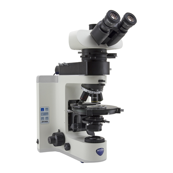
Overview Manual version DIOPTER EYEPIECE ADJUSTMENT RING PHOTO / TV PORT OBSERVATION HEAD ALC SYSTEM DIC PRISM SLOT NOSEPIECE OBJECTIVES CONTROL STAGE KEYBOARD CONDENSER CONDENSER HEIGHT ADJUSTMENT KNOB FIELD MAIN DIAPHRAGM SWITCH FOCUS LOCK LEVER INTENSITY ADJUSTMENT DIAL Page 4... - Page 5 LIGHT PATH SELECTOR LEVER FILTER HOLDER ALC SOCKET SLIDE HOLDER APERTURE DIAPHRAGM FINE FOCUS KNOB CONDENSER CENTERING SCREWS X-Y STAGE MOVEMENT KNOBS TENSION ADJUSTMENT COLLAR COARSE FOCUS KNOB Page 5...
- Page 6 Motorised version Only the parts related to the motor are highlighted; all the other components of the microscope remain unchan- ged compared to the manual version. NOSEPIECE X-AXIS MOVEMENT KNOB (MANUAL MOVEMENT) STAGE FOCUS MOTOR Page 6...
- Page 7 STAGE CONNECTION CABLE X-AXIS MOVEMENT KNOB (MANUAL MOVEMENT) NOSEPIECE ROTATION BUTTONS Page 7...
-
Page 8: Unpacking
Unpacking The microscope is housed in a moulded Styrofoam container. Remove the tape from the edge of the container and lift the top half of the container. Take some care to avoid that the opti- cal items (objectives and eyepieces) fall out and get damaged. Using both hands (one around the arm and one around the base), lift the microscope from the container and put it on a stable desk. -
Page 9: Motorised Version
Motorised version Once opened the box, the microscope parts (motorised version) are the following: ⑥ ⑤ ⑦ ① ② ③ ⑧ ④ ⑨ ⑧ ⑪ ⑩ ① Frame ⑦ ALC system (M-1030) ( Optional) ② Objectives ⑧ Microscope power supply ③... -
Page 10: Assembling The Microscope
Assembling the microscope 1. Put the microscope stand on a solid ta- ble. Insert M-1030 attachment provi- ded) above the stand, using the 2mm Al- len wrench to tighten the screw. (Fig.1) Fig. 1 2. Connect the cable of the ALC (Automa- tic Light Control) system to the socket pla- ced on the right side of the frame. - Page 11 5. Insert the condenser under the stage: position until it is well inserted into its holder (under the condenser there is a pin that must fully enter the guide of the holder). (Fig. 5) 6. Lock condenser fixing knob ①. ①...
-
Page 12: Only For Motorised Version
Only for motorised version 10. Assemble the stage in the same way as the manual version. Check the perfect alignment of the rear part of the stage with the rear arm of the frame. An imperfect alignment could lead to an incorrect functioning of the system. -
Page 13: Summary Of Brightfield Observation Procedures
8. Summary of brightfield observation procedures (Used commands) (Chapter) Bring on “1” the main switch Main switch ON-OFF key on the keyboard Turn on the microscope and adjust light intensity. Light adjustment dial Slide holder Put a slide on the stage Revolving nosepiece Insert 10X objective into the light path Focus the specimen Coarse and fine focusing knobs Observation head... -
Page 14: Use Of The Microscope
Use of the microscope General switch on To activate the transmitted light illuminator, insert the plug of the external power supply into the mains so- cket and turn the main switch ①, located on the left side of the stand, to the position “1”. (Fig. 12) ①... -
Page 15: Adjust The Observation Head
Adjust the observation head Loosen the lock-screw ①, turn the observation head to a comfortable position for observation, and then lock the lock-screw. (Fig. 15) ① Fig. 15 Adjust the interpupillary distance Hold the right and left parts of the observation head using both hands and adjust the interpupillary distan- ce by turning the two parts until one circle of light can be seen. -
Page 16: Use Of Eyeshields
Use of eyeshields • Use with eyeglasses Fold rubber eyeshields with both hands. Folded eyeshields avoid scratching the lenses of eyeglas- ses. (Fig. 18) • Use without eyeglasses Raise eyeshields and observe at the microscope placing eyes to the shields, avoiding external light to disturb the observation. -
Page 17: Stage
9.10 Stage Stage accepts standard slides 26 x 76 mm, thickness 1,2 mm with coverslide 0,17mm. (Fig. 22) It is possible to place two slides side by side on the stage. 1. Open the spring arm of the slide holder ① and place from the front the slide on the stage. -
Page 18: Use Of Oil Immersion Objective
IRIS DIAPHRAGM 70-80% 30-20% Fig. 26 FIELD OF VIEW 9.12 Use of oil immersion objective 1. Focus the specimen with a low power objective. 2. Lower the stage (remembering to lock the coarse upper limit knob). 3. Put a drop of oil (provided) on the area of the specimen to be observed. -
Page 19: Use Of Alc System
9.13 Use of ALC system 1. Adjust the desired brightness through the eyepie- ces using the light intensity dial (chapter 9.3). 2. Press the MEM key ① to store this setting (Fig. ② 28). The light on the microspoe will turn off for some seconds, the will turn on again. -
Page 20: Use Of Universal Condenser For Brightfield / Darkfield / Phase Contrast
10. Use of universal condenser for brightfield / darkfield / phase contrast Universal condenser provided with B-1000 allows observation in brightfield, darkfield and phase contrast. Fig. 32 Fig. 33 Fig. 34 Fig. 35 Fig. 36 Observation mode Condenser turret position Brightfield BF (Fig. 32) Darkfield DF (Fig. 33) Phase contrast 10x 10/20 (Fig. -
Page 21: Phase Contrast Observation (Ph)
10.3 Phase contrast observation (PH) 1. Center the condenser as already described at page 14. 2. Rotate the condenser turret to insert the “10/20” UPPER PART position OF CENTERING 3. Insert 10x objective into the light path. TELESCOPE 4. Place a specimen on the stage and focus. 5. -
Page 22: Use Of Green Filter
10.4 Use of green filter • Green filter is used to increase the contrast of the image during phase contrast observation. • Place the filter on the field lens of the microscope and begin the observation. (Fig. 41) • For brightfield or darkfield observation it advisa- ble to remove the green filter from the light path. -
Page 23: Dic Observation
11. DIC observation The microscope allows the observation in Differential Interferential Contrast (DIC) with two different methods: Koehler DIC and Nomarski DIC. The Koehler DIC method is the simplest both from the point of view of installation and from the point of view of use, while the Nomarski DIC method provides for a more complex setup. -
Page 24: Nomarski Dic Transmitted Light
Nomarski DIC transmitted light The observation in Nomarski DIC in transmitted light requires the kit consisting of the following accessori- es: Universal condenser ① (containing the dedicated ③ DIC prisms according to the objectives in use), AA- nalyzer for transmitted light ②, DIC slider ③. (Fig. ①... - Page 25 3. Remove the slide from the stage. 4. Turn the polarizer knob Ⓗ under the condenser to achieve maximum darkening of the eyepieces, and then tighten the polarizer locking screw Ⓖ. (Fig. 49) Ⓗ Ⓖ Fig. 49 5. Once the maximum darkening is found, remo- ve the slider from the nosepiece, remove the analyzer from the dummy slider and insert it into ⑥...
-
Page 26: Microphotography
12. Microphotography ② 12.1 Installing the “C” mount adapter 1. Loosen the clamping screw ① on the trinocular port and remove the dust cap ②. (Fig. 45) 2. Screw the C-mount adapter ③ to the camera ④and insert the round dovetail of the C-mount into the empty hole of the trinocular port, then tighten the clamping screw ①. -
Page 27: Maintenance
13. Maintenance To think about when and after using the microscope • The microscope should always be kept vertically when moving it and be careful so that no moving parts, such as the eyepieces, fall out. • Never mishandle or impose unnecessary force on the microscope. •... -
Page 28: Troubleshooting
14. Troubleshooting Review the information in the table below to troubleshoot operating problems. PROBLEM CAUSE SOLUTION I. Optical Section: LED operates, but field of view re- Power supply is unplugged. Connect mains dark. Brightness is too low Set brightness to a proper level The edge of the field of view is The incident illuminator is not cor- Change the angle of the incident illuminator... - Page 29 IV. Viewing tube assembly The field of view of the two eyes is The interpupillar distance is not Adjust the interpupillar distance different correct The dioptric correction is not right Adjust the dioptric correction The viewing technique is not cor- When look into the objective, do not stare rect, and the operator is straining at the specimen but look at the whole field...
-
Page 30: Equipment Disposal
Equipment disposal Art.13 Dlsg 25 July 2005 N°151. “According to directives 2002/95/EC, 2002/96/EC and 2003/108/EC relating to the reduction in the use of hazardous substances in electrical and electronic equipment and waste disposal.” The basket symbol on equipment or on its box indicates that the product at the end of its useful life should be col- lected separately from other waste. - Page 31 OPTIKA S.r.l. ® Via Rigla, 30 - 24010 Ponteranica (BG) - ITALY Tel.: +39 035.571.392 info@optikamicroscopes.com - www.optikamicroscopes.com OPTIKA Spain spain@optikamicroscopes.com OPTIKA USA usa@optikamicroscopes.com OPTIKA China china@optikamicroscopes.com OPTIKA India india@optikamicroscopes.com OPTIKA Central America camerica@optikamicroscopes.com...
- Page 32 Serie B-1000 MANUALE DI ISTRUZIONI Modello B-1000 B-1000BF B-1000PH B-1000TI-2 B-1000TI-3 B-1000TI-5 B-1000TI-10 Ver. 2.1 2019...
- Page 33 Sommario Avvertenza Simboli Informazioni sulla sicurezza Uso previsto Descrizione dello strumento Versione manuale Versione motorizzata Disimballaggio Assemblaggio Versione manuale Versione motorizzata Assemblaggio del microscopio Solo per versione motorizzata Sommario delle procedure di osservazione in campo chiaro Uso del microscopio Accensione generale Tastierino di controllo Regolazione della luminosità...
-
Page 34: Simboli
Avvertenza Questo microscopio è uno strumento scientifico di alta precisione, progettato per durare a lungo con una minima manutenzione; la realizzazione è secondo i migliori standard ottici e meccanici, per poter essere utilizzato quotidianamente. Vi ricordiamo che questo manuale contiene informazioni importanti per la sicurezza e per la manutenzione dello strumento, e deve quindi essere messo a disposizione di coloro che lo utilizzeranno. -
Page 35: Descrizione Dello Strumento
Descrizione dello strumento Versione manuale ANELLO OCULARE REGOLAZIONE DIOTTRICA USCITA FOTO/ VIDEO TESTA DI OSSERVAZIONE SISTEMA ALC ALLOGGIAMENTO PRISMA DIC REVOLVER OBIETTIVI TASTIERINO DI TAVOLINO CONTROLLO CONDENSATORE MANOPOLA REGOLAZIONE ALTEZZA CONDENSATORE DIAFRAMMA DI INTERRUTTORE CAMPO GENERALE LEVA BLOCCO DI MESSA A MANOPOLA FUOCO REGOLAZIONE... - Page 36 SELETTORE RIPARTIZIONE LUCE PORTA FILTRO CONNETTORE FERMAVETRINI DIAFRAMMA DI APERTURA MANOPOLA MICROMETRICA DI VITI DI MESSA A FUOCO CENTRAGGIO CONDENSATORE MANOPOLE TRASLAZIONE REGOLAZIONE FRIZIONE MANOPOLA MACROMETRICA DI MESSA A FUOCO Pagina 37...
-
Page 37: Versione Motorizzata
Versione motorizzata Vengono indicate solo le parti relative alle motorizzazioni; tutte le altre componenti del microscopio rimangono invariate rispetto alla versione manuale. REVOLVER MANOPOLA TRASLAZIONE ASSE X (MOVIMENTO MANUALE) TAVOLINO MOTORE DI MESSA A FUOCO Pagina 38... - Page 38 CAVO DI CONNESSIONE TAVOLINO MANOPOLA TRASLAZIONE ASSE Y (MOVIMENTO MANUALE) TASTI ROTAZIONE REVOLVER Pagina 39...
-
Page 39: Disimballaggio
Disimballaggio Il microscopio si trova in un imballaggio di polistirolo espanso stampato. Dopo aver tolto il nastro adesivo da tutti gli imballi, sollevare la metà superiore dell’imballaggio. Fare attenzione a non far cadere o danneggiare i com- ponenti ottici (obiettivi e oculari). Estrarre il microscopio dal suo imballaggio con entrambe le mani (una intorno al braccio e una intorno alla base) e appoggiarlo su un piano stabile. - Page 40 Versione motorizzata Una volta aperto l’imballo, le parti del microscopio (versione motorizzata) sono le seguenti: ⑥ ⑤ ⑦ ① ② ③ ④ ⑨ ⑧ ⑪ ⑩ ① Stativo ⑦ Sistema ALC (M-1030) (Opzionale) ② Obiettivi ⑧ Alimentatore microscopio ③ Tavolino ⑨...
-
Page 41: Assemblaggio Del Microscopio
Assemblaggio del microscopio 1. Posizionate il microscopio su un piano stabile. Inserire il dispositivo M-1030 (se fornito) al di sopra dello stativo, e fissatelo stringendo la vite con la brugola da 2 mm in dotazione. (Fig.1) Fig. 1 2. Collegare il cavo dal sistema ALC (Au- tomatic Light Control) - Page 42 5. Inserire il condensatore sotto il tavolino. Con- trollare che sia correttamente inserito nel suo al- loggiamento (sotto il condensatore si trova uno spinotto che deve entrare completamente nella guida del supporto del condensatore). (Fig. 5) 6. Serrare la vite di fissaggio del condensatore ①. ①...
-
Page 43: Solo Per Versione Motorizzata
Solo per versione motorizzata 10. Montare il tavolino allo stesso modo della ver- sione manuale. Verificare il perfetto allinea- mento della parte posteriore del tavolino con il braccio posteriore dello stativo. Un non per- fetto allineamento potrebbe portare ad un non corretto funzionamento del sistema. -
Page 44: Sommario Delle Procedure Di Osservazione In Campo Chiaro
Sommario delle procedure di osservazione in campo chiaro (Comandi usati ) (Capitolo) Portare su “1” l’interruttore generale Interruttore generale Tasto ON-OFF sul tastierino Accendere il microscopio e regolare l’intensità luminosa. Rotella regolazione intensità Fermavetrini Posizionare un preparato sul tavolino. Revolver Inserire l’obiettivo 10x nel percorso ottico Manopole macro e micrometrica di Mettere a fuoco il preparato... -
Page 45: Uso Del Microscopio
Uso del microscopio Accensione generale Per attivare l’illuminatore in luce trasmessa, inserire la spina dell’alimentatore esterno nella presa di rete ed portare l’interruttore principale ①, posto sul lato sinistro dello stativo, nella posizione “1”. (Fig. 12) ① Fig. 12 Tastierino di controllo L’illuminazione del B-1000 può... -
Page 46: Regolazione Della Testa Di Osservazione
Regolazione della testa di osservazione Allentare la vite di fissaggio ①, truotate la testa in po- sizione confortevole per l’osservazione, poi stringere la vite di fissaggio. (Fig. 15) ① Fig. 15 Regolazione della distanza interpupillare Tenere la parte destra e sinistra della testa d’osser- vazione usando entrambe le mani e regolare la di- stanza interpupillare ruotando le due parti fino ad ot- tenere la visione di un unico cerchio di luce. -
Page 47: Uso Dei Paraocchi In Gomma
Uso dei paraocchi in gomma • Uso con occhiali da vista Abbassare i paraocchi in gomma con entrambe le mani. La presenza dei paraocchi abbassati evita di graffiare le lenti degli occhiali. (Fig. 18) • Uso senza occhiali da vista Rialzare i paraocchi ed osservare al microscopio ap- poggiando gli occhi ai paraocchi, in modo da evitare che la luce esterna arrivi a disturbare l’occhio. -
Page 48: Tavolino
9.10 Tavolino Il tavolino accetta vetrini standard 26 x 76 mm, spes- sore 1,2 mm e coprioggetto 0,17mm. (Fig. 22) È possibile alloggiare due vetrini affiancati sul tavo- lino. ① 1. Allargare il braccio movibile del fermapreparati ① e posizionare frontalmente i vetrini sul tavolino. 2. -
Page 49: Uso Di Un Obiettivo Ad Immersione
DIAFRAMMA 70-80% AD IRIDE 30-20% Fig. 26 CAMPO VISIVO 9.12 Uso di un obiettivo ad immersione 1. Mettere a fuoco con un obiettivo a basso ingran- dimento. 2. Abbassare il tavolino (avendo cura di avere im- postato il blocco di messa a fuoco). 3. -
Page 50: Uso Del Sistema Alc
9.13 Uso del sistema ALC 1. Regolare la luminosità desiderata agli oculari uti- lizzando la rotellina di regolazione del microsco- pio (parag. 9.3). ② 2. Premere il tasto MEM ① (Fig. 28). La luce al mi- croscopio si spegne per qualche secondo e poi ①... -
Page 51: Condensatore Universale Per Campo Chiaro / Scuro / Contrasto Di Fase
10. Condensatore universale per campo chiaro / scuro / contrasto di fase Il condensatore universale in dotazione al modello B-1000 consente l’osservazione in campo chiaro, campo scuro e contrasto di fase. Fig. 32 Fig. 33 Fig. 34 Fig. 35 Fig. 36 Modo di osservazione Posizione torretta condensatore Campo chiaro... -
Page 52: Osservazione In Contrasto Di Fase (Ph)
10.3 Osservazione in contrasto di fase (PH) PARTE SUPERIORE 1. Centrare il condensatore come descritto a pag. DEL TELESCOPIO 2. Ruotare la torretta del condensatore per inserire la posizione “10/20”. 3. Inserire l’obiettivo 10x nel percorso ottico. 4. Posizionare un campione sul tavolino e mettere a fuoco. -
Page 53: Uso Del Filtro Verde
10.4 Uso del filtro verde • Il filtro verde viene utilizzato per aumentare il contrasto dell’immagine durante l’osservazione in contrasto di fase. • Appoggiare il filtro sulla lente di campo del micro- scopio (Fig. 41) ed iniziare l’osservazione. • Per l’osservazione in campo chiaro o in campo scuro si consiglia di rimuovere il filtro dal percor- so ottico. -
Page 54: Osservazione In Dic
11. Osservazione in DIC Il microscopio consente di effettuare l’osservazione in Contrasto Interferenziale Differenziale (DIC) con due di- verse metodiche: Koehler DIC e Nomarski DIC. La metodica Koehler DIC è la più semplice sia dal punto di vista dell’installazione sia dal punto di vista dell’uti- lizzo, mentre la metodica Nomarski DIC prevede una messa a punto più... -
Page 55: Nomarski Dic Luce Trasmessa
11.2 Nomarski DIC luce trasmessa L’osservazione in Nomarski DIC in luce trasmessa richiede il kit composto dai seguenti accessori: Con- densatore universale ① (contenente i prismi DIC ③ dedicati agli obiettivi in uso), Analizzatore per luce trasmessa ②, slitta DIC ③. (Fig. 46) ①... - Page 56 3. Rimuovere il vetrino dal tavolino. 4. Ruotare la rotella del polarizzatore Ⓗ sotto il con- densatore per ottenere il massimo oscuramento agli oculari, quindi serrare la vite di bloccaggio del polarizzatore Ⓖ. (Fig. 49) Ⓗ Ⓖ Fig. 49 5. Una volta trovato il massimo oscuramento estrar- re la slitta dal revolver, rimuovere l’analizzatore dalla slitta vuota ed inserirlo nel prisma DIC.
-
Page 57: Microfotografia
12 Microfotografia ② 12.1 Montaggio dell’adattatore passo “C” 1. Allentare la vite di bloccaggio ① sul tubo trinocu- lare e rimuovere il tappo antipolvere ②. (Fig. 53) 2. Avvitare l’adattatore passo “C” ③ alla telecame- ra ④ e installare l’attacco rotondo del passo C nel foro vuoto del tubo trinoculare, quindi riavvita- re la vite di serraggio ①. -
Page 58: Manutenzione
13 Manutenzione Prima e dopo l’utilizzo del microscopio • Tenere il microscopio sempre in posizione verticale quando lo si sposta. • Assicurarsi inoltre che le parti mobili, ad esempio gli oculari, non cadano. • Non maneggiare senza precauzioni e non adoperare inutile forza sul microscopio. •... -
Page 59: Guida Alla Risoluzione Dei Problemi
14 Guida alla risoluzione dei problemi Consultare le informazioni riportate nella tabella seguente per risolvere eventuali problemi operativi. PROBLEMA CAUSA SOLUZIONE I. Sezione Ottica: L’illuminazione è accesa ma il campo L’alimentatore è scollegato. Collegarlo visivo è scuro. La luminosità è troppo bassa Regolarla ad un livello adeguato I bordi del campo visivo sono vignet- Il revolver non è... - Page 60 III. Sezione Elettrica Il LED non si accende. Lo strumento non viene alimentato Verificare il collegamento del cavo di ali- mentazione La luminosità è insufficiente La luminosità è regolata bassa Regolare la luminosità La luce lampeggia Il cavo di alimentazione non è col- Verificare il collegamento del cavo legato bene IV.
-
Page 61: Smaltimento
Smaltimento Ai sensi dell’articolo 13 del decreto legislativo 25 luglio 2005 n°151. “Attuazione delle direttive 2002/95/CE, 2002/96/ CE e 2003/108/CE, relative alla riduzione dell’uso di sostanze pericolose nelle apparecchiature elettriche ed elet- troniche, nonché allo smaltimento dei rifiuti”. Il simbolo del cassonetto riportato sulla apparecchiatura o sulla sua confezione indica che il prodotto alla fine del- la propria vita utile deve essere raccolto separatamente degli altri rifiuti. - Page 62 OPTIKA S.r.l. ® Via Rigla, 30 - 24010 Ponteranica (BG) - ITALY Tel.: +39 035.571.392 info@optikamicroscopes.com - www.optikamicroscopes.com OPTIKA Spain spain@optikamicroscopes.com OPTIKA USA usa@optikamicroscopes.com OPTIKA China china@optikamicroscopes.com OPTIKA India india@optikamicroscopes.com OPTIKA Central America camerica@optikamicroscopes.com...
Need help?
Do you have a question about the B-1000 Series and is the answer not in the manual?
Questions and answers