Table of Contents
Advertisement
Quick Links
Advertisement
Table of Contents

Related Manuals for Owandy Radiology I-MAX 3D


Summary of Contents for Owandy Radiology I-MAX 3D
- Page 1 EN USER MANUAL I-MAX 3D NIMXEN020I June 2022...
- Page 3 @ 86 kV ref. IEC 60601-1-3 Par. 7.1 and update of inherent filtration at chapter 6 (additional filtration value was removed) Added new chapter 7 – Removable part list Added the acquisition mode selection between 97, 98 Owandy Radiology SAS...
- Page 4 3D Single Jaw, 3D Maxillary Teeth and 3D Mandibular Teeth exams 11.04.22 Update laser labels 20, 21 Update system labels Update exposure times 27,28 Update laser and sensor characteristics 20.06.22 Adjusted mistake at par. Dental impression/Dental scan Owandy Radiology SAS...
- Page 5 User Manual - Revision history NIMXEN020I THIS PAGE IS INTENTIONALLY LEFT BLANK Owandy Radiology SAS...
-
Page 7: Table Of Contents
FUNCTIONS, MODELS AND VERSIONS ................ 22 TECHNICAL CHARACTERISTICS ............23 DIMENSIONS ........................29 TUBE LOADING CURVES, ANODE HEATING AND COOLING CURVES ..... 32 PC REQUIREMENTS ....................... 34 6.3.1 PC MINIMUM CHARACTERISTICS ..............34 6.3.2 PC SUGGESTED CHARACTERISTICS .............. 35 Owandy Radiology SAS... - Page 8 User Manual - Contents NIMXEN020I SOFTWARE ........................35 I-MAX 3D – PC COMMUNICATION ................. 35 REFERENCE STANDARD ....................36 CBCT CONDITIONS OF OPERATION ................38 6.7.1 REFERENCE PLANE ..................38 CTDI INFORMATION ......................39 6.8.1 MEASURE CONDITIONS ..................39 6.8.2 MEASUREMENT PROCEDURE .................
- Page 9 15. ERROR MESSAGES ................95 16. MAINTENANCE ..................98 17. PANORAMIC IMAGE ASSESSMENT ............ 99 17.1 PROPER POSITIONING OF THE PATIENT ..............100 17.2 PATIENT POSITIONING ERRORS ................102 17.2.1 TURNED HEAD ....................102 17.2.2 TILTED HEAD ....................103 Owandy Radiology SAS...
- Page 10 17.2.4 BACKWARD ANGULATION OF THE HEAD ............. 105 17.2.5 TONGUE EFFECT ..................... 106 17.2.6 SPINE EFFECT ....................107 Manufacturer OWANDY RADIOLOGY SAS has the right to modify products or their specifications to improve performances, quality or reproducibility Product and its accessories specifications may change without advice.
-
Page 11: Introduction
I-MAX 3D is an electrical medical device and can only be used under the supervision of a physician or of highly qualified personnel, with necessary knowledge of X-ray protection. The user is liable for legal compliance in relation to the installation and operation of the device. -
Page 12: Specification Of The Intended Use
SPECIFICATION OF THE INTENDED USE 2.1 Application and medical purpose I-MAX 3D is an extra-oral dental panoramic and CBCT (aka CBVT) X-ray unit to take either two dimensional (panoramic, TMJ and sinus exams) or three dimensional radiographic exams of teeth, jaw and oral structures. -
Page 13: Applied Parts
NIMXEN020I 2.2 Applied parts During normal use, I-MAX 3D is in contact with the patient via the handle, the chin rest and bite, the temple clamp and the head strips for 3D exams, classified as Type B applied parts. Typical doses delivered to the patient during extra-oral... -
Page 14: Panoramic Mode
17.84 19.27 20.71 22.14 23.58 25.01 26.44 27.88 29.31 30.74 32.18 33.03 33.76 34.39 19.63 21.20 22.78 24.36 25.93 27.51 29.09 30.66 32.24 33.82 35.39 36.34 37.13 37.83 12.5 22.30 24.09 25.89 27.68 29.47 31.26 33.05 34.84 36.64 38.43 40.22 41.29 42.20 42.99 Owandy Radiology SAS... - Page 15 PANORAMIC EXAM in the table below: Exam Ratio 0.55 Half-panoramic 0.85 Low Dose 0.90 Ortho Rad panoramic 0.33 Frontal dentition Bitewing L or R 0.24 0.47 Bitewing L and R 0.71 0.65 Sinus Owandy Radiology SAS...
-
Page 16: Mode
9.25 9.94 10.63 11.32 12.01 12.70 13.39 14.08 14.77 15.46 16.15 16.85 17.54 The air kerma for TMJs 3D exams can be calculated using the ratio vs 3D mode in the table below: Exam Ratio TMJ 3D Owandy Radiology SAS... -
Page 17: Safety Information
SAFETY INFORMATION Warning Please read this chapter thoroughly. Owandy Radiology designs and manufactures its devices in compliance with safety requirements; furthermore, it supplies all information necessary for correct use, and warnings related to dangers associated with X-ray generating units. Owandy Radiology cannot be held liable for: •... -
Page 18: Warnings
Before performing any maintenance operation, disconnect the unit from the power supply. I-MAX 3D is an electric medical device and so can only be used under the supervision of suitably qualified medical personnel, with necessary knowledge of X-ray protection. -
Page 19: Precautions While Using Laser Centring Devices
In reference to this directive, the lasers present on the I-Max 3D are parts of class 1. A warning label (See picture below) is affixed to I-Max 3D to indicate a laser in class 1 is mounted internally and caution is advised. -
Page 20: Protection Against Radiation
2 m both from the X-ray source and from the patient, as shown in the following figure. Minimum distance from X-ray source 2 m Protected area Figure 1 Owandy Radiology SAS... -
Page 21: Pediatric Use: Summary
HTTPS://WWW.IAEA.ORG/RESOURCES/RPOP/RESOURCES/TRAINING-MATERIAL#3 Device specific features and instructions 3.2.1.3 I-MAX 3D provides as standard with all units, the following specific design features and instructions that enable safer use of our device with pediatric patients: Design features important to pediatric imaging Paragraph Adult/Child exam modality: child selection adapts exposure current (mA) 9.4 and 10.1.1... -
Page 22: Information About Electromagnetic Compatibility
I-MAX 3D should not be used adjacent to or stacked with other equipment; if adjacent use is necessary, I-MAX 3D has to be observed to verify if it operates in a normal way. Interference may occur in the vicinity of equipment marked with the symbol... -
Page 23: Electromagnetic Emissions
3.3.1 Electromagnetic emissions In accordance with the IEC 60601-1-2 Ed4 standard, I-MAX 3D is suitable for use in the electromagnetic environment specified below. The customer or user of the system must ensure that it is used in the said environment. -
Page 24: Electromagnetic Immunity
3.3.2 Electromagnetic immunity In accordance with the IEC 60601-1-2 Ed4 standard, I-MAX 3D is suitable for use in the electromagnetic environment specified below. The customer or user of the system must ensure that it is used in the said environment. -
Page 25: Cybersecurity Measures
Like all computer-based systems, I-MAX 3D might be exposed to Cybersecurity threats. I-MAX 3D is equipped with hardware provisions that make sure that no unwanted X-ray exposure, laser radiation or motorized movements can be activated even in case of cyber-attack or software failure. -
Page 26: Environmental Risks And Disposal
Illegal disposal of the product by the owner of the equipment will result in administrative sanctions, as provided for by applicable regulations. Owandy Radiology SAS... -
Page 27: Symbols Used
User Manual – Safety information NIMXEN020I 3.6 Symbols used In this manual and on I-MAX 3D itself, apart from the symbols indicated on the keyboard, the following icons are also used: Symbols Description Device with type B applied parts Some parts of the device contain materials and liquids that, at the end of the unit's lifecycle, must be disposed of at appropriate disposal centres. - Page 28 Directives Exposure enabled status (the corresponding green LED is on) X-Ray emission (the corresponding yellow LED is on) Electronic instructions for use symbol for medical devices, according to EN ISO 15223-1: 2016 Emergency Button identification Owandy Radiology SAS...
-
Page 29: Cleaning And Disinfection
2% glutaraldehyde. Note To ensure a greater level of hygiene the handles of the equipment are covered with a special antibacterial paint which, thanks to the emission of silver ions, reduces the development of micro-organisms. Owandy Radiology SAS... -
Page 30: Description
User Manual – Description NIMXEN020I DESCRIPTION 5.1 Identification labels and laser labels 5.1.1 Position of Identification labels Owandy Radiology SAS... -
Page 31: Warning And Caution Labels
User Manual – Description NIMXEN020I 5.1.2 Warning and caution labels Laser symbol label Laser WARNING label WARNING label Owandy Radiology SAS... -
Page 32: Functions, Models And Versions
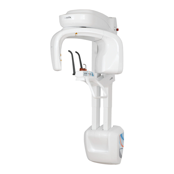
User Manual – Description NIMXEN020I 5.2 Functions, models and versions I-MAX 3D, manufactured by Owandy Radiology, is a complete panoramic X-ray system that can perform the following exams: • Panoramic adult or child exams, with 3 sizes and 3 types of biting for a total of 18 combinations with automatic selection;... -
Page 33: Technical Characteristics
User Manual – Technical characteristics NIMXEN020I TECHNICAL CHARACTERISTICS General features Type I-MAX 3D Manufacturer Owandy Radiology Class Class I with type B applied parts according to IEC 60601-1 classification. Protection degree IPX0 standard device Line voltage 99-132V 198-264 V Rated line voltage... - Page 34 (*) In case the 80x80 limitation is set, the values will change to: 3D Full Dentition 80 mm x 80 mm (Diameter x Height); 3D Single Jaw (Mandibular and Maxillary) 80 mm x 50 mm (Diameter x Height) Owandy Radiology SAS...
- Page 35 The declared image magnification value is valid after proper software calibration. Note I-MAX 3D is based on a standard dentition and ascending rami shape. This shape, based on statistical studies, establishes a form for the dentomaxillofacial complex, adopted as "standard".
- Page 36 See Figure 2 Cooling By convection Leakage radiation at 1 m < 0.5 mGy/h @ 86 kV - 12.5 mA - 3s duty cycle 1/16 Tube-head maximum thermal capacity 310kJ Figure 2: Tube-head target angle (view from the bottom) Owandy Radiology SAS...
- Page 37 Optical power on the working surface < 0.39 mW Laser class Class 1 laser product according to IEC standard 60825-1:2014 3D Digital sensor Detector type CMOS flat panel Sensitive Area (H x L) 139.2 : 144.0 x 118.6 : 119.6 mm Owandy Radiology SAS...
- Page 38 < 95% without condensation Minimum atmospheric pressure for transport 630 hPa and storage Note The handles of the equipment are covered with a special antibacterial paint which, thanks to the emission of silver ions, reduces the development of micro-organisms. Owandy Radiology SAS...
-
Page 39: Dimensions
User Manual – Technical characteristics NIMXEN020I 6.1 Dimensions Figure 3: I-MAX 3D dimensions - Wall mounted version Owandy Radiology SAS... - Page 40 User Manual – Technical characteristics NIMXEN020I 797 (31.4") 400 (15.8") 100 (3.9") 953 (37.5") Figure 4: I-MAX 3D dimensions - Wall mounted with floor support version Owandy Radiology SAS...
- Page 41 User Manual – Technical characteristics NIMXEN020I 962 (37.9") 790 (31.1") 996 (39.2") 953 (37.5") Figure 5: I-MAX 3D dimensions - Floor mounted version Owandy Radiology SAS...
-
Page 42: Tube Loading Curves, Anode Heating And Cooling Curves
User Manual – Technical characteristics NIMXEN020I Tube loading curves, anode heating and cooling curves Tube "CEI OPX 105-12" (0.5 IEC 336) Tube loading curves Anode heating and cooling curves Owandy Radiology SAS... - Page 43 User Manual – Technical characteristics NIMXEN020I Tube head cooling curve Owandy Radiology SAS...
-
Page 44: Pc Requirements
PC to be used with the machine must comply with the standard IEC 60950-1:2005. Note on Monitor characteristics: the PC and the monitor are not supplied with the equipment. In order to properly view images taken with I-MAX 3D, the PC monitor must have the following minimum characteristics: •... -
Page 45: Pc Suggested Characteristics
968 kB/slice. 6.4 I-MAX 3D – PC communication The communication between I-MAX 3D and computer is carried out with a LAN connection based on a TCP/IP protocol and a point to point connection between the detector and computer using an UDP protocol on a gigabit Ethernet. -
Page 46: Reference Standard
User Manual – Technical characteristics NIMXEN020I 6.5 Reference standard Medical electrical equipment for extra-oral dental radiography I-MAX 3D complies with: IEC 60601 1: 2005 (3rd ed.) Medical electrical equipment - Part 1: General requirements for basic safety and essential performance IEC 60601 1: 2005 (3rd ed.) + Am1:2012... - Page 47 2011/65/EU, 2006/42/EC. Classifications I-MAX 3D is an electrical medical X-ray device classified as class I type B according to 60601-1, with continuous operation at an intermittent load. According to 93/42/EEC Medical Devices Directive, the equipment is classified as class II B.
-
Page 48: Cbct Conditions Of Operation
The Figure 6 shows the position of the reference plane and its location with respect to the chin rest, the focal spot and the irradiated volume by the X-ray cone beam. Each exam has a proper chin support that gives the proper reference plane offset. Figure 6 Owandy Radiology SAS... -
Page 49: Ctdi Information
The dose detector is placed in the phantom in one of the positions at a time. The default values for adult and normal size (84 kV – 5 mA) are selected. An exposure is performed. The dose measure is recorded. Owandy Radiology SAS... -
Page 50: Measured Values
(84 kV). CDTI Value Relative to Conditions of Operation CDTI 100,PERIPHERAL,MAX 60 kV (minimum value) 0.93 86 kV (maximum value) 1.00 Maximum deviation from the nominal values given in the preceding tables is ± 25%. Owandy Radiology SAS... -
Page 51: Dose Profile
User Manual – Technical characteristics NIMXEN020I 6.7.4 Dose profile In the following graph the dose profile is displayed along a line z perpendicular to the tomographic plane measured in the center to the Dose Phantom. dose Owandy Radiology SAS... -
Page 52: Removable Part List
"▼" on the front of the chin support itself. Specific positioner which allows to perform the TMJ Positioner open/closed mouth TMJ exam. Dedicated chin support Maxillary-Sinus Chin Support ensuring a perfect coverage of the Maxillary Sinus area. Owandy Radiology SAS... -
Page 53: Quality Assurance Program
QuickVision installation media: a shortcut on the desktop will be created • kV meter (NOT provided by Owandy Radiology SAS): used to measure exposure parameters. All the tools are provided with the unit, except kV meter. The 3D quality phantom is provided as standard with 110-120V units. -
Page 54: Functioning Of The Indicator Lights
At the end of the check, switch OFF the unit. Laser correctly aligned Laser misaligned Figure 9 In case the test fails, repeat it checking that there is no mechanical interference. If misalignment is still present, call technical assistance. Owandy Radiology SAS... -
Page 55: Panoramic Image Quality Check
Open QuickVision software and open the patient "Quality Test". If not present, create a new patient (Name: "Quality"; Family name: "Test"). Select the "Mouth" icon. From the "ACQ" toolbar, select the GUI icon to open the virtual keyboard. Owandy Radiology SAS... - Page 56 Mount the centering tool on the support plate and place it on the chin rest support. Centering tool Support plate Figure 10: Support plate and centering tool positioning On the main menu of the virtual interface, select "Test" exam, the following image will be displayed: Owandy Radiology SAS...
- Page 57 Select the "Ruler" icon and measure the distance between the two external spheres; this value must be 173mm ± 2mm. In case the test fails, call technical assistance. 10. Record the tests results in the log book at paragraph 8.4.1. Owandy Radiology SAS...
-
Page 58: Log Book
User Manual – Quality assurance program NIMXEN020I 8.4.1 Log book Dimension Symmetry 2 mm Acceptance range-> 171 - 175 mm Date Measured value Measured value Owandy Radiology SAS... -
Page 59: Image Quality Check
In the first page of the QC tool the user can configure the test, by specifying the following information: • Device • Phantom used • Tester information • Practitioner information • Type of test (Acceptance/Constancy) • Local regulation. Once the test is set up, it can be started by clicking on "Start test". Owandy Radiology SAS... - Page 60 "Export test report". The report will be exported in two file formats: .pdf and .csv. Record the measurements in the logbook provided at paragraph 8.5.12. Note In case you find any value out of the acceptable range, please call your service representative for a system inspection. Owandy Radiology SAS...
-
Page 61: Test Image Acquisition
From the virtual keyboard, click on the "Setting" icon and verify that the "apply anti-artefact filter" is disabled. Figure 12 On the main menu of the virtual interface, select "Test" exam, the following image will be displayed Select "3D" exam. Owandy Radiology SAS... - Page 62 3D Quality phantom Support plate Figure 13 On the PVC insertion is present a position reference; this reference must be positioned towards the keyboard side. Position reference Figure 14 Owandy Radiology SAS...
-
Page 63: Nyquist Frequency
"Image noise" cell of the QC log book at paragraph 8.5.12 • Contrast: verify that the displayed value is greater or equal to 400. Report this value in the "Low contrast resolution" cell of the QC log book at paragraph 8.5.12. Owandy Radiology SAS... -
Page 64: Spatial Resolution
(5 - Figure 11). Verify that the Slice thickness value displayed is in the range from 15.3mm to 18.7mm (nominal 17.0mm). Report these values in the "Slice thickness" cell of the QC log book at paragraph 8.5.12. Owandy Radiology SAS... -
Page 65: Homogeneity
In the last section an index is displayed summarizing the quality of the 3D image with respect to the given dose. It is computed starting from the quality parameters previously measured: Contrast to Noise Ratio, and Dose at the isocentre. The index is expressed in 1/(mGy•cm Owandy Radiology SAS... -
Page 66: Log Book
User Manual – Quality assurance program NIMXEN020I 8.5.12 Log book Owandy Radiology SAS... -
Page 67: Dosimetry Test (Paragraph For Authorised Personnel)
In the "Sensor centring parameters" panel set the following exposure parameters: 60 kV, 2 mA, 3 s. Press the X-ray button to take an exposure and verify that the measured values are in the acceptance limits listed in the Table at point 6. Owandy Radiology SAS... - Page 68 If the values are still out of range, perform the test using the invasive method as described in the Service Manual • If the values are still out of range, call technical assistance. Record the test results in the log book at paragraph 7.6.1. Owandy Radiology SAS...
-
Page 69: Log Book
User Manual – Quality assurance program NIMXEN020I 8.6.1 Log book Parameter set -> 60 kV, 2mA, 3s 86 kV, 12.5 mA, 3s Acceptance range-> 55.2-64.8 kV 2.85-3.15 s 79.1-92.8 kV 2.85-3.15 s Date kV measured Time measured kV measured Time measured Owandy Radiology SAS... -
Page 70: General Instructions For Use
5. Place the proper chin rest relating to the current exam selection. If the chin rest is correct, the white LED will become steady ON, otherwise it will blink quickly. 6. Position the patient. 7. Press ">O<" button to set up the equipment in the start exam position; the green LED lights Owandy Radiology SAS... -
Page 71: Emergency Button
The emergency button only stops the vertical column movement. In case of an emergency column situation, press the emergency button to stop the movement. If columns don't move, check that the emergency button is not pressed; rotate the button to release it. Owandy Radiology SAS... -
Page 72: Positioning The Chin Support
User Manual – General instructions for use NIMXEN020I Positioning the chin support I-MAX 3D is equipped with different types of supports: • a standard chin rest fitted with a bite or a removable appendix for edentulous patients • a tiniest chin rest fitted with a bite or a removable appendix for edentulous patients •... - Page 73 3D Single Jaw - Maxillary 3D Maxillary Teeth - Frontal / Thin chin support Premolar / Molar 2D Sinus 3D Sinus Thin chin support with 3D TMJ edentulous patients appendix 2D TMJ C/O 2D TMJ Single phase TMJ positioner Owandy Radiology SAS...
- Page 74 User Manual – General instructions for use NIMXEN020I Temple clamps must be always used to block the patient's head. For 3D exams, the head strip shall also be used. Temple clamps Head strip for 3D exams Owandy Radiology SAS...
-
Page 75: Keyboard - Description And Functions
User Manual – General instruction for use NIMXEN020I Keyboard - Description and functions Figure 17 shows a general view of I-MAX 3D control Interface. Figure 17 - Keyboard Label Description The up/down movement of the column is controlled by the corresponding keys. - Page 76 Temple clamps closing/release knob. Chin rest control LED: • White fixed, the chin rest is correct for the selected exam • White blinking, the chin rest is not present or not correct for the selected exam Owandy Radiology SAS...
-
Page 77: Graphical User Interface - Description And Functions
Test mode may also be useful when the equipment is used on child patients, to show them how the equipment works before running the exam. Owandy Radiology SAS... - Page 78 Panoramic, Bitewing, TMJ and Sinus. Second selection: define the exam modality. Exam type selection is done in three steps for 3D exams: First selection: select the 3D program. Second selection: select the appropriate F.OV. Third selection: define the anatomical region. Owandy Radiology SAS...
-
Page 79: Main Gui Area Functions
"3" exposure parameters. Selecting the area indicated in the red circle, it is possible to see all the available exams. Clicking the area indicated in the red circle it is possible to reduce the Exam selection area. Owandy Radiology SAS... -
Page 80: Digital Sensor
I-MAX 3D is equipped with a CMOS flat panel: suitable for 2D and 3D imaging. I-MAX 3D control system checks the consistency of safety measures that allow for correct use of the digital sensor; in particular to prevent acquisition when the image management and processing system is not ready to receive the image, it displays the message "Sensor not ready". -
Page 81: Making An Exam
X-rays. Warning Since the X-ray button is a "dead man's switch", its release before the end of the exposure, immediately stops the X-ray emission and the arm rotation. Error 362 or Error 760 will be displayed. Owandy Radiology SAS... - Page 82 (Figure 1). Note I-MAX 3D assumes that the digital sensor is ready: if this is not the case, the blue light indicator of "Computer connection" status (5 - Figure 17) start blinking slowly. To reset the message on I-MAX 3D, press "OK" on the GUI and follow the instruction provided (if on the equipment keyboard the green light indicator of "Machine Ready"...
-
Page 83: Anatomic / Manual Exposure
MANUAL: with the possibility to vary the kV and mA values already set. Note The exam parameters set as the default are values to be taken as the starting point. Users can optimise the parameters according to their needs. Owandy Radiology SAS... -
Page 84: Anatomic Exposure
Default exposure values in 2D TMJ mode Adult Patient Child Patient (10,6 seconds) (10,6 seconds) Small Medium Large Exposure values in 3D Full Dentition, 3D Extended Volume and 3D Airways Adult Patient Child Patient (7 seconds) (7 seconds) Small Medium Large Owandy Radiology SAS... - Page 85 Users can optimise the parameters according to their needs. Note The type of biting does not affect the kV and mA values, but it affects the position of the focus layer, by adapting rotation movement to the patient's anatomy. Owandy Radiology SAS...
-
Page 86: Manual Exposure
A parameter can be modified by pressing the increase key and the decrease key of the parameter repeatedly. The kV value can vary between 60 and 86 kV, with 2 kV steps. The mA value can vary between 2 and 12.5 mA according to the R20 scale. Owandy Radiology SAS... -
Page 87: Image Processing Window
The Image Processing menu, if activated, will be displayed at the end of the acquisition in order to customize the default image post-processing settings. The feature can be either enabled or disabled through the corresponding option available under Settings. Figure 19 Owandy Radiology SAS... - Page 88 The button Save will apply the current setting to the corresponding button and will set the filter as default in acquisition (Figure 21). The button 4 is set as default to load the original image (without post-processing) and it can be fully customized as above described. Figure 21 Owandy Radiology SAS...
-
Page 89: D Exams
Users can optimise the parameters according to their needs. Note The new I-MAX 3D is based on a standard dentition and ascending rami shape. This shape, based on statistical studies, establishes a form for the dentomaxillofacial complex, adopted as "standard". I-MAX 3D follows a rototranslation path which maintains the magnification factor as stated in the Technical Characteristics of each type of exam as constant along this "standard"... - Page 90 In particular, in accordance with IEC 60601-2-63, the maximum deviation (including the correction according to the above curve and instrumental doubt) is within ±10% for the kV, while for the tube current it is within ±15%. Owandy Radiology SAS...
-
Page 91: Standard Panoramic
Panoramic exam, reducing the rays' emission time. This exam is used, for instance, during treatment continuation phases or where a lack of pathologies of the same joint is already known. Follow the instructions for normal panoramic exams for patient positioning. Owandy Radiology SAS... -
Page 92: Ortho Rad Dentition
This exam is normally used when it is already known that the patient has a problem on the bite-sectors of the arch, so it is possible to reduce patient irradiation. Follow the instructions for normal panoramic for patient positioning. Owandy Radiology SAS... -
Page 93: Tmj C/O
A single acquisition is made to obtain 2 images representing the right and left condyle of the temporo-mandibular arch (TMJ) with closed mouth or open mouth. RIGHT condyle LEFT condyle exposure exposure Figure 23: TMJ Single phase 12.10 Sinus The image is taken on the maxillary sinus area. Owandy Radiology SAS... -
Page 94: D Exams
80x50 mm FOV. Two acquisition modes are available: Standard definition (175 µm voxel size) for a standard resolution 3D acquisition, and High definition (87.5 µm voxel size) for the enhancement of the finest details. Owandy Radiology SAS... -
Page 95: Maxillary Teeth
Warning The portion of image volume in the "extended" area is reconstructed using a partial set of projections. Compared to the central part of the volume, such image portion can have lower definition in the anatomical details. Owandy Radiology SAS... -
Page 96: Dental Impression / Dental Model Scan
1. Cast scan (Denture) 2. Dental impression (Silicone) 3. Plaster model To perform the scan program, place the support plate on the chin rest and place the dental impression/dental model in the centre of the plate: Owandy Radiology SAS... -
Page 97: Metal Artefact Reduction Filter (Mar)
The same filter may be also applied/removed retroactively at any time through the option “new reconstruction” available for exams already archived to the database. Note MAR-processed images should be always compared with the original unprocessed images. Non-processed image MAR-processed image Figure 25 Owandy Radiology SAS... -
Page 98: New Reconstruction
To run a new reconstruction of a 3D study select its icon inside the exam list of the patient folder and right click on it. Then select "New Reconstruction". Figure 26 The following window will be displayed: Figure 27 Owandy Radiology SAS... - Page 99 MAR label as shown in figure: Figure 28 Note After having run a first reconstruction, if a further reconstruction is launched with or without the option as in Figure 27, the new 3D study will overwrite the previous reconstructed study. Owandy Radiology SAS...
-
Page 100: Patient Positioning
Warning During the patient positioning, make sure the equipment cannot collide with any object in the room. Note If columns don't move, check that the emergency button is not pressed. Rotate the button to release it. Owandy Radiology SAS... - Page 101 - external auditory meatus - with the bottom edge of the orbital fossa 47 - Ala-tragus line: plane that identifies a line that ideally connects the anterior nasal spine and the centre of the external auditory meatus. Figure 29: Reference lines Owandy Radiology SAS...
- Page 102 Figure 30: 2D Panoramic / 3D Full Dentition patient positioning Description Label Description Label Sagittal medial line Temple clamps closing/release knob Frankfurt line Laser knob Tiniest chin rest Figure 31: 2D Sinus / 3D Sinus / 3D TMJ / 3D Maxillary patient positioning Owandy Radiology SAS...
- Page 103 Frankfurt line Laser knob TMJ positioner Figure 32: 2D TMJ closed mouth patient positioning Description Label Description Label Sagittal medial line Temple clamps closing/release knob TMJ positioner Laser knob Figure 33: 2D TMJ open mouth patient positioning Owandy Radiology SAS...
-
Page 104: Exams
Mid sagittal plane must be centered and vertical. • Camper plane (ala-tragus plane) must be horizontal. • Patient’s incisors must be positioned into the reference notch of the bite. • Patient must stay motionless during the examination. Owandy Radiology SAS... -
Page 105: Error Messages
Zero position optical sensor never activated Timeout on movement Hardware key board (U.I.C.) Code Error description 270 / 271 Hardware key fault X-Ray Controls Code Error description RX button pressed on start-up or before exam RX button released during emission Owandy Radiology SAS... - Page 106 One or more keycodes are pressed Button >O< pressed during movements DSPU Code Error description 1003 DSPU hardware error detected MCU – DSPU communication error 1004 1005 CAN Bus hardware error 1007 Flat panel temperature too high Owandy Radiology SAS...
- Page 107 Image buffer allocation failure PC driver interface (OSP / VSP) Code Error description 1401 3D sensor frame lost during exam 1402 3D sensor configuration failure 1403 Software watchdog error 1404 3D sensor does not detect X-rays during exam Owandy Radiology SAS...
-
Page 108: Maintenance
Besides the above controls, the Service Engineer will also check the following during preventive maintenance: Frequency Type of check Annually Correct equipment centring Annually Check technical factors Annually Perform sensor calibration Annually Check that the fixing screws are tightened Owandy Radiology SAS... -
Page 109: Panoramic Image Assessment
Figure 34 Anatomic structure Palatal plane Maxillary sinus Maxilla and maxillary tuberosity Temporo mandibular condyle Ascending ramus of the TMJ Coronoid process (overlap with maxilla) Mandibular canal Chin foramen Anterior nasal spine Nasal cavities Ioid bone (normally duplicated) Owandy Radiology SAS... -
Page 110: Proper Positioning Of The Patient
6. Spine should be well stretched, this is normally obtained by asking the patient to step forward, making sure that all other conditions are unchanged. If not properly extended, the spine will cause the appearing of a lower exposed area (clearer) in the front part of the image. Owandy Radiology SAS... - Page 111 Any flaring of dentition may not allow crowns and apices of both arches to fit in the image layer at the same time. For these patients, you must purposely move him/her further forward in order to move the apices into the image layer. Owandy Radiology SAS...
-
Page 112: Patient Positioning Errors
Problem The patient's head is turned to one side (left or right) in the mid-sagittal plane. Effects Condyles are different in size. The ramous on one side is much wider that the other one. Asymmetric spine compensation. Owandy Radiology SAS... -
Page 113: Tilted Head
User Manual – Panoramic image assessment NIMXEN020I 17.2.2 Tilted head Figure 38 Problem The patient's head is tilted to one side. Effects One condyle appears higher than the other one and the inferior border of the mandible is slanting. Owandy Radiology SAS... -
Page 114: Downward Angulation Of The Head
The shadow of the hyoid bone is typically superimposed on the anterior mandible. Condyles may be cut off at the top of the radiograph. Pre-molars are severely overlapped. Severe curvature of the occlusal plane. Owandy Radiology SAS... -
Page 115: Backward Angulation Of The Head
The hard palate is superimposed over the apices of the maxillary teeth. Both condyles may be off the edges of the image area. The upper incisors can be blurred. Flattening of the occlusal plane. Owandy Radiology SAS... -
Page 116: Tongue Effect
The patient’s tongue was not held closely to the roof of the mouth during the exposure. Effects A dark air space between the dorsum of the tongue hard soft palates (palatoglossal air spaces) obscures the apical region of the maxillary teeth. Owandy Radiology SAS... -
Page 117: Spine Effect
User Manual – Panoramic image assessment NIMXEN020I 17.2.6 Spine effect Figure 42 Problem The patient is slumped. Effects The spinal column isn't well stretched causing a ghost image of the spine superimposed in the centre of the image. Owandy Radiology SAS... - Page 118 User Manual NIMXEN020I MAINTENANCE LOGBOOK Installation: Date Technician Maintenance: Date Technician Cause Maintenance: Date Technician Cause Maintenance: Date Technician Cause Maintenance: Date Technician Cause Maintenance: Date Technician Cause Maintenance: Date Technician Cause Maintenance: Date Technician Cause Owandy Radiology SAS...
- Page 119 User Manual NIMXEN020G Owandy Radiology SAS...
Need help?
Do you have a question about the I-MAX 3D and is the answer not in the manual?
Questions and answers