Table of Contents
Advertisement
Quick Links
Advertisement
Table of Contents

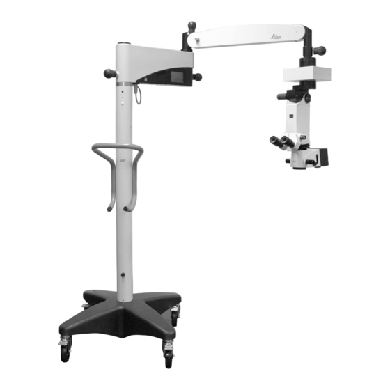
Summary of Contents for Leica Microsystems M620 F20
- Page 1 Leica M620 F20 User Manual 10 714 371 - Version 01...
- Page 2 Leica surgical microscope in an optimum way. For valuable information about Leica Microsystems products and services and the address of your nearest Leica representative, please visit our website, www.leica-microsystems.com...
-
Page 3: Table Of Contents
Chapter overview 1 Introduction 2 Product identification 3 Safety notes 4 Design 5 Functions 6 Controls 7 Preparation before surgery 8 Operation 9 Components and accessories 10 Care and maintenance 11 Disposal 12 What to do if ...? 13 Technical data Leica M620 F20 / Ref. - Page 4 Contents Introduction Operation About this user manual/installation manual .....3 Positioning the microscope ........26 1.2 Symbols in this user manual ........3 8.2 Adjusting the focus ..........27 8.3 Adjusting the magnification ........27 Product identification 8.4 Adjusting the illumination ........27 Optional product features .........3 8.5 Switching from main to spare illumination .....28 8.6 Setting the illumination type and Safety notes...
-
Page 5: Introduction
Introduction Introduction Product identification The model and serial numbers of your product are located on the About this user manual/ identification label on the underside of the horizontal arm. installation manual Enter this data in your user manual and always refer to it when you contact us or the service workshop regarding any questions In this user manual the surgical microscope Leica M620 F12 is you may have. -
Page 6: Safety Notes
• The power cord must be mechanically secured with the "Power Input" socket to prevend accidental disconnection. • All parts of the Leica M620 F20 surgical microscope shall not be serviced or maintained while in use with a patient. • Lamps shall not be changed while in use with a patient. -
Page 7: Directions For The Operator Of The Instrument
• Operate the system only with all equipment in its proper closer than 30 cm (12 inches) to any part of the Leica M620 F20, position (all covers fitted, doors closed). - Page 8 • Other accessories, provided that these have been expressly necessary for the surgery. Infants, patients with aphakia (whose approved by Leica Microsystems as being technically safe in eyelens has not been replaced by an artificial lens with a UV this context.
-
Page 9: Dangers Of Use
Safety notes Dangers of use CAUTION Surgical microscope can move without warning! WARNING Always secure the footbrakes when you are not moving Risk of injury from downward movement of surgical the system. microscope! Never change the accessories or attempt to rebalance CAUTION the microscope while it is above the field of operation. - Page 10 Safety notes WARNING Danger of fatal electric shock! Disconnect the power cable from the instrument power socket before changing fuses. CAUTION Risk of burns! The lamp and illumination mount get very hot. Check that the lamp and illumination mount have cooled before you remove the lamp.
-
Page 11: Signs And Labels
Safety notes Signs and labels Leica M620 F20 / Ref. 10 714 371 / Version 01... - Page 12 Safety notes ANVISA Registration Grounding label number (only Brazil) INMETRO label (only Brazil) OCP 0004 Transport position (F20 floor stand) Mandatory label - read the user manual carefully before operating the System weight label product. (F20) Web address for 270 KG electronic version of the user manual.
-
Page 13: Design
Design Design Functions Illumination The illumination of the surgical microscope Leica M620 is a Halogen light system with 2 bulbs (1), where the second is only a spare. Both bulbs are located in the optics carrier. In case of a failure of the lamp in use, the other lamp can be selected with the slide button (3) for quick-change lamp mount (2). -
Page 14: Balancing System
Functions Balancing System Footbrakes With a balanced surgical microscope Leica M620 F20 you can move Footbrakes are attached to each of the four wheels on the stand. the optics carrier in any position without tilting or falling down. The wheel is engaged and released with the footbrake engage/ After balancing, all movements during operation only need a minor release lever (1). -
Page 15: Controls
Controls Controls Lamp housing Control unit 1 Main switch 2 Control Panel 1 Lamp replacement cover 2 Slide button for quick-change lamp mount Tilt head/focus unit 3 Socket for potential equalization for connecting the Leica M620 F20 to a potential equalization device. This is part of the customer's building installation. -
Page 16: Footswitch (Standard Configuration)
Controls Footswitch (standard Stand configuration) 1 XY adjustment 2 Microscope illumination darker 3 No Function 4 Microscope illumination on/off 5 Zoom up/down 6 Focus bottom and top 7 Microscope illumination brighter See page for alternative footswitches and free programming of footswitches. 1 Articulation brakes 2 Rotary knob for balancing 3 Retaining pin... -
Page 17: Optics Carrier
Controls Optics carrier User interface of the control panel For information on operating the control panel, see page "Main" menu 1 Insert for filter (UV protection filter GG475, protection filter 5×) 2 Jalousie blind rotary knob for fade out/fade in 6° illumination 3 Handles 1 Illumination lighter/darker 2 Zoom up/down... -
Page 18: Preparation Before Surgery
Preparation before surgery Preparation before surgery Transport the surgical microscope and secure it in its installation location Transporting the surgical Pull the power plug from the socket and wrap the power cable around the handle. microscope Hook in the footswitch to the clip. Step on the footbrake release lever (4) to release the footbrakes. -
Page 19: Positioning The Surgical Microscope At The Operating Table
Preparation before surgery Positioning the surgical microscope at the operating table CAUTION Danger of fatal electrical shock! The surgical microscope may be connected to a grounded socket only. Carefully move the surgical microscope to the operating table Adjust the tilt of the footswitch. Rotate the footrest in or out. using the handle and position it for the forthcoming operation. -
Page 20: Installing Binocular Tube, Eyepiece And Objective
Preparation before surgery Installing binocular tube, eyepiece Fitting the objective protection glass and objective Screw the holder for the objective protection glass (2) into the objective and position it such that the mark (3) faces A range of options enables the surgical microscope to be matched backwards. -
Page 21: Fitting Adapters For Accessories
Preparation before surgery Fitting adapters for accessories Installing the beam splitter/stereo adapter Loosen the clamping screw (1). Push the beam splitter/stereo adapter into the dovetail ring. Tighten the clamping screw. Beam splitter with 50/50 % observation, Insert the filter holder (2). alternative: beam splitter with 70/30 % observation Stereo adapter for accessories For installing accessories with M600 interface (dovetail ring) under... -
Page 22: Adjusting The Second-Observer Tube
Preparation before surgery Adjusting the second-observer Fitting the adapter tube Insert the adapter in the beam splitter. Tighten the rotary ring (1). Install stereo attachment/second observer tube Insert the stereo attachment/second observer tube into the beam splitter. Tighten the rotary ring (1). Stereo attachment for second observer The dual stereo attachment can be attached to the left or right on the beam splitter and turned as required. -
Page 23: Fitting Documentation Accessories
Preparation before surgery Mount adapter with included screws (see manual of assistant's stereomicroscope). Focus assistant's image with sterile-operable focus drive (2). Moving assistant's stereomicroscope to left/right: Remove sterile handles (3) with hub. Loosen clamping screw (1) and turn the assistant's stereomicroscope. -
Page 24: Selecting Documentation Accessories
Preparation before surgery Selecting documentation accessories Zoom Video Adapter Zoom Video Adapter TV adapter TV adapter Photo/TV Photo/TV TV adapter TV adapter Photo/TV Photo/TV Zoom Video Zoom Video TV adapter TV adapter dual attachment dual attachment Adapter dual attachment dual attachment Adapter 35 mm 35 mm... -
Page 25: Adjusting The Eyebase And Eyepoint
Preparation before surgery Adjusting the eyebase and Adjusting the parfocality eyepoint Rotary ring for adjusting the diopter settings Adjusting the diopter settings Adjust the diopter settings accurately for each eye separately; only this method will ensure that the image will stay in focus throughout the entire zoom range (parfocal). -
Page 26: Changing Accessories Of The Surgical Microscope And Balancing The Swing Arm
Preparation before surgery 7.10 Changing accessories of the Mounting accessories surgical microscope and Outfit the microscope and all accessories for use. balancing the swing arm WARNING Risk of injury from downward movement of surgical microscope! Never change the accessories or attempt to rebalance the microscope while it is above the field of operation. -
Page 27: Preparing The Surgical Microscope For Use
Preparation before surgery 7.12 Preparing the surgical microscope 7.13 Checking the function of the for use lamp Switch on the power switch (2). Switch on the microscope using the power switch. The main lamp will light up. Move the slide button for the quick-change lamp mount (3) to the other side. -
Page 28: Operation
Operation Operation 7.15 Reversing + and – directions of the XY unit Positioning the microscope The "XY-reverse" function must be assigned to the Adjusting the mid position: footswitch (see page 30). Press the "Reset XY-unit" key (4). Use the "XY-reverse" button (1) on the footswitch to reverse the The XY unit will move to its middle position. -
Page 29: Adjusting The Focus
Operation Adjusting the illumination Auto reset: The "auto reset" function can be deactivated on the control (in accordance with factory settings) panel in the service area. WARNING Light that is too intense can damage the retina! Hold both handles and move the microscope to its uppermost Observe the warning messages in the "Safety Notes"... -
Page 30: Switching From Main To Spare Illumination
Operation Switching from main to spare Setting the illumination type and illumination working distance WARNING Failure of the illumination can be dangerous for the patient! If the main illuminator fails, switch to the auxiliary illuminator immediately. If a lamp is defective, the warning symbol for a defective lamp flashes on the "Main"... -
Page 31: Operating The Control Panel
Press arrow keys "+" or "–" in "Light" (1). The service area is password-protected. The brightness will change as required. Please contact your Leica Microsystems service Press arrow keys "+" or "–" in "Zoom" (2). representative. The magnification will change as desired. - Page 32 Operation Custom settings for the operating surgeon Press the "Back" (10) button. The configuration process will be canceled and the "PROGStart In the main menu under "User", press the button under which Values" menu will reappear. the settings are to be stored, 1-4, for at least 3 seconds. The field (12) indicates the user (1-4) for whom the settings are The "PROG-Start Values"...
-
Page 33: Decomissioning
Setting Auto Reset This function can be disabled in the service area. Please contact your Leica Microsystems service representative. When the "Auto Reset" function is enabled, all drives move to their reset positions and the illumination is turned off when the surgical microscope is moved to its uppermost position. -
Page 34: Components And Accessories
Components and accessories Components and accessories Observer side Main surgeon Main surgeon Assistant 2 Assistant 1 Assistant 1 Assistent 2 Assistent 1 Assistent 1 1 Inverter 7 Binocular tube 2 Laser filter 8 Eyepiece 3 Stereo adapter 9 Adapter 4 Beam splitter aœ... - Page 35 Components and accessories Item No. Image Component/accessory Description Oculus SDI • Inverter Laser filter 2 beams • Third-party product, purchased only from third parties Stereo adapter • For assembling a beam splitter Beam splitter 50/50 • The two interfaces can be used as assistant ports Beam splitter 70/30 and documentation ports.
- Page 36 Components and accessories Item No. Image Component/accessory Description Binocular tube, 180°, variable Binocular tube, 45° • Optional for use on the assistant attachment Eyepiece, 10× Eyepiece, 8.33× Eyepiece, 12.5× Leica ToricEyePiece • Facilitates adjustments to the angle of toric intraocular lenses via an integrated scale •...
-
Page 37: Patient-Side
Components and accessories Patient-side Lens Leica RUV800 Oculus BIOM Protective glass with holder aœ Stereo Assistant Microscope with adapter Leica M620 F20 / Ref. 10 714 371 / Version 01... - Page 38 Components and accessories Item No. Image Component/accessory Objective APO WD175 Objective APO WD200 Objective f = 175 mm Objective f = 200 mm Objective f = 225 mm Leica RUV800 WD175 • For observing the fundus of the Leica RUV800 WD200 patient's eye •...
-
Page 39: Video Accessories For Leica M620 F20
Components and accessories Combination options on the patient side Stereo Assistant – – – – – – – ✔ ✔ Microscope Leica RUV800 – – – – – ✔ ✔ Oculus BIOM – – – – ✔ ✔ ✔ ✔ Protective glass –... -
Page 40: Load Table
Components and accessories Load table You can find the value for the max. load in the "Technical Data" chapter, page Equipment Leica M620 F20 serial number ............ Max. load for stand from microscope interface... kg Installation Group Art. No. Description Weight Quantity Total Assistant... - Page 41 Components and accessories Installation Group Art. No. Description Weight Quantity Total Accessories for front 10448554 Leica ToricEyePiece 0.10 kg section of the eye Accessories for rear 10448555 Leica RUV800 WD175, complete 0.53 kg section of the eye 10448556 Leica RUV800 WD200, complete 10448392 Oculus SDI 0.72 kg...
-
Page 42: Care And Maintenance
Care and maintenance 10 Care and maintenance 10.2 Cleaning the control panel 10.1 Care instructions CAUTION Damage to the control panel! • Put a dust cover over the instrument during breaks in work. The light of the instrument may be harmful. Risk of eye •... -
Page 43: Care And Maintenance Of The Leica Footswitch
Care and maintenance 10.4 Care and Maintenance of the 10.6 Changing the bulb Leica Footswitch CAUTION Risk of burns! After each operation, clean the Leica footswitch in warm or hot The lamp and illumination mount get very hot. water (but below 60 °C). It is basically maintenance-free. In case of Check that the lamp and illumination mount have defects, please contact the responsible service organization. -
Page 44: Notes On Reprocessing Of Resterilizable Products
It is recommended to perform the reprocessing of a product immediately following its use. 10.8.1 General Products Preparation for cleaning Reusable products supplied by Leica Microsystems (Schweiz) AG, Remove the product from the surgical microscope. Medical Division, such as rotary knobs, objective protective glasses and capping pieces. Cleaning: manual Equipment: Running water, detergent, alcohols, microfiber cloth. - Page 45 10.8.3 Sterilization table The following table gives an overview of the available sterilizable components to the surgical micrsocopes of Leica Microsystems (Schweiz) AG, Medical Devision. Permissible sterilization methods Art.
-
Page 46: Disposal
Disposal 11 Disposal The respective applicable national laws must be observed for disposal of the products, with the involvement of corresponding disposal companies. The unit packaging is to be recycled. 12 What to do if ...? If electrically operated functions do not work properly, always check these points first: •... -
Page 47: Tv, Photography
Specimen is out of focus. The specimen is not precisely focused. Focus precisely, use graticule if necessary. If your instrument has a malfunction that is not described here, contact your Leica Microsystems representative. Leica M620 F20 / Ref. 10 714 371 / Version 01... -
Page 48: Technical Data
Technical data 13 Technical data 13.3 Stand Castors 4× Ø 100 mm 13.1 Electrical data Footbrakes 4× integrated in castors Total weight Approx. 270 kg with max. load Power socket Swing arm brakes 3 mechanical articulation brakes with F20 floor stand Central on the horizontal arm brake knob 100-240 VAC, 50/60 Hz... -
Page 49: Control Unit
Technical data 13.5 Control unit 13.7 Ambient conditions Connection sockets for +10 °C to +40 °C • Power cable +50 °F to +104 °F • Footswitch 30 % to 75 % rel. humidity • Zero point adjustment 780 mbar to 1013 mbar atmospheric pressure Display Storage... -
Page 50: Standards Fulfilled
Safety IEC 60601-1; EN 60601-1; UL60601-1; CAN/CSA C22.2 NO 60601-1-14:2014. • Electromagnetic compatibility IEC 60601-1-2; EN 60601-1-2. • The Medical Division, within Leica Microsystems (Schweiz) AG, Place the wedge (1) in front of the threshold. holds the management system certificates for the international... -
Page 51: Dimensions
Technical data 13.11 Dimensions 1145 Transport position (dimensions in mm) Leica M620 F20 / Ref. 10 714 371 / Version 01... - Page 52 Technical data max. 1444 max. 1444 608 x 608 max. 155° max. 155° 360° max. 180° max. 180° max. 180° max. 180° Leica M620 F20 (unit of measure mm) Leica M620 F20 / Ref. 10 714 371 / Version 01...
- Page 53 Leica M620 F20 / Ref. 10 714 371 / Version 01...
- Page 54 10 714 371en/01 • Copyright © by Leica Microsystems (Schweiz) AG, Medical Division, CH-9435 Heerbrugg, 2022 • 01.2022 • Subject to change. LEICA and the Leica Logo are registered trademarks of Leica Microsystems IR GmbH. Leica Microsystems (Schweiz) AG · Max Schmidheiny Strasse 201 · CH-9435 Heerbrugg T +41 71 726 3333 ·...


Need help?
Do you have a question about the M620 F20 and is the answer not in the manual?
Questions and answers